. 2015 Jul 28;10(7):e0132921.
doi: 10.1371/journal.pone.0132921. eCollection 2015.

Tolerance and Cross-Tolerance following Toll-Like Receptor (TLR)-4 and -9 Activation Are Mediated by IRAK-M and Modulated by IL-7 in Murine Splenocytes

Affiliations

Tolerance and Cross-Tolerance following Toll-Like Receptor (TLR)-4 and -9 Activation Are Mediated by IRAK-M and Modulated by IL-7 in Murine Splenocytes

Mark W Julian et al. PLoS One. .

Abstract

Objective: Immune suppression during critical illness predisposes to serious infections. We sought to determine the mechanisms regulating tolerance and cross-tolerance to common pro-inflammatory danger signals in a model that recapitulates the intact in vivo immune response.

Materials and methods: Flt3-expanded splenocytes obtained from wild-type or matching IRAK-M knockout (IRAK-M-/-), C57BL/6, male mice (8-10 weeks old) were treated repeatedly or alternately with either LPS or CpGA DNA, agonists of Toll-like receptor (TLR)-4 and -9, respectively, over successive 24-hour periods. Supernatants were collected following each 24-hour period with cytokine release (ELISA) and splenocyte IRAK-M expression (Western blot) determined. Tolerance and cross-tolerance were assessed in the absence or presence of programmed death receptor (PD)-1 blocking antibody or IL-7 pre-treatment.

Main results: Splenocytes notably exhibited both tolerance and cross-tolerance to subsequent treatments with either LPS or CpGA DNA. The character of tolerance and cross-tolerance in this model was distinct following initial LPS or CpGA treatment in that TNFα and IFNγ release (not IL-10) were suppressed following LPS; whereas, initial CpGA treatment suppressed TNFα, IFNγ and IL-10 release in response to subsequent stimulation (LPS or CpGA). Tolerance and cross-tolerance were unrelated to IL-10 release or PD-1 but were attenuated in IRAK-M-/- splenocytes. IL-7 significantly suppressed IRAK-M expression and restored TNFα and IFNγ production without influencing IL-10 release.

Conclusions: In summary, acute immune tolerance and cross-tolerance in response to LPS or CpGA were distinct in that LPS selectively suppressed pro-inflammatory cytokine responses; whereas, CpGA suppressed both pro- and anti-inflammatory responses. The induction of tolerance and cross-tolerance in response to common danger signals was mechanistically unrelated to IL-10 or PD-1 but was directly influenced by IRAK-M expression. IL-7 reduced IRAK-M expression and attenuated immune tolerance induced by either LPS or CpGA, and thus may be useful for reversal of immune tolerance in the setting of critical illness.


Conflict of interest statement

Competing Interests: The authors have declared that no competing interests exist.

Figures

Fig 1
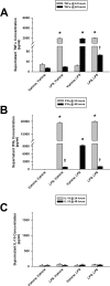
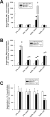
Fig 1. Repeated exposure to TLR-4 ligand, LPS, induces tolerance to splenocyte pro-inflammatory cytokine release.
The presented data was derived from at least 4 independent experiments, each in triplicate. (A) LPS (10 ng/ml) induced significant TNFα release from cultured Flt3L-expanded mouse splenocytes (1 × 10⁶ cells/ml) 24 hours post-treatment. In response to a subsequent LPS treatment, TNFα release was dramatically reduced 24 hours later. (B) Similarly, significant splenocyte IFNγ release was observed 24 hours following LPS treatment. IFNγ release was markedly decreased 24 hours after a successive LPS treatment. This release, however, was very similar to the sustained IFNγ release observed over the subsequent 24-hour period in the absence of follow-up treatment (LPS, Vehicle). (C) Splenocytes failed to demonstrate any IL-10 release in response to LPS treatment [*p < 0.01, relative to time-matched untreated (Vehicle, Vehicle) controls; p < 0.01, compared to the associated LPS treatment group at 24 hours (LPS, LPS) and the time-matched LPS treatment alone group (Vehicle, LPS), and p < 0.01, relative to the time-matched untreated (Vehicle, Vehicle) control and the associated LPS treatment group at 24 hours (LPS, Vehicle)].
Fig 2
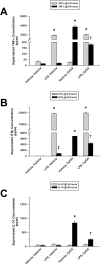
Fig 2. Tolerance to splenocyte cytokine release results from repeated exposures to TLR-9 ligand, CpGA DNA.
The presented data was derived from at least 4 independent experiments, each in triplicate. (A) 24 hours post-treatment, CpGA DNA (1 μM) significantly induced Flt3L-expanded mouse splenocyte (1 × 10⁶ cells/ml) TNFα release. This effect was notably attenuated 24 hours after subsequent treatment with CpGA DNA. (B) Likewise, splenocyte exposure to CpGA DNA yielded marked release of IFNγ by 24 hours post-treatment. Successive treatment with CpGA DNA resulted in a significant reduction in IFNγ release which was observed to be comparable to the sustained IFNγ release typically demonstrated during the subsequent 24 hours when untreated (CpGA, Vehicle). (C) Splenocyte IL-10 release in response to repeated treatments with CpGA DNA yielded a pattern similar to that observed with IFNγ release [*p < 0.01, compared to time-matched untreated (Vehicle, Vehicle) controls; p < 0.05, relative to the associated CpGA treatment group at 24 hours (CpGA, CpGA) and the time-matched CpGA treatment alone group (Vehicle, CpGA); p < 0.01, compared to the time-matched untreated (Vehicle, Vehicle) control and the associated CpGA treatment group at 24 hours (CpGA, Vehicle), and § p < 0.01, relative to the associated CpGA treatment group at 24 hours (CpGA, CpGA) only].
Fig 3
Fig 3. Cross-tolerance to splenocyte cytokine release results from Sequential exposures to LPS followed by CpGA DNA.
The presented data was derived from at least 4 independent experiments, each in triplicate. (A) Flt3L-expanded splenocyte (1 × 10⁶ cells/ml) TNFα release was dramatically diminished 24 hours following treatment with CpGA DNA (1 μM) after a prior 24-hour exposure to LPS (10 ng/ml). (B) Similarly, following a previous 24-hour exposure to LPS, splenocyte IFNγ release was significantly suppressed 24 hours after treatment with CpGA DNA. (C) Consistently, splenocyte IL-10 release demonstrated the same pattern of cross-tolerance in response to CpGA DNA at 24 hours post-treatment after an earlier 24-hour exposure to LPS [*p < 0.01, compared to time-matched untreated (Vehicle, Vehicle) controls; p < 0.05, relative to the time-matched CpGA treatment alone group (Vehicle, CpGA), and p < 0.01, compared to the time-matched untreated (Vehicle, Vehicle) control and the associated LPS treatment group at 24 hours (LPS, Vehicle)].
Fig 4
Fig 4. Sequential exposures to CpGA DNA followed by LPS lead to cross-tolerance to splenocyte cytokine release.
The presented data was derived from at least 4 independent experiments, each in triplicate. (A) Following a prior 24-hour exposure to CpGA DNA (1 μM), TNFα release from Flt3L-expanded mouse splenocytes (1 × 10⁶ cells/ml) was significantly reduced 24 hours after subsequent treatment with LPS (10 ng/ml). (B) Likewise, splenocyte IFNγ release was markedly diminished 24 hours after treatment with LPS following a previous 24-hour exposure to CpGA DNA. (C) Surprisingly, splenocyte IL-10 release was significantly and notably augmented in response to LPS at 24 hours post-treatment after an earlier 24-hour exposure to CpGA DNA [*p < 0.01, relative to time-matched untreated (Vehicle, Vehicle) controls; p < 0.05, compared to the time-matched LPS treatment alone group (Vehicle, LPS), and p < 0.01, relative to the time-matched untreated (Vehicle, Vehicle) control and the associated CpGA treatment group at 24 hours (CpGA, Vehicle)].
Fig 5
Fig 5. Splenocyte membrane expressions of both PD-1 and PD-L1 increase following TLR-4/TLR-9 stimulation.
The presented data was derived from at least 3 independent experiments. Data represents the percentage of grouped splenocyte populations demonstrating both PD-1 and PD-L1 membrane expression by flow cytometry analysis 48 hours after successive 24-hour exposures to the ligand treatment combinations detailed on the X-axis. (A) Representative flow cytometry scatter plots demonstrating that the percentage of both PD-1- and PD-L1-expressing splenocytes increased after successive stimulation with either LPS or CpGA DNA compared to Vehicle, Vehicle (Veh, Veh)-treated controls. (B) Both PD-1 (left) and PD-L1 (right) membrane expression levels were significantly increased in monocytes and dendritic cells after either LPS or CpGA DNA exposure. (C) Likewise, following either LPS or CpGA DNA treatment, T cells demonstrated elevated membrane expression levels of both PD-1 (left) and PD-L1 (right) identical to those observed in the monocytes/dendritic cells [PD-1 expression: *p < 0.05, compared to the untreated (Vehicle, Vehicle) control group (white bar); PD-L1 expression: *p < 0.05, relative to the untreated (Vehicle, Vehicle) control group (white bar). Following an initial given treatment (i.e., LPS or CpGA DNA), there was no statistical difference among the 3 secondary treatment groups (shaded bars) for either PD-1 (left) or PD-L1 (right).].
Fig 6
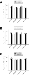
Fig 6. Tolerance and cross-tolerance to splenocyte cytokine release is unaffected by blocking PD-1.
The presented data was derived from at least 3 independent experiments, each in triplicate. Data represents splenocyte cytokine release 48 hours after successive 24-hour exposures to the ligand treatment combinations detailed on the X-axis. (A) Pre-treatment (30 minutes) of Flt3L-expanded splenocytes (1 × 10⁶ cells/ml) with an mouse anti-PD-1 blocking antibody (or corresponding isotypic control antibody, PD-1C) (5 μg/ml) prior to the secondary 24-hour ligand treatment had no effect upon resultant TNFα release regardless of the treatment combination employed. (B) Similarly, anti-PD-1 pre-treatment did not alter resultant splenocyte IFNγ release for any of the treatment combinations used. (C) Consistently, resultant splenocyte IL-10 release was unaffected for all treatment combinations by anti-PD-1 pre-treatment.
Fig 7
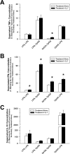
Fig 7. IL-7 pre-treatment restores responsiveness to subsequent ligand exposures attenuating tolerance and cross-tolerance.
The presented data was derived from at least 3 independent experiments, each in triplicate. Data represents splenocyte cytokine release (as a percent of the matching no tolerance/cross-tolerance [i.e., (Vehicle, LPS) or (Vehicle, CpGA), respectively] secondary ligand exposure alone) 48 hours after successive 24-hour exposures to the ligand treatment combinations detailed on the X-axis. (A) Following a 24-hour exposure to CpGA DNA (1 μM), pre-treatment (30 minutes) of Flt3L-expanded mouse splenocytes (1 × 10⁶ cells/ml) with human recombinant IL-7 (25 ng/ml) prior to the subsequent ligand treatment significantly improved responsiveness to CpGA DNA-induced TNFα release 24 hours post-treatment. (B) Regardless of the treatment combination employed, tolerance and cross-tolerance to splenocyte IFNγ release in response to the secondary ligand exposure was attenuated by IL-7 pre-treatment. (C) Pre-treatment with IL-7 had no significant effect upon splenocyte IL-10 release resulting from the secondary ligand exposure [*p < 0.05, compared to the matching secondary treatment alone (white bar)].
Fig 8
Fig 8. IRAK-M-/- splenocytes demonstrate diminished tolerance and cross-tolerance through improved responsiveness to subsequent ligand exposures.
The presented data was derived from at least 3 independent experiments, each in triplicate. Data represents splenocyte cytokine release [as a percent of secondary ligand exposure in matching wild-type (WT) splenocytes] 48 hours after successive 24-hour exposures to the ligand treatment combinations detailed on the X-axis. (A) Following a 24-hour exposure to CpGA DNA (1 μM), Flt3L-expanded mouse IRAK-M-/- splenocytes (1 × 10⁶ cells/ml) showed significantly improved responsiveness to subsequent CpGA DNA-induced TNFα release at 24 hours post-treatment which was further augmented by IL-7 (25 ng/ml) pre-treatment (30 minutes). (B) Regardless of the treatment combination employed, tolerance and cross-tolerance to IRAK-M-/- splenocyte IFNγ release in response to the secondary ligand exposure was significantly attenuated. This observation was unaffected by IL-7 pre-treatment. (C) IRAK-M-/- splenocytes demonstrated no enhancement in IL-10 responsiveness to the secondary ligand exposure regardless of IL-7 pre-treatment [*p < 0.01, relative to the matching wild-type secondary treatment (white bar), and p < 0.01, compared to the matching CpGA DNA secondary treatment in the absence of IL-7 (black bar)].
Fig 9
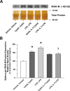
Fig 9. Elevated splenocyte IRAK-M expression in response to repeated TLR-4 or TLR-9 ligand exposures is suppressed by IL-7 pre-treatment.
The presented data is representative of at least 3 independent experiments. (A) Representative photomicrograph of a Western blot demonstrating the changes observed in IRAK-M expression 48 hours after successive 24-hour repeated ligand exposures (with or without a 30 minute IL-7 pre-treatment). (B) Relative band density of splenocyte IRAK-M expression was dramatically elevated for both successive treatment combinations and notably reduced with intervening IL-7 pre-treatment [*p < 0.05, relative to the untreated (Vehicle, Vehicle) control group, and p < 0.05, compared to the corresponding LPS treatment combination in the absence of IL-7 (LPS, LPS)].

Similar articles

Cited by

References

    1. Dobrovolskaia MA, Medvedev AE, Thomas KE, Cuesta N, Toshchakov V, Ren T, et al. Induction of in vitro reprogramming by Toll-Like Receptor (TLR)2 and TLR4 agonists in murine macrophages: Effects of TLR “homotolerance” versus “heterotolerance” on NF-kappa B signaling pathway components. J Immunol. 2003; 170:508–519. - PubMed
    1. Draisma A, Pickkers P, Bouw MP, van der Hoeven JG. Development of endotoxin tolerance in humans in vivo. Crit Care Med. 2009; 37:1261–1267. 10.1097/CCM.0b013e31819c3c67 - DOI - PubMed
    1. Castegren M, Skorup P, Lipcsey M, Larsson A, Sjolin J. Endotoxin tolerance variation over 24 h during porcine endotoxemia: association with changes in circulation and organ dysfunction. PLoS One. 2013; 8:e53221 10.1371/journal.pone.0053221 - DOI - PMC - PubMed
    1. Leentjens J, Kox M, Koch RM, Preijers F, Joosten LA, van der Hoeven JG, et al. Reversal of immunoparalysis in humans in vivo; a double-blind, placebo-controlled, randomized pilot study. Am J Respir Crit Care Med. 2012; 186:838–845. 10.1164/rccm.201204-0645OC - DOI - PubMed
    1. Dalpke AH, Lehner MD, Hartung T, Heeg K. Differential effects of CpG-DNA in Toll-like receptor-2/-4/-9 tolerance and cross-tolerance. Immunology. 2005; 116:203–212. - PMC - PubMed

Publication types

MeSH terms

LinkOut - more resources