. 2016 Feb;23(2):323-32.
doi: 10.1038/cdd.2015.100. Epub 2015 Jul 24.

Kremen1 and Dickkopf1 control cell survival in a Wnt-independent manner

Affiliations

Kremen1 and Dickkopf1 control cell survival in a Wnt-independent manner

F Causeret et al. Cell Death Differ. 2016 Feb.

Abstract

In multicellular organisms, a tight control of cell death is required to ensure normal development and tissue homeostasis. Improper function of apoptotic or survival pathways can not only affect developmental programs but also favor cancer progression. Here we describe a novel apoptotic signaling pathway involving the transmembrane receptor Kremen1 and its ligand, the Wnt-antagonist Dickkopf1. Using a whole embryo culture system, we first show that Dickkopf1 treatment promotes cell survival in a mouse model exhibiting increased apoptosis in the developing neural plate. Remarkably, this effect was not recapitulated by chemical Wnt inhibition. We then show that Dickkopf1 receptor Kremen1 is a bona fide dependence receptor, triggering cell death unless bound to its ligand. We performed Wnt-activity assays to demonstrate that the pro-apoptotic and anti-Wnt functions mediated by Kremen1 are strictly independent. Furthermore, we combined phylogenetic and mutagenesis approaches to identify a specific motif in the cytoplasmic tail of Kremen1, which is (i) specifically conserved in the lineage of placental mammals and (ii) strictly required for apoptosis induction. Finally, we show that somatic mutations of kremen1 found in human cancers can affect its pro-apoptotic activity, supporting a tumor suppressor function. Our findings thus reveal a new Wnt-independent function for Kremen1 and Dickkopf1 in the regulation of cell survival with potential implications in cancer therapies.


Figures

Figure 1
Figure 1
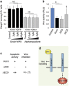
Dickkopf1 acts as a survival factor in a Wnt-independent manner. (a) Ablated PGK:Cre;Dbx1DTA embryos cultured in the absence or presence of either recombinant Dkk1 or endo-IWR1 and stained by TUNEL. Top lane is a dorsal view (anterior is up, scale bar: 100 μm). Lower lane corresponds to cryostat sections (collected at the level of the dashed line) of the embryos shown above (scale bar: 100 μm). The neural plate is surrounded by a dashed line to allow a better visualization. (b and c) Histogram (mean±S.D.) and cumulative distributions showing the number of TUNEL+ cells per section counted in ablated embryos untreated (35 sections from five animals), Dkk1-treated (25 sections from three animals at 0.2 μg/ml, 36 sections from four animals at 0.5 μg/ml and 34 sections from four animals at 1 μg/ml) or Endo-IWR1-treated (27 sections from three animals at 10 μM); *P<0.01 using Student's t-test and P<0.003 using Kolmogorov–Smirnov test, **P<0.003 using Student's t-test and P<0.001 using Kolmogorov–Smirnov test, NS: nonsignificant
Figure 2
Figure 2
Kremen1 has a pro-apoptotic activity. (ac) HEK293T cells overexpressing Krm1 (a), Krm2 (b) or Lrp6 (c) are identified by nuclear GFP expression (green). Activated Caspase-3 is revealed by immunostaining (red); scale bar: 10 μm. (d) Histogram representing the proportion of activated Caspase-3+ cells among GFP+ cells in the experiments shown in (ac) and normalized to 1 (mean±S.D.); *P<0.001 using Student's t-test. (eg) HEK293T cells transfected with HA-tagged Krm1 full length (e), lacking its intracellular domain (f) or lacking its extracellular domain (g). Cells were immunostained for HA (red) and activated Caspase-3 (green); scale bar: 10 μm. (h) Quantification of the proportion of activated Caspase-3+ cells among HA+ cells normalized to 1 (mean±S.D.) in the experiments shown in (fh); *P<0.001 using Student's t-test
Figure 3
Figure 3
Kremen1 is a dependence receptor for Dickkopf1 (a and b) HEK293T cells co-transfected with HA-tagged Krm1 (red) and either GFP (a) or Dkk1 (b) and immunostained for activated Caspase-3 (green); scale bar: 10 μm. (c) Histogram of the proportion of activated Caspase-3+ cells among Krm1-expressing cells upon co-transfection with increasing amounts of GFP or various Krm1 ligands and normalized to 1 (mean±S.D.); *P<0.001 and §P<0.01 using Student's t-test. (d) Quantification of the activated Caspase-3 fluorescence relative to HA fluorescence per cell measured for n=139, 58, 120 and 63 Caspase-3+ cells. Each dot represents one cell, log2 scale, *P<0.001 using Kolmogorov–Smirnov test. (e) Quantification of the proportion of activated Caspase-3+ cells among Krm1-HA+ cells normalized to 1 (mean±S.D.) in the absence or presence of 0.1, 0.2, 0.5 and 1 μg/ml recombinant Dkk1; *P<0.001 using Student's t-test. (f) Quantification of the proportion of activated Caspase-3+ cells among Krm1-HA+ cells cultured in a medium previously conditioned by GFP- or Dkk1-transfected cells, normalized to 1 (mean±S.D.); *P<0.001 using Student's t-test. (g) Quantification of the proportion of activated Caspase-3+ cells among Krm1-HA+ cells seeded on a carpet of GFP or Dkk1-transfected cells. §P<0.01 using Student's t-test
Figure 4
Figure 4
Kremen1 acts in a Wnt-independent manner. (a) Quantification of the proportion of activated Caspase-3+ cells among HA+ cells following co-transfection with either Krm1 and GFP (black bars) or Krm1 and Dkk1 (gray bars). Inhibition of the Wnt pathway using increasing doses of endo-IWR1 does not affect Krm1-induced apoptosis. Activation of the Wnt pathway with increasing doses of Azakenpaullone has no effect on Dkk1-mediated rescue of Krm1-induced apoptosis, NS: nonsignificant. (b) Luciferase assay indicating the relative activity of the Wnt-signaling pathway in cells transfected with Krm1 or Krm1 truncations, compared with control cells; mean±S.D., *P<0.001 using Student's t-test, §P<0.05 compared with control or with Krm1 using Student's t-test. (c) Summary of apoptotic and anti-Wnt activities of Krm1 and its truncation mutants as shown in Figures 2h and 4b. (d) A model summarizing our findings: Krm1 mediates Wnt inhibition in the presence of Dkk1 and triggers apoptosis in its absence
Figure 5
Figure 5
Kremen1 apoptotic activity is not common among vertebrates. (a) Protein-sequence comparison of the intracellular domain of Krm1 in mouse (amino acids 414–473), chick (393–450), xenopus (396–452) and zebrafish (410–465). Conservation of residues is indicated below according to the nomenclature of the ClustalX2 software. (bd) HEK293T cells transfected with cKrm1 (b), xKrm1 (c), or zKrm1 (d) are identified by GFP expression (green). Activated Caspase-3 immunostaining (in red) is negative in all three conditions; scale bar in (b): 10 μm. (e) Histogram representing the proportion of activated Caspase-3+ cells among GFP+ cells in the experiments shown in (bd) and normalized to 1 (mean±S.D.); * P<0.001 using Student's t-test. (f and g) Cryosections of chick spinal cord electroporated with HA-tagged mKrm1 (f) or cKrm1 (g). Transfected cells are shown in red (HA immunolabeling) and apoptotic cells in green (TUNEL staining); scale bar in (f): 20 μm. (h) Histogram representing the number of apoptotic cells counted per 100 μm of electroporated ventricular zone in the experiments shown in (f and g) (mean±S.D.); *P<0.001 using Student's t-test
Figure 6
Figure 6
Kremen1 C-terminal domain is responsible for apoptotic signaling. (a) Quantification of the proportion of activated Caspase-3+ cells among HA+ cells following transfection of mKrm1, Krm1 bearing a C-terminal Flag tag or point mutants S472G and D473N, normalized to 1 (mean±S.D.); *P<0.001 using Student's t-test. (bd) HEK293T cells transfected with HA-tagged mKrm1 (b), cKrm1 (c), or a modified cKrm1 bearing S and D residues at the C terminus (d). HA+-transfected cells are shown in red and activated Caspase-3+ cells in green; scale bar in (b): 10 μm. (e) Quantification of the proportion of activated Caspase-3+ cells among HA+ cells in the experiments shown in (bd) normalized to 1 (mean±S.D.); *P<0.001 using Student's t-test. (f) Luciferase assay indicating the relative activity of the Wnt-signaling pathway (mean±S.D.) in cells transfected with wild type or mutant mouse and chicken Krm1 constructs; *P<0.001 using Student's t-test. NS: nonsignificant. (g) Phylogenetic tree of various vertebrates for which a krm1 sequence was found. Length of the branches is arbitrary. Species in bold are those for which we tested Krm1 apoptotic activity. RefSeq accession numbers are given when available. The two human sequences are generated by alternative splicing. *indicate cases where several sequences were found but with identical C termini. #Sloth and armadillo sequences were manually reconstructed using Ensembl. §Opossum and platypus sequences are referenced in UniProtKB under the accession numbers H9H6Q3 and F7FX81, respectively. Protein sequences of the C termini were aligned. Residues identical to the mouse sequence are highlighted in green. Residues that differ from the mouse sequence are indicated in red when located at positions that are critical for Krm1 apoptotic activity and in yellow otherwise. The red circle on the phylogenetic tree represents the stage at which the apoptotic activity of Krm1 was most likely acquired (lineage of the placental mammals). (h) Histogram representing the proportion of activated Caspase-3+ cells among GFP+ cells following transfection of mKrm1 or a modified version bearing the same C terminus as the human long isoform, normalized to 1 (mean±S.D.); *P<0.001 using Student's t-test
Figure 7
Figure 7
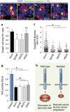
Somatic mutations found in human cancers can affect Krm1 pro-apoptotic activity. (ad) HEK293T cells expressing HA-tagged mKrm1 (a) or cancer mutants S419F (b) S437L (c) and I453V (d). Immunostaining revealed HA+ cells in red and activated Caspase-3 in green; scale bar in (a): 10 μm. (e) Quantifications the proportion of activated Caspase-3-positive cells among HA-positive cells in the experiments shown in (ad), normalized to 1 (mean±S.D.); *P<0.001 using Student's t-test; NS: nonsignificant. (f) Quantification of the activated Caspase-3 fluorescence relative to HA fluorescence per cell measured for n=171 (Krm1), 107 (S419F), 110 (S437L) and 89 (I453V) cells. Each dot represent one cell; *P<0.001 using Kolmogorov–Smirnov test. (g) Luciferase assay indicating the relative activity of the Wnt-signaling pathway in cells transfected with wild-type Krm1 or cancer mutants; *P<0.001 using Student's t-test; NS: nonsignificant. (h) Proposed model: Krm1 apoptotic activity favors the elimination of abnormal cells; mutations affecting its ability to activate Caspase-3 may confer a selective advantage to cancer cells

Similar articles

Cited by

References

    1. 1Goldschneider D, Mehlen P. Dependence receptors: a new paradigm in cell signaling and cancer therapy. Oncogene 2010; 29: 1865–1882. - PubMed
    1. 2Mehlen P, Rabizadeh S, Snipas SJ, Assa-Munt N, Salvesen GS, Bredesen DE. The DCC gene product induces apoptosis by a mechanism requiring receptor proteolysis. Nature 1998; 395: 801–804. - PubMed
    1. 3Llambi F, Causeret F, Bloch-Gallego E, Mehlen P. Netrin-1 acts as a survival factor via its receptors UNC5H and DCC. EMBO J 2001; 20: 2715–2722. - PMC - PubMed
    1. 4Thibert C, Teillet MA, Lapointe F, Mazelin L, Le Douarin NM, Mehlen P. Inhibition of neuroepithelial patched-induced apoptosis by sonic hedgehog. Science 2003; 301: 843–846. - PubMed
    1. 5Tauszig-Delamasure S, Yu LY, Cabrera JR, Bouzas-Rodriguez J, Mermet-Bouvier C, Guix C et al. The TrkC receptor induces apoptosis when the dependence receptor notion meets the neurotrophin paradigm. Proc Natl Acad Sci USA 2007; 104: 13361–13366. - PMC - PubMed

Publication types