Review
. 2016;16(6):430-54.
doi: 10.2174/1389557515666150722100610.

Progress in Small Molecule Therapeutics for the Treatment of Retinoblastoma

Affiliations
Review

Progress in Small Molecule Therapeutics for the Treatment of Retinoblastoma

Eleanor M Pritchard et al. Mini Rev Med Chem. 2016.

Abstract

While mortality is low for intraocular retinoblastoma patients in the developed world who receive aggressive multimodal therapy, partial or full loss of vision occurs in approximately 50% of patients with advanced bilateral retinoblastoma. Therapies that preserve vision and reduce late effects are needed. Because clinical trials for retinoblastoma are difficult due to the young age of the patient population and relative rarity of the disease, robust preclinical testing of new therapies is critical. The last decade has seen advances towards identifying new therapies including the development of animal models of retinoblastoma for preclinical testing, progress in local drug delivery to reach intraocular targets, and improved understanding of the underlying biological mechanisms that give rise to retinoblastoma. This review discusses advances in these areas, with a focus on discovery and development of small molecules for the treatment of retinoblastoma, including novel targeted therapeutics such as inhibitors of the MDMX-p53 interaction (nutlin-3a), histone deacetylase (HDAC) inhibitors, and spleen tyrosine kinase (SYK) inhibitors.


Conflict of interest statement

Conflict of Interest: The authors have no conflicts of interest to declare

Figures

Figure 1
Figure 1. Ocular Drug Delivery Routes
Systemic delivery relies on penetration of the blood retinal barrier (BRB) to bring drug to the posterior segment of the eye. Local delivery routes include intra-arterial (perfusion of the ophthalmic artery), intravitreal (direct bolus injection into the vitreous), topical (application of drug to the exterior of the eye) and periocular (injection into the periocular space just outside the globe), including subconjunctival (injection below the conjunctiva).
Figure 2
Figure 2. Chemical structures of standard of clinical retinoblastoma therapeutics
(a) carboplatin, (b) vincristine, (c) etoposide, (d) topotecan, (e) doxorubicin, (f) idarubicin and (g) melphalan
Figure 3
Figure 3. Use of intra-arterial infusion of melphalan to treat retinoblastoma is associated with both high rate of tumor response and prevalent, acute ocular toxicities
(a) Retinoblastoma tumor before and (b) 3 weeks after one 3 mg dose of intra-arterial melphalan (reproduced with permission from Abramson et al., 2008).[62] (c–d) Real-time retinal observations during intra-arterial melphalan infusion in a nonhuman primate model reveals retinal artery precipitates.[69]
Figure 4
Figure 4. Chemical structures of candidate retinoblastoma therapeutics investigated in preclinical studies
(a) oubain (b) digoxin (c) nutlin-3a (d) BAY-61-3606 (e) R406 (f) R788 (g) entinostat (h) paclitaxel (i) calcitriol (j) combretastatin A4 (CA-4P) (k) fluphenazine (l) chlorpromazine (m) arsenic trioxide
Figure 5
Figure 5. Inhibition of the MDMX/MDM2-p53 interaction by nutlin-3a kills retinoblastoma cells in vitro and in vivo
(a) p53-MDMX/MDM2 pathway as a target in retinoblastoma (modifed from Brennan et al. [24] and Nalepa et al[189]) (b) Pharmacokinetics of nutlin-3a following oral administration (filled circles, solid lines) or subconjunctival administration (empty squares, dotted lines). Concentration of nutlin-3a measured at 0.5, 1, 2, 4 and 8 hours in plasma (red lines) and vitreous (black lines). Concentration vs. time plot was used to fit a 2-compartment model to determine the area under the curve (AUC) and calculate the ratio of the AUC in vitreous/plasma for each route of delivery.[24] (c) Kaplan Meier survival curves for orthotopic xenograft mice receiving no treatment (black line) compared to mice receiving standard of care vincristine/etoposide/carboplatin (VCR/ETO/CBP) (blue line) and compared to those receiving subconjunctival nutlin-3a/systemic topotecan (nutlin-3a/TPT) (red line).[24] (d) Representative xenogen images for orthotopic xenografts rats treated with subconjunctival nutlin-3 and topotecan for 5 days (reproduced with permission from Laurie et al[134])
Figure 6
Figure 6. SYK inhibition as a therapeutic target for retinoblastoma
(a) Immunohistochemistry of retinoblastoma tissue and normal retina tissue H&E (left, purple) and anti-SYK (right, brown) showing increased expression of SYK in retinoblastoma tissue but not normal retina (reproduced with permission from Zhang et al[31]) (b) Survival curves of orthoptopic xenografts (SJRB001X) mice receiving subconjunctival BAY-61-3606 in combination with systemic topotecan (red line, n=20) or untreated (black line). Bay-61-3606 in combination with topotecan improved treatment outcome (reproduced with permission from Zhang et al[31]). (c) Pharmacokinetic behavior of R406 administered following subconjunctival delivery of R406. Concentration of R406 measured at 0.5, 1.0, 2.0, and 4.0 hours in the plasma (dashed line, empty circles) and vitreous (solid line, filled circles) (reproduced with permission from Pritchard et al[54]). (d) Kaplan Meier plot of event free survival (EFS) of orthoptopic xenografts (SJ-39) mice receiving subconjunctival R406 in combination with systemic topotecan (dashed black line, n=20) or untreated (solid gray line, n=10). Due to insufficient intraocular exposure, subconjunctival R406 does not improve provide efficacy in this model (reproduced with permission from Pritchard et al[54]). (e) Downstream targets in the SYK pathway.
Figure 7
Figure 7. Paclitaxel kills Y79 retinoblastoma cells in culture and reduces tumor burden in a transgenic mouse model of retinoblastoma
(a) Dose dependent tumor burden reduction in transgeneic mice treated with paclitaxel (doses of paclitaxel represent milligrams in 20 µL of 100% DMSO administered via subconjunctival injection). Tumor burden determined by taking the ratio of tumor area to total globe area of the histology cross-section with the largest tumor focus. Reductions in tumor burden for saline- and DMSO-treated control eyes were not statistically significant (reproduced with permission from Suárez et al[157]) (b-c) representative histopathologic examination of enucleated globes of transgenic retinoblastoma mice (b) left untreatedor (c) treated with 0.25 mg paclitaxel administered via subconjunctival injection (reproduced with permission from Suárez et al[157])
Figure 8
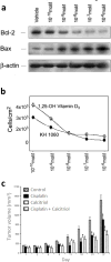
Figure 8. Vitamin D analogs inhibit tumor cell growthin vitroand in preclinical models of retinoblastoma
(a) Representative immunoblot analysis of apoptosis-related proteins in Y79 cells incubated for 72 hr with vitamin D analog 1,25-(OH)2D3 shows dose-dependent increase of Bax and dose-dependent decrease of Bcl-2 (reproduced with permission from Wagner et al[163]). (b) Dose response curves of Y79 cells treated with vitamin D analogs (1,25-(OH)2D3 (empty diamonds) and the synthetic analogue KH1060 (filled diamonds) (reproduced with permission from Wagner et al[163]). (c) In vivo tumor volumes in a subcutaneous Y79 xenograft mouse model of retinoblastoma during 5 weeks of treatment with cisplatin, calcitriol, combination therapy and control. Combination therapy (calcitriol + cisplatin) significantly inhibited tumor growth compared to controls (reproduced with permission from Kulkarni et al[125]).
Figure 9
Figure 9. Inhibition of angiogenesis by anti-VEGF antibody (Bevacizumab) or small molecule angiogenesis inhibitor (CA-4P) inhibits tumor growth in murine models of retinoblastoma
(a) Expression of vascular endothelial growth factor (VEGF) in culture medium of Y-79 cells incubated at 37°C in a hypoxic chamber and in a normoxic chamber (reproduced with permission from Lee et al[168]) (b) Inhibition of angiogenesis by treatment with an anti-VEGF antibody (Bevacizumab) produces dose-dependent inhibition of tumor growth in a xenograft model of retinoblastoma in a xenograft model (asterisks and dagger denote a statistically significant difference from the control group) (reproduced with permission from Lee et al[168]). (c) Dose dependent tumor reduction in eyes treated with CA-4P normalized to untreated control eyes reduction (reproduced with permission from Escalona-Benz et al[170]). (d) Representative H&E histopathology sections of an eye treated with 15 mg CA-4P and (e) an untreated control eye (reproduced with permission from Escalona-Benz et al[170]).

Similar articles

Cited by

References

    1. Federico S, Brennan R, Dyer MA. Childhood Cancer and Developmental Biology: A Crucial Partnership. Curr. Top. Dev. Biol. 2011;94:1–13. - PMC - PubMed
    1. Chintagumpala M, Chevez-Barrios P, Paysse EA, Plon SE, Hurwitz R. Retinoblastoma: Review of Current Management. The Oncologist. 2007;12:1237–1246. - PubMed
    1. Shields JA, Shields CL. Treatment of Retinoblastoma with Photocoagulation. Trans. Pa. Acad. Ophthalmol. Otolaryngol. 1990;42:951–954. - PubMed
    1. Shields JA, Shields CL, De Potter P. Photocoagulation of Retinoblastoma. Int. Opthalmol. Clin. 1993;33:95–99. - PubMed
    1. Shields CL, Santos CM, Diniz W, Gündüz K, Mercado G, Cater JR, Shields JA. Thermotherapy for Retinoblastoma. Arch. Ophthalmol. 1999;117:885–893. - PubMed

Publication types

MeSH terms