

Effect of piracetam and nimodipine on full-thickness skin burns in rabbits
- PMID: 26192365
- PMCID: PMC7949967
- DOI: 10.1111/iwj.12478
Effect of piracetam and nimodipine on full-thickness skin burns in rabbits
Abstract
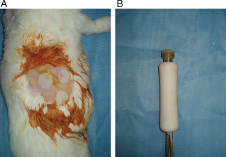
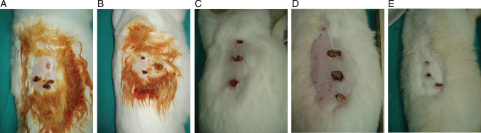
The potential of several drugs for full-thickness skin burns has been investigated, but the treatment of such burns remains a challenge in plastic surgery. The present study was designed to determine the effect of systemic and topical administration of piracetam and nimodipine on full-thickness skin burn wound healing. A total of 36 New Zealand male rabbits were divided into six groups. Full-thickness skin burns were produced in all the groups, except the control group. Piracetam was administered systemically (piracetam-IV) and topically (piracetam-C) for 14 days, and nimodipine was administered systemically (nimodipine-IV) and topically (nimodipine-C) over the burn wounds for 14 days. The sham group underwent burn injury but was not administered any drug. After 21 days, gross examination and histopathological analysis were performed and the results were compared statistically. Nimodipine-C and nimodipine-IV had no effect on burn wound healing. However, both piracetam-IV and piracetam-C significantly enhanced the healing of the full-thickness skin burn wounds, although the latter was more effective, useful and practical in burn wound healing. The histopathological features of the wounds in the piracetam-C group were closer to those of the control group than those of the other groups. Piracetam-C rather than piracetam-IV may promote full-thickness burn wound healing in rabbits.
Keywords: Burn; Full-thickness skin burn; Nimodipine; Piracetam; Wound.
© 2015 Medicalhelplines.com Inc and John Wiley & Sons Ltd.
Conflict of interest statement
None.
Figures

Similar articles
-
Preclinical assessment of safety and efficacy of intravenous delivery of autologous adipose-derived regenerative cells (ADRCs) in the treatment of severe thermal burns using a porcine model.Burns. 2018 Sep;44(6):1531-1542. doi: 10.1016/j.burns.2018.05.006. Epub 2018 Jun 27. Burns. 2018. PMID: 29958745
-
[Clinical characteristics and treatment of diabetic patients with superficial partial-thickness burn on feet].Zhonghua Shao Shang Za Zhi. 2019 Jan 20;35(1):25-30. doi: 10.3760/cma.j.issn.1009-2587.2019.01.006. Zhonghua Shao Shang Za Zhi. 2019. PMID: 30678398 Chinese.
-
[Effects and mechanism of rat epidermal stem cells treated with exogenous vascular endothelial growth factor on healing of deep partial-thickness burn wounds in rats].Zhonghua Shao Shang Za Zhi. 2020 Mar 20;36(3):195-203. doi: 10.3760/cma.j.cn501120-20191125-00441. Zhonghua Shao Shang Za Zhi. 2020. PMID: 32241045 Chinese.
-
Dressings for superficial and partial thickness burns.Cochrane Database Syst Rev. 2008 Oct 8;(4):CD002106. doi: 10.1002/14651858.CD002106.pub3. Cochrane Database Syst Rev. 2008. Update in: Cochrane Database Syst Rev. 2013 Mar 28;(3):CD002106. doi: 10.1002/14651858.CD002106.pub4. PMID: 18843629 Updated. Review.
-
The Use of Acellular Fish Skin Grafts in Burn Wound Management-A Systematic Review.Medicina (Kaunas). 2022 Jul 9;58(7):912. doi: 10.3390/medicina58070912. Medicina (Kaunas). 2022. PMID: 35888631 Free PMC article. Review.
Cited by
-
Novel pharmacotherapy for burn wounds: what are the advancements.Expert Opin Pharmacother. 2019 Feb;20(3):305-321. doi: 10.1080/14656566.2018.1551880. Epub 2018 Dec 5. Expert Opin Pharmacother. 2019. PMID: 30517046 Free PMC article. Review.
References
-
- Kumar V, Abbas AK, Aster JC. Robbins basic pathology, 9th edn. Philadelphia, PA: Elsevier Health Sciences, 2012.
-
- Najmi M, Shariatpanahi ZV, Tolouei M, Amiri Z. Effect of oral olive oil on healing of 10‐20% total body surface area burn wounds in hospitalized patients. Burns 2015;41:493–6. - PubMed
-
- Li J, Zhang YP, Zarei M, Zhu L, Sierra JO, Mertz PM, Davis SC. A topical aqueous oxygen emulsion stimulates granulation tissue formation in a porcine second‐degree burn wound. Burns 2015;41:1049–57. - PubMed
-
- Arslan K, Karahan O, Okuş A, Unlü Y, Eryılmaz MA, Ay S, Sevinc B. Comparison of topical zinc oxide and silver sulfadiazine in burn wounds: an experimental study. Ulus Travma Acil Cerrahi Derg 2012;18:376–83. - PubMed
MeSH terms
Substances
LinkOut - more resources
Full Text Sources
Medical
