Assessment of Liver Fibrosis with Diffusion-Weighted Magnetic Resonance Imaging Using Different b-values in Chronic Viral Hepatitis
- PMID: 26183515
- PMCID: PMC5588272
- DOI: 10.1159/000434682
Assessment of Liver Fibrosis with Diffusion-Weighted Magnetic Resonance Imaging Using Different b-values in Chronic Viral Hepatitis
Abstract
Objective: To examine the effectiveness of apparent diffusion coefficient (ADC) values and to compare the reliability of different b-values in detecting and identifying significant liver fibrosis.
Subjects and methods: There were 44 patients with chronic viral hepatitis (CVH) in the study group and 30 healthy participants in the control group. Diffusion-weighted magnetic resonance imaging (DWI) was performed before the liver biopsy in patients with CVH. The values of ADC were measured with 3 different b-values (100, 600, 1,000 s/mm2). In addition, liver fibrosis was classified using the modified Ishak scoring system. Liver fibrosis stages and ADC values were compared using areas under the receiver-operating characteristic (ROC) curve.
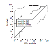
Results: The study group's mean ADC value was not statistically significantly different from the control group's mean ADC value at b = 100 s/mm2 (3.69 ± 0.5 × 10⁻³ vs. 3.7 ± 0.3 × 10⁻³ mm2/s) and b = 600 s/mm2 (2.40 ± 0.3 × 10⁻³ vs. 2.5 ± 0.5 × 10⁻³ mm2/s). However, the study group's mean ADC value (0.99 ± 0.3 × 10⁻³ mm2/s) was significantly lower than that of the control group (1.2 ± 0.1 × 10⁻³ mm2/s) at b = 1,000 s/mm2. With b = 1,000 s/mm2 and the cutoff ADC value of 0.0011 mm2/s for the diagnosis of liver fibrosis, the mean area under the ROC curve was 0.702 ± 0.07 (p = 0.0015). For b = 1,000 s/mm2 and the cutoff ADC value of 0.0011 mm2/s to diagnose significant liver fibrosis (Ishak score = 3), the mean area under the ROC curve was 0.759 ± 0.07 (p = 0.0001).
Conclusion: Measurement of ADC values by DWI was effective in detecting liver fibrosis and accurately identifying significant liver fibrosis when a b-value of 1,000 s/mm2 was used.
© 2015 S. Karger AG, Basel.
Figures


Similar articles
-
[Comparative study on clinical and pathological changes of liver fibrosis with diffusion-weighted imaging].Zhonghua Yi Xue Za Zhi. 2009 Jul 7;89(25):1757-61. Zhonghua Yi Xue Za Zhi. 2009. PMID: 19862980 Chinese.
-
Does steatosis affect the performance of diffusion-weighted MRI values for fibrosis evaluation in patients with chronic hepatitis C genotype 4?Turk J Gastroenterol. 2017 Jul;28(4):283-288. doi: 10.5152/tjg.2017.16640. Epub 2017 Jun 7. Turk J Gastroenterol. 2017. PMID: 28594328
-
Feasibility of diagnosing and staging liver fibrosis with diffusion weighted imaging.Chin Med Sci J. 2008 Sep;23(3):183-6. doi: 10.1016/s1001-9294(09)60036-5. Chin Med Sci J. 2008. PMID: 18853855
-
Diagnosis of liver fibrosis and cirrhosis with diffusion-weighted imaging: value of normalized apparent diffusion coefficient using the spleen as reference organ.AJR Am J Roentgenol. 2010 Sep;195(3):671-6. doi: 10.2214/AJR.09.3448. AJR Am J Roentgenol. 2010. PMID: 20729445
-
Diffusion-weighted magnetic resonance imaging for the assessment of liver fibrosis in chronic viral hepatitis.PLoS One. 2021 Mar 4;16(3):e0248024. doi: 10.1371/journal.pone.0248024. eCollection 2021. PLoS One. 2021. PMID: 33662022 Free PMC article. Clinical Trial.
Cited by
-
Multiparametric MR mapping in clinical decision-making for diffuse liver disease.Abdom Radiol (NY). 2020 Nov;45(11):3507-3522. doi: 10.1007/s00261-020-02684-3. Epub 2020 Aug 5. Abdom Radiol (NY). 2020. PMID: 32761254 Free PMC article. Review.
-
The Role of Signal Transducer and Activator of Transcription 5 and Transforming Growth Factor-β1 in Hepatic Fibrosis Induced by Chronic Hepatitis C Virus Infection in Egyptian Patients.Med Princ Pract. 2018;27(2):115-121. doi: 10.1159/000487308. Epub 2018 Jan 31. Med Princ Pract. 2018. PMID: 29402841 Free PMC article.
-
Diffusion weighted magnetic resonance imaging of liver: Principles, clinical applications and recent updates.World J Hepatol. 2017 Sep 18;9(26):1081-1091. doi: 10.4254/wjh.v9.i26.1081. World J Hepatol. 2017. PMID: 28989564 Free PMC article. Review.
-
The clinical value of the apparent diffusion coefficient of liver magnetic resonance images in patients with liver fibrosis compared to healthy subjects.J Family Med Prim Care. 2018 Nov-Dec;7(6):1501-1505. doi: 10.4103/jfmpc.jfmpc_299_18. J Family Med Prim Care. 2018. PMID: 30613549 Free PMC article.
-
Diffusion-weighted MRI of the liver: challenges and some solutions for the quantification of apparent diffusion coefficient and intravoxel incoherent motion.Am J Nucl Med Mol Imaging. 2021 Apr 15;11(2):107-142. eCollection 2021. Am J Nucl Med Mol Imaging. 2021. PMID: 34079640 Free PMC article. Review.
References
-
- Taouli B, Tolia AJ, Losada M, et al. Diffusion-weighted MRI for quantification of liver fibrosis: preliminary experience. AJR Am J Roentgenol. 2007;189:799–806. - PubMed
-
- Vere CC, Streba CT, Streba L, et al. Lipid serum profile in patients with viral liver cirrhosis. Med Princ Pract. 2012;21:566–568. - PubMed
-
- Berg T, Sarrazin C, Hinrichsen H, et al. Does non-invasive staging of fibrosis challenge liver biopsy as a gold standard in chronic hepatitis C? Hepatology. 2004;39:1456–1457. - PubMed
-
- Afdhal NH, Nunes D. Evaluation of liver fibrosis: a concise review. Am J Gastroenterol. 2004;99:1160–1174. - PubMed
-
- Kugelmas M. Liver biopsy. Am J Gastroenterol. 2004;99:1416–1417. - PubMed
MeSH terms
LinkOut - more resources
Full Text Sources
Other Literature Sources
Medical

