. 2015 Jul 16;6(7):e1818.
doi: 10.1038/cddis.2015.182.

AIP1-mediated actin disassembly is required for postnatal germ cell migration and spermatogonial stem cell niche establishment

Affiliations

AIP1-mediated actin disassembly is required for postnatal germ cell migration and spermatogonial stem cell niche establishment

J Xu et al. Cell Death Dis. .

Abstract

In mammals, spermatogonial stem cells (SSCs) arise from early germ cells called gonocytes, which are derived from primordial germ cells during embryogenesis and remain quiescent until birth. After birth, these germ cells migrate from the center of testicular cord, through Sertoli cells, and toward the basement membrane to form the SSC pool and establish the SSC niche architecture. However, molecular mechanisms underlying germ cell migration and niche establishment are largely unknown. Here, we show that the actin disassembly factor actin interacting protein 1 (AIP1) is required in both germ cells and Sertoli cells to regulate this process. Germ cell-specific or Sertoli cell-specific deletion of Aip1 gene each led to significant defects in germ cell migration after postnatal day 4 or 5, accompanied by elevated levels of actin filaments (F-actin) in the affected cells. Furthermore, our data demonstrated that interaction between germ cells and Sertoli cells, likely through E-cadherin-mediated cell adhesion, is critical for germ cells' migration toward the basement membrane. At last, Aip1 deletion in Sertoli cells decreased SSC self-renewal, increased spermatogonial differentiation, but did not affect the expression and secretion levels of growth factors, suggesting that the disruption of SSC function results from architectural changes in the postnatal niche.


Figures
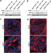
Figure 1
Figure 1
Reduction of AIP1 level and increase of F-actin level as the results of Aip1 deletion in either Sertoli cells or germ cells. (a and b) Western blot analysis of AIP1 protein levels in isolated THY1+ germ cells from geAip1−/− testes at P7 (a) and isolated Sertoli cells from seAip1−/− testes at P7 (b). The control for (a) is THY1+ germ cells from Aip1fl/+ testes and it is compared with THY1+ germ cells from geAip1−/−. The control for (b) is Sertoli cells from Aip1fl/fl testes and it is compared with Sertoli cells from seAip1−/− (c and d) Representative confocal images of phalloidin staining (red) of tissue sections from the P7 controls, geAip1−/− (c) and seAip1−/− (d) testes. Cell nuclei were stained with DAPI (blue). Scale bars: 10 μm
Figure 2
Figure 2
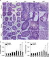
Severe spermatogenesis defects in geAip1−/− and seAip1−/− mice. (a) HE staining of tissue sections of testes from the controls, geAip1−/− and seAip1−/− mice at the indicated postnatal dates. No significant morphological defects can be seen in geAip1−/− and seAip1−/− testes at P2. At P7, germ cells in both geAip1−/− and seAip1−/− testes displayed abnormal positioning of a significant portion of germ cells in the central region of testicular cord, whereas almost all the germ cells in the control testes at P7 would have their migration and positioned themselves adjacent to the basement membrane. At P14, geAip1−/− and seAip1−/− testes displayed distinct phenotype. geAip1−/− testes at P14 displayed apparent reduction in the number of spermatocytes, whereas seAip1−/− testes showed no significant reduction in the number of spermatocytes as compared with the control. At P21 and P35, in which round spermatids and spermatozoa begin to form, respectively, geAip1−/− testes exhibited marked reduction in the number of spermatids (at P21) and spermatozoa (at P35), indicating that spermatogenesis were effectively blocked at this stage. Similarly, seAip1−/− testes also displayed severe reduction of spermatids and spermatozoa. And both geAip1−/− and seAip1−/− males are sterile. Arrows point to germ cells that were positioned in the central region of the testicular cord and had not reached the basement membrane. Scale bars: 10 μm. (b and c) Ratios of testis/body weight were significantly reduced in the P21 and P35 geAip1−/− and seAip1−/− mice as compared with the controls. The strong size reduction in both geAip1−/− and seAip1−/− testes at P21 and P35 are consistent with the severe defects of spermatogenesis occurring in both knockout mice. Data are presented as means±S.E.M.; n≥3 in each group; **P<0.01, ***P<0.001
Figure 3
Figure 3
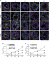
AIP1 is required in both germ cells and Sertoli cells for germ cells migration during postnatal testis development. (a) Germ cells from geAip1−/− and seAip1−/− testes displayed significant defects in their migration as compared with their respective control. Control germ cells (labeled by MVH staining, green) at P2 mostly remained in the central of each tubule, whereas migratory germ cells (labeled by PLZF, green) were observed at P4–P5. By P6 or P7, most of the germ cells had reached and contacted the basement membrane of the testicular cord. In comparison, germ cells in the geAip1−/− and seAip1−/− testes showed similar localization at P2 and P4 as that of the controls, but displayed significant migration delay starting from P5 or P6. GATA4 staining labels the Sertoli cells (red). White arrows point to examples of germ cells that had not reached the basement membrane. Scale bar: 10 μm. (b and c) Number of germ cells that were located at either the basal or central region of the testicular cord was quantified in geAip1−/− (b) and seAip1−/− (c) testes from P2 to P7 with respect to their controls. Each image was taken from a 5-μm-thick tissue section. A total of 51 tissue cross-sections were analyzed for each genotype at a certain time point (from three mice, except for P2 geAip1−/−). Data are presented as means±S.E.M.; *P<0.05, **P<0.01, ***P<0.001
Figure 4
Figure 4
Aip1 deletion caused ectopic actin accumulations in the cell cortical regions and within lamellipodial protrusions during postnatal testis development. (a and b) Immunostaining of MVH (green) and β-actin (red) of testis tissue sections from P5 control, geAip1−/− and seAip1−/− mice. The cells numbered 1–5 in the controls are wild-type germ cells migrating toward the basement membrane. Each arrow points to the migration direction and the polarized protrusion of the numbered germ cell. The cells numbered 6–12 are Aip1-deleted germ cells containing brightly stained actin patches in cell cortical regions. Arrowheads point to Aip1-ablated Sertoli cells with ectopic actin patches. Cell nuclei were stained with DAPI (blue). Scale bars: 10 μm
Figure 5
Figure 5
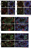
Aip1 deletion in either germ cells or Sertoli cells caused alterations in E-cadherin distribution pattern in both germ cells and Sertoli cells. (a and b) Co-immunostaining of E-cadherin (red) and PLZF (green) of testis sections from the P7 control, geAip1−/− (a) and seAip1−/− (b) mice. (c and d) Co-immunostaining of E-cadherin (red) and SOX9 (Sertoli cell marker, green) of testis sections from the P7 control, geAip1−/− (c) and seAip1−/− (d) mice. White arrows point to regions of Sertoli cells where E-cadherin level is upregulated, and the asterisk (*) signs mark germ cells whose E-cadherin distribution pattern is affected. Cell nuclei were stained with DAPI (blue). Scale bars: 10 μm
Figure 6
Figure 6
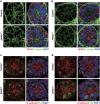
The distribution patterns of N-cadherin and β1-integrin were not significantly affected. (a and b) Co-immunostaining of β1-integrin (green) and MVH (red) of testis sections from the P7 control, geAip1−/− (a) and seAip1−/− (b) mice. (c and d) Co-immunostaining of N-cadherin (red) and PLZF (green) of testis sections from the P7 control, geAip1−/− (c) and seAip1−/− (d) mice. Scale bars: 10 μm
Figure 7
Figure 7
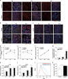
Aip1 deletion in Sertoli cells resulted in increase of spermatogonial differentiation and decrease of SSC self-renewal. (a) Co-immunostaining of OCT4 and PLZF in P4, P7 and P9 seAip1−/− and control testes. The result was quantified in (d). (b) Co-immunostaining of MVH and PLZF in P7, P9 and P12 seAip1−/− and control testes, with results quantified in (e) and (f). (c) Co-immunostaining of activated caspase-3 and PLZF in P7, P9 and P12 seAip1−/− and control testes, with results quantified in (g). (dg) Quantification of SSC (d; PLZF and OCT double positive), undifferentiated spermatogonia (e; labeled by PLZF), germ cells (f; labeled by MVH) and apoptotic spermatogonia (g; PLZF and activated caspase-3 double positive) per cross-section of each testicular cord for both seAip1−/− and control mice at indicated postnatal dates. A total of 51 tissue cross-sections were analyzed for each genotype at a certain time point (from three mice, except for P12 in c and g). (h and i) Relative expression levels of c-Kit (h) and Stra8 (i) from RT-PCR analysis. (j and k) Increased differentiation as demonstrated by flow cytometry analysis of the c-Kit+ cells. (j) Quantification of percentage of c-Kit+ cell from flow cytometry data. (k) Scale bars: 50 μm. Data are presented as means±S.E.M.; *P<0.05, **P<0.01, ***P<0.001
Figure 8
Figure 8
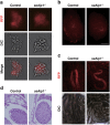
SSCs transplanted from defective niche maintained stem cell function and spermatogenesis potential both in vivo and in vitro. To trace the donor germ cells, especially in transplantation assay, seAip1−/− mice were crossed to Rosa26-mT/mG mice, a dual color reporter line, which possesses loxP sites on either side of a membrane-targeted tdTomato (mT) cassette. After breeding with cre transgenic mice, the resulting offspring would have the mT cassette deleted and the downstream membrane-targeted EGFP (mG) cassette expressed. (a) Representative THY1+ germ cell clumps from testes of seAip1−/−:Rosa26-mT/mG and Aip1fl/fl:Rosa26-mT/mG mice that had been passaged for 16 times (2.5 months). (b, c) Representative images of recipient testes containing mT-labeled colonies (b) and mT-labeled seminiferous tubules from recipient testes (c). (d) Normal spermatogenesis shown by HE staining of the recipient testis that was transplanted with cells from seAip1−/−:Rosa26-mT/mG and Aip1fl/fl:Rosa26-mT/mG. Scale bars: 10 μm in (a), 1 mm in (b), 0.2 mm in (c) and 20 μm in (d)

Similar articles

Cited by

References

    1. 1Oatley JM, Brinster RL. The germline stem cell niche unit in mammalian testes. Physiol Rev 2012; 92: 577–595. - PMC - PubMed
    1. 2Saitou M, Yamaji M. Primordial germ cells in mice. Cold Spring Harb Perspect Biol 2012; 4: 1–19. - PMC - PubMed
    1. 3Huckins C, Clermont Y. Evolution of gonocytes in the rat testis during late embryonic and early postnatal life. Arch Anat Histol Embryol 1968; 1: 341–354. - PubMed
    1. 4Clermont Y, Perey B. Quantitative study of the cell population of the seminiferous tubules in immature rats. Am J Anat 1957; 100: 241–267. - PubMed
    1. 5Culty M. Gonocytes, the forgotten cells of the germ cell lineage. Birth Defects Res C Embryo Today 2009; 87: 1–26. - PubMed

Publication types

MeSH terms