

Alternatively spliced tissue factor synergizes with the estrogen receptor pathway in promoting breast cancer progression
- PMID: 26179105
- PMCID: PMC4560996
- DOI: 10.1111/jth.13049
Alternatively spliced tissue factor synergizes with the estrogen receptor pathway in promoting breast cancer progression
Abstract
Background: Procoagulant full-length tissue factor (flTF) and its minimally coagulant alternatively spliced isoform (asTF), promote breast cancer (BrCa) progression via different mechanisms. We previously showed that flTF and asTF are expressed by BrCa cells, resulting in autoregulation in a cancer milieu. BrCa cells often express hormone receptors such as the estrogen receptor (ER), leading to the formation of hormone-regulated cell populations.
Objective: To investigate whether TF isoform-specific and ER-dependent pathways interact in BrCa.
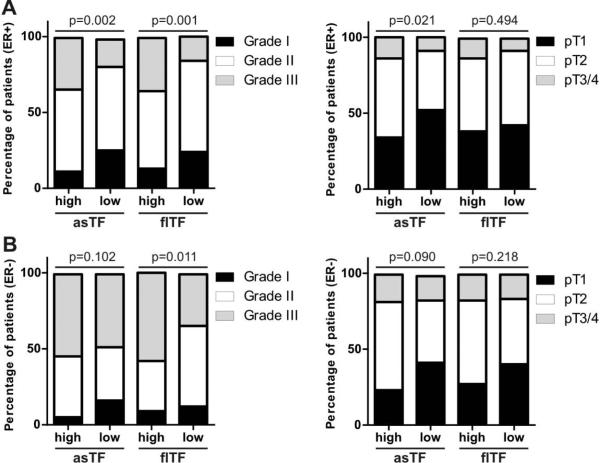
Methods: Tissue factor isoform-regulated gene sets were assessed using ingenuity pathway analysis. Tissues from a cohort of BrCa patients were divided into ER-positive and ER-negative groups. Associations between TF isoform levels and tumor characteristics were analyzed in these groups. BrCa cells expressing TF isoforms were assessed for proliferation, migration and in vivo growth in the presence or absence of estradiol.
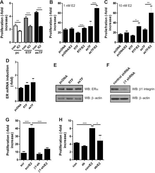
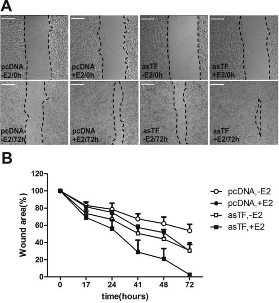
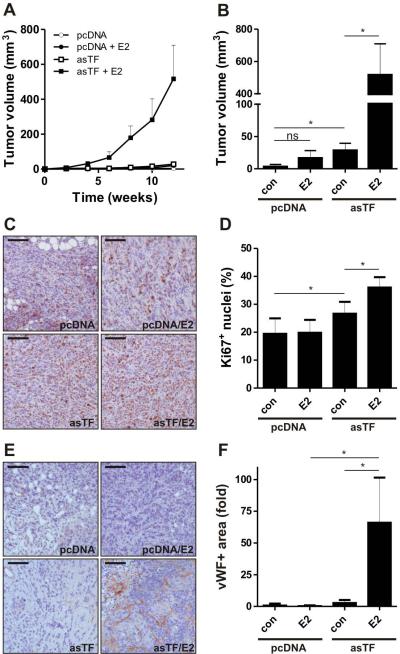
Results: Ingenuity pathway analysis pointed to similarities between ER- and TF-induced gene expression profiles. In BrCa tissue specimens, asTF expression was associated with grade and stage in ER-positive but not in ER-negative tumors. flTF was only associated with grade in ER-positive tumors. In MCF-7 cells, asTF accelerated proliferation in the presence of estradiol in a β1 integrin-dependent manner. No synergy between asTF and the ER pathway was observed in a migration assay. Estradiol accelerated the growth of asTF-expressing tumors but not control tumors in vivo in an orthotopic setting.
Conclusion: Tissue factor isoform and estrogen signaling share downstream targets in BrCa; the concomitant presence of asTF and estrogen signaling is required to promote BrCa cell proliferation.
Keywords: blood coagulation; cell movement; cell proliferation; integrin beta1; tumors.
© 2015 International Society on Thrombosis and Haemostasis.
Figures
Similar articles
-
Interplay between alternatively spliced Tissue Factor and full length Tissue Factor in modulating coagulant activity of endothelial cells.Thromb Res. 2017 Aug;156:1-7. doi: 10.1016/j.thromres.2017.05.028. Epub 2017 May 25. Thromb Res. 2017. PMID: 28570958 Free PMC article.
-
Alternatively spliced tissue factor promotes breast cancer growth in a β1 integrin-dependent manner.Proc Natl Acad Sci U S A. 2013 Jul 9;110(28):11517-22. doi: 10.1073/pnas.1307100110. Epub 2013 Jun 25. Proc Natl Acad Sci U S A. 2013. PMID: 23801760 Free PMC article.
-
Expression of flTF and asTF splice variants in various cell strains and tissues.Mol Med Rep. 2019 Mar;19(3):2077-2086. doi: 10.3892/mmr.2019.9843. Epub 2019 Jan 10. Mol Med Rep. 2019. PMID: 30664196 Free PMC article.
-
Alternatively spliced tissue factor. A crippled protein in coagulation or a key player in non-haemostatic processes?Hamostaseologie. 2010 Aug;30(3):144-9. Hamostaseologie. 2010. PMID: 20680231 Review.
-
Splice variants of Tissue Factor and integrin-mediated signaling.Thromb Res. 2012 May;129 Suppl 2:S34-7. doi: 10.1016/j.thromres.2012.02.027. Epub 2012 Mar 17. Thromb Res. 2012. PMID: 22425320 Review.
Cited by
-
The coagulome and the oncomir: impact of cancer-associated haemostatic dysregulation on the risk of metastasis.Clin Exp Metastasis. 2018 Apr;35(4):237-246. doi: 10.1007/s10585-018-9875-0. Epub 2018 Feb 28. Clin Exp Metastasis. 2018. PMID: 29492795 Review.
-
Prognostic values of tissue factor and its alternatively splice transcripts in human gastric cancer tissues.Oncotarget. 2017 May 16;8(32):53137-53145. doi: 10.18632/oncotarget.17942. eCollection 2017 Aug 8. Oncotarget. 2017. PMID: 28881799 Free PMC article.
-
Tissue Factor and Cancer: Regulation, Tumor Growth, and Metastasis.Semin Thromb Hemost. 2019 Jun;45(4):385-395. doi: 10.1055/s-0039-1687894. Epub 2019 May 16. Semin Thromb Hemost. 2019. PMID: 31096306 Free PMC article. Review.
-
Identifications of novel mechanisms in breast cancer cells involving duct-like multicellular spheroid formation after exposure to the Random Positioning Machine.Sci Rep. 2016 May 27;6:26887. doi: 10.1038/srep26887. Sci Rep. 2016. PMID: 27230828 Free PMC article.
-
Interplay between alternatively spliced Tissue Factor and full length Tissue Factor in modulating coagulant activity of endothelial cells.Thromb Res. 2017 Aug;156:1-7. doi: 10.1016/j.thromres.2017.05.028. Epub 2017 May 25. Thromb Res. 2017. PMID: 28570958 Free PMC article.
References
-
- Lumachi F, Brunello A, Maruzzo M, Basso U, Basso SM. Treatment of estrogen receptor-positive breast cancer. Curr Med Chem. 2013;20:596–604. - PubMed
-
- Masood S. Estrogen and progesterone receptors in cytology: a comprehensive review. Diagn Cytopathol. 1992;8:475–91. - PubMed
-
- Kumar V, Chambon P. The estrogen receptor binds tightly to its responsive element as a ligand-induced homodimer. Cell. 1988;55:145–56. - PubMed
-
- Pratt WB. The hsp90-based chaperone system: involvement in signal transduction from a variety of hormone and growth factor receptors. Proc Soc Exp Biol Med. 1998;217:420–34. - PubMed
Publication types
MeSH terms
Substances
Grants and funding
LinkOut - more resources
Full Text Sources
Other Literature Sources
Medical
Miscellaneous
