Review
. 2015 Aug;227(2):194-213.
doi: 10.1111/joa.12337.

Synaptic-like vesicles and candidate transduction channels in mechanosensory terminals

Affiliations
Review

Synaptic-like vesicles and candidate transduction channels in mechanosensory terminals

Guy S Bewick. J Anat. 2015 Aug.

Abstract

This article summarises progress to date over an exciting and very enjoyable first 15 years of collaboration with Bob Banks. Our collaboration began when I contacted him with (to me) an unexpected observation that a dye used to mark recycling synaptic vesicle membrane at efferent terminals also labelled muscle spindle afferent terminals. This observation led to the re-discovery of a system of small clear vesicles present in all vertebrate primary mechanosensory nerve terminals. These synaptic-like vesicles (SLVs) have been, and continue to be, the major focus of our work. This article describes our characterisation of the properties and functional significance of these SLVs, combining our complementary skills: Bob's technical expertise and encyclopaedic knowledge of mechanosensation with my experience of synaptic vesicles and the development of the styryl pyridinium dyes, of which the most widely used is FM1-43. On the way we have found that SLVs seem to be part of a constitutive glutamate secretory system necessary to maintain the stretch-sensitivity of spindle endings. The glutamate activates a highly unusual glutamate receptor linked to phospholipase D activation, which we have termed the PLD-mGluR. It has a totally distinct pharmacology first described in the hippocampus nearly 20 years ago but, like the SLVs that were first described over 50 years ago, has since been little researched. Yet, our evidence and literature searches suggest this glutamate/SLV/PLD-mGluR system is a ubiquitous feature of mechanosensory endings and, at least for spindles, is essential for maintaining mechanosensory function. This article summarises how this system integrates with the classical model of mechanosensitive channels in spindles and other mechanosensory nerve terminals, including hair follicle afferents and baroreceptors controlling blood pressure. Finally, in this time when there is an imperative to show translational relevance, I describe how this fascinating system might actually be a useful therapeutic drug target for clinical conditions such as hypertension and muscle spasticity. This has been a fascinating 15-year journey in collaboration with Bob who, as well as having an astute scientific mind, is also a great enthusiast, motivator and friend. I hope this exciting and enjoyable journey will continue well into the future.

Keywords: baroreceptors; glutamate; lanceolate endings; mechanosensory terminal; metabotropic glutamate receptor; muscle spindle; synaptic-like vesicles.


Figures

Fig 1
Fig 1
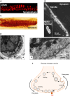
Synaptic-like vesicles (SLVs) in muscle spindle annulospiral endings. (A) The upper drawing is a reconstruction of a serially sectioned cat muscle spindle showing the incoming myelinated afferent axon arriving from below, as it then branches and eventually loses its myelin sheath to deliver a series of characteristically annulospiral endings wrapping around intrafusal muscle fibres. Scale bar: 100 μm. The red box delineates an area of terminal typically sampled to reveal the clusters of 50-nm-diameter, clear ‘synaptic-like’ vesicles within. Shown below is one such section. The regular array of contractile proteins is seen at the top, with the paler, floccular sensory nerve terminal seen below. The most obvious SLV clusters are indicated with arrowheads, but closer inspection shows that SLVs are scattered throughout. Note that the clusters are not all focussed towards the muscle fibre, i.e. they do not appear to be truly ‘synaptic’. SLVs are as likely to be clustered adjacent to terminal membrane facing away from the muscle fibre (e.g. cluster indicated by the right-most arrowhead) as towards it. (B) An historical quantification (for younger readers: 1 Å = 10−10 m, i.e. 10 Å = 1 nm) of the diameters of all vesicles within primary sensory endings revealed a range of diameters and a mix of clear and dense-cored vesicles. However, by far the most abundant population is about 500 Å, or 50 nm. (C) Top: fluorescent labelling of motor nerve terminals stimulated in RH414, a prototype styryl pyridinium dye used in the development of the more commonly used dye, FM1-43. During this work with Bill Betz and Steve Fadul (University of Colorado Health Sciences Center, Denver), we showed dye internalisation occurred by endocytosis with recaptured vesicle membrane. This is when we first noticed (Bottom) the characteristic labelling of the annulospiral endings of muscle spindle primary afferent terminals in the same muscle (rat lumbrical muscle). Spindle labelling occurred even if the muscle was unloaded (i.e. not stretched) and in the presence of tetrodotoxin (TTX) to block afferent discharge. Thus, electrical and mechanical activity were not required to get labelling, suggesting at least a basal level of SLV endocytosis occurs at rest. From Bewick et al. (2005) with permission.
Fig 2
Fig 2
Muscle spindle primary afferents express synaptic vesicle-associated proteins and exhibit endocytosis. Immunoreactivity in muscle spindles for the ubiquitous synaptic vesicle proteins (A) synaptophysin (cat) (B) synapsin I (rat: courtesy of Arild Njå, University of Oslo) and Ca2+ -binding protein (C) calretinin (cat). Note the insert showing labelling for synapsin I in a motor nerve terminal of the same muscle in (B). (D) Evidence of endocytosis in an annulospiral terminal from a cat muscle spindle. (a) Transverse section showing the underlying nuclear bag intrafusal fibres (dark area, upper left), partially enclosed by the sensory terminal (lighter area, lower right). The arrow indicates the area of interest shown at higher magnification in (b). There are several things to note at this point. First, note the presence of a coated pit (arrow) typical of clathrin-mediated endocytosis during membrane recovery of synaptic vesicles. This is typical of membrane recovery, rather than exocytosis. Second, the pit is of approximately 50 nm diameter. Finally, the membrane recovery is occurring on the side of the terminal away from the muscle fibre, i.e. such membrane recovery can occur all over the surface of the terminal. (C) Simplified schema of vesicle recycling from exocytosis (vesicle fusion and neurochemical release), through endocytosis, via specialised budding proteins, and subsequent refilling with neurotransmitter/modulator, then docking ready for re-release. (B–D) From Bewick & Banks (2015), with permission.
Fig 3
Fig 3
Characteristics of FM1-43 labelling of muscle spindle annulospiral primary sensory endings. (A) FM1-43 labelling of a primary afferent terminal in a live rat lumbrical muscle, ex vivo. (B) Maximum intensity projections of a confocal series of optical slices in a rat lumbrical spindle at low magnification (a) and an expansion of the rectangular area is also shown (b). (c) Labelling occurs spontaneously (resting length), but is increased fourfold by stretching (stretch) during incubation (2 h, 10 μm). The muscle was returned to resting length briefly two–three times each 30 min, before being re-pinned at the maximum length. (C) FM1-43 internalisation (a) is strongly inhibited (b) by inorganic salts that block Ca2+ channels, in this case Co2+ . This is quantified below (c, top). Note that 10 mm Ca, which blocks some stretch-sensitive channels, has no effect on FM1-43 internalisation, suggesting it is not entering through the mechanosensory channels, but rather by SLV endocytosis. (D) This conclusion of vesicle-mediated labelling is further reinforced by the release of dye from labelled terminals. This is in sharp contrast to labelling by the dye permeating through the pore of the open mechanosensory channels, which is irreversible. (a) A labelled terminal (Start) shows little dye loss during 5 min rest (Rest). However, 5 min of vibration (200 Hz, 50 μm) applied to the pole of the spindle with a blunt vibrating probe elicits a marked reduction of intensity, i.e. dye loss (Stim1). The rate of destain returns to basal levels on returning to rest, but resumes on a second vibration (Stim2). This indicates FM1-43 is being lost by SLV exocytosis, and at a rate proportional to the mechanical activity. (b,c) The vibration-evoked destaining was quantified and was markedly reduced in 0 mm Ca2+. Thus, destaining (i.e. SLV exocytosis) is Ca2+ -sensitive, which is another parallel with synaptic vesicle turnover. (B–D) From Bewick et al. (2005) with permission.
Fig 4
Fig 4
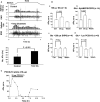
FM1-43 does not block stretch-evoked spindle firing and evidence that endogenous glutamate secretion from SLVs regulates spindle stretch sensitivity. (A) 2 h in 5 μm FM1-43 does not inhibit stretch-evoked spindle firing in rat 4th lumbrical muscles, indicating the dye does not block the mechanosensory channels in muscle spindles and, therefore, terminal labelling is unlikely to be due to dye permeation through the channels expressed in these fully differentiated mature terminals. (B) Immunogold labelling for glutamate in muscle spindle primary afferents. (a) Transverse ultrathin section through an intrafusal fibre (i) with two paler terminal profiles (t) on its surface, labelled with immunogold for glutamate. (ii) Higher magnification of the rectangular area in (i) showing the high density of gold particles in vesicle (v) containing areas of the terminal compared with surrounding structures, including the intrafusal muscle fibre. For this technique fixation is milder, to preserve antigenicity, so vesicle preservation is not as clear in these sections. (b) Quantification of gold particle density of two different spindle Ia primary terminals compared with other tissues in the same rat (dark and light grey bars, respectively) and no primary controls (small mid-tone grey bars). In both cases, glutamate-like immunoactivity (gold particle density) was at least twice that in non-glutamatergic tissues, such as glial cell processes, intra- and extrafusal muscle fibres and motor neurone dendrites. It was also at least as much as in putative central terminals of I afferents on motor neurone dendrites. In one case (dark bars) it was equivalent to that in cerebellar mossy fibre terminals of the cerebellum (both are glutamatergic synapses). No primary controls show negligible labelling. IF, intrafusal fibres; XF, extrafusal fibres. From Bewick et al. (2005), with permission. (C) Spindle primary endings label heavily for the vesicular glutamate transporter vGluT1, indicating the endogenous glutamate is loaded into SLVs (from Wu et al. , with permission). (D) Inhibition of glutamate re-uptake with TBOA greatly increases stretch-evoked firing from rat lumbrical muscle spindles over a 2–3-h period, in a reversible manner. This indicates the extracellular accumulation of endogenously secreted glutamate makes the ending more sensitive to stretch. *P < 0.05, ***P < 0.001 vs pre-drug control firing. (E) Latrotoxin application, which causes uncontrolled exocytosis in spindles, substantially increases stretch-evoked spindle firing in rat 4th lumbricals by 1 h of application, presumably as glutamate exocytosis is greatly increased. Over the next few hours, firing to a standard stretch slowly declines, becoming inhibited from 210 min (3.5 h) of toxin incubation. This presumably reflects SLV, and hence glutamate, depletion. Bungarotoxin was added to block interference by the activation of the intrafusal fibres by fusimotor neurones. Red bar = bungarotoxin application. Yellow bars = statistically significant in comparison to t − 60 min (pre-drug control) at (*) P < 0.01. Thus, t + 60 min (latrotoxin peak excitation), t + 210–270 min (latrotoxin inhibition).
Fig 5
Fig 5
Spindle stretch sensitivity is regulated by an atypical glutamate receptor with the pharmacology of the hippocampal PLD-mGluR. (A) (Top to bottom). A trapezoid profile of the stretch applied to the rat 4th lumbrical muscles. Muscles are stretched by 1 mm, which represents ∼10% increase in length, for 5 s before returning to the original length. The ‘spike rate’ is shown in 100-ms windows, revealing a rapid increase in firing for this particular muscle during the stretch to a new length (dynamic response). Firing then settles to a slightly lower plateau rate at the new length, until released to the original length, when firing stops. The ‘afferent discharge’ is shown in the raw electroneurogram, which is recorded from the whole muscle nerve, and thus represents the firing from all the 8–12 spindles found in this lumbrical muscle. Below this, the same muscle response is shown following 1 h incubation in 1 mm glutamate. This approximately doubles the firing rate for the same stretch. The very bottom histogram shows quantification for n = 6 preparations. This increase in stretch-sensitivity is entirely reversible (not shown). (B) The reversible increase in firing rate with 100 μm glututamate (a). (b) This response cannot be blocked by antagonists of all the 11 cloned glutamate receptors (kynurenate: all three iGluRs; MCPG/CPPG: all eight mGluRs), even when applied together. This indicates glutamate is not acting through any of the cloned receptors. (c) Glutamate excitability is totally blocked by RS 3,5-DHPG, an agonist at group I mGluRs but an antagonist of the PLD-mGluR first reported in the hippocampus. (d) Glutamate excitability is blocked somewhat more potently by PCCG-13, a selective antagonist specifically developed for this receptor. These experiments indicate exogenous glutamate is activating the PLD-mGluR to regulate spindle stretch-sensitivity. (C) Quantification of stretch-evoked firing in a single rat 4th lumbrical muscle during prolonged PCCG-13 application. When applied alone, without exogenous glutamate, high concentrations (10 μm) of the selective PLD-mGluR antagonist PCCG-13 can totally abolish stretch-evoked responses, over a period of 4–6 h. This effect is entirely reversible. This illustrates that blocking the activation of the PLD-mGluR by endogenous glutamate secretion from SLVs means the ending cannot sustain a sensitivity to stretch, i.e. endogenous glutamate-mediated activation of the receptor is necessary to maintain its stretch sensitivity. From Bewick et al. (2005), with permission.
Fig 6
Fig 6
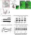
Baroreceptor terminals have SLV, internalise and release FM1-43, and exhibit glutamate sensitivity for stretch-evoked firing. (A) Baroreceptor terminals on the aortic arch of the rat (lower image) have a high density of SLVs, when viewed at the EM level. D, aortic depressor nerve; LCC, left common carotid artery; LSC, left subclavian artery. (B) Aortic baroreceptor terminals take up and release FM1-43. Adapted from Krauhs (1979), with permission. (a) Schematic of the anatomical position of the aortic baroreceptors in humans. Inset, FM1-43 labelling of baroreceptor terminals in mouse. (b) Higher magnification of FM1-43-labelled baroreceptor terminals shown in the inset in (a). Terminal depolarisation with 60 mm K+ stimulates FM1-43 release, indicating dye internalisation and release is due to SLV recycling. (C) Responses of various working heart brainstem preparation outputs to topical glutamate application to the baroreceptors. (a) Glutamate application to the aortic baroreceptors increases firing rate of the aortic depressor nerve (ADN). ∫ADN, integrated ADN activity. (b) Increasing (arrow) perfusion pump pulse pressure (PP) in the aorta evokes a marked increase in ADN firing. Topical PCCG-13, the selective PLD-mGluR antagonist, onto the baroreceptor greatly reduces the evoked response, an effect that can be washed out. Subsequent exogenous glutamate application increases pressure-evoked ADN firing again. (c) Topical glutamate application to the baroreceptors produces a dramatic decrease in heart rate, and sympathetic nerve firing. Thus, enhancing baroreceptor sensitivity causes reflex inhibition likely to induce a reduction in peripheral blood pressure. bpm, beats per minute; HR, heat rate; ∫SNA, integrated sympathetic nerve activity. (d) Just as topical glutamate application to the aortic baroreceptors increases sympathoinhibition, PCCG-13 reduces it. These experiments show the PLD-mGluR on baroreceptor terminals can be a suitable target for regulating sympathoinhibition, which controls peripheral blood pressure.
Fig 7
Fig 7
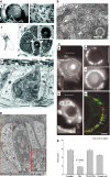
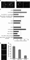
Hair follicle lanceolate mechanosensory endings of the anterior skin of the mouse pinna have SLVs, secretion-associated proteins and glutamate. (A) (a) The mouse pinna preparation pinned, anterior skin face down, in a Sylgard-lined dish filled with Liley’s solution, and set up for electrophysiological recording. The whole posterior skin has been removed, as well as a large area of elastic cartilage and adipose tissue (at) from the cleared area (ca). By folding the cleared area back, access was gained to the hair shafts, allowing two or three within the vibrated area (va) to be mechanically displaced by a fire-polished glass capillary (not shown). The nerves (n) are branches of the mandibular division of the trigeminal and are set up for differential recording of the neurogram using recording (re) and indifferent (i.e.) suction electrodes. (b) Brightfield image of mouse pinna skin viewed from the dermal side, showing several hair follicles. The bases of the hair shafts are clearly seen; each shaft is partly surrounded by a sebaceous gland that appears dark. Scale bar: 100 μm. (c) Diagram of the structure and location of the innervation of a hair follicle. The lanceolate ending consists of the group of terminals forming the palisade-like structure immediately below the lobular sebaceous gland (from Bannister, 1976). The dashed line indicates the typical plane of section for subsequent images for fluorescence and light microscopy. (d) Semi-thin (1 μm) cross-section through a hair follicle (hf) at the level of the sebaceous gland (sg) and lanceolate ending, as indicated in (c). The lanceolate ending surrounds the follicle (arrows), and terminals appear as dark structures alternating between lighter accessory cells, shown in greater detail in the inset at top right. Mouse pinna, Toluidine Blue; scale bar (main image) indicates 20 μm. (e) EM of an ultrathin cross-section through a lanceolate ending, showing a single, darkly stained, sensory terminal (st) almost completely enclosed by pale-staining glial cell (gc) processes. Note the numerous 50-nm-diameter vesicle profiles in the terminal axoplasm (white arrows). Mouse pinna; scale bar indicates 0.5 μm. (B) SLVs shown in higher magnification (white arrows), and labelling with FM1-43 produces a characteristic circle of lanceolate endings around a central hair shaft. (C) Immunohistochemical and genetic identification of the sensory terminals and glial cells of lanceolate nerve endings. (a) Anti-neurofilament protein (NFP)-like immunoreactivity is localized in structures identified as preterminal axons (pa) and sensory terminals in a mouse pinna follicle. Several terminals are shown enlarged in the inset. Epifluorescence; scale bar indicates 10 μm. (b) Structures identified as sensory terminals also react strongly with anti-synapsin I antibody. Mouse pinna, epifluorescence; scale bar indicates 20 μm. (c) SynaptopHluorin fluorescence shows the expression of the v-SNARE synaptobrevin in the lanceolate terminals in a very similar pattern to NFP and synaptophysin. Mouse pinna, epifluorescence; scale bar as in (B). (d) Anti-S-100 antibody, in contrast, labels paired structures identified as glial cells (gc) and their processes in a mouse pinna follicle. Pairing of the processes is particularly apparent in the enlarged inset and is distinct from the unpaired processes seen in (a). Epifluorescence; scale bar indicates 10 μm. (e) A follicle from rat pinna double-labelled with antibodies against synaptophysin (red) and S-100 (green). Where the ending is precisely orthogonal within the section (white arrows), individual red profiles can be seen clearly to be almost entirely enclosed by paired green profiles, identified as sensory terminals and glial cell processes, respectively. Laser-scanning confocal microscopy; scale bar indicates 5 μm. (D)Lanceolate sensory terminals are enriched in glutamate. An EM of a thin section of a sensory lanceolate terminal (st) is shown with enclosing glial cells (gc) immunogold labelled to show glutamate-like immunoreactivity. A portion is enlarged in the inset, showing gold particles more clearly. (E) A histogram (means ± SEM) summarising the quantitative assessment of glutamate-like immunoreactivity. From Banks et al. (2013), with permission.
Fig 8
Fig 8
Glutamate regulates SLV recycling by activating the PLD-mGluR. (A) Like spindle afferent terminals, lanceolate endings spontaneously label with FM1-43. Exogenous glutamate increases, while (B) PCCG-13 decreases, dye internalisation. (C) Top to bottom. Histogram summarising the effects of various GluR ligands on FM1-43 internalisation. Light grey bars – the glutamate-mediated increase is blocked by PCCG-13 but not the cocktail of classical iGluR and mGluR antagonists (4-CPG, CPPG, kynurenate). Dark grey bars – MCPG (group I and group II antagonist) does not significantly inhibit FM1-43 internalisation, while glutamate increases it, and PCCG-13 produces a dose-dependent decrease. The other antagonist cocktails produce little if any effect on labelling. (D) FIPI, the PLD inhibitor, also causes a dose-dependent decrease in FM1-43 uptake/SLV endocytosis. These effects are quantified in (E). These data indicate that, as for spindle firing, the PLD-mGluR regulates SLV endocytic uptake of FM1-43. From Banks et al. (2013), with permission.
Fig 9
Fig 9
Anti-ENaC and anti-ASIC 2 subunit immunoreactivity localises to sensory terminals of rat muscle spindles. (A) Double-immunofluorescent labelling of the sensory regions of rat muscle spindles, comparing anti-ENaC subunit with anti-synaptophysin reactivities. Upper panels (red): anti-ENaC α, β, γ or δ immunoreactivity; lower panels (green): anti-synaptophysin immunoreactivity of the corresponding spindles. Control: anti-ENaC antibody replaced with non-immune rabbit IgG. Immunoreactivity was clearly visible with antibodies to the α, β and γ subunits, but there was little reaction with the anti-ENaC δ antibody; this is in contrast to the control where no specific reactivity was discernible. (B) Double-immunofluoresent labelling of the sensory regions of rat muscle spindles, comparing anti-ASIC2 with anti-synaptophysin reactivities. Upper panels (red): anti-ASIC2 immunoreactivity; lower panels (green): anti-synaptophysin immunoreactivity of the corresponding spindles. Anti-ASIC2 immunoreactivity was evident on sensory terminals in contrast to controls (anti-ASIC2 antibody blocked with peptide, or replaced with non-immune goat IgG) where specific immunoreactivity of the sensory terminals was not visible. From Simon et al. (2010), with permission.
Fig 10
Fig 10
Spindles and lanceolate endings express SK2 Ca2+ -activated K+ channels and ASIC2. (A) SK2-like immunoactivity (red) is present in preterminal axons and the terminals, identified by SLV-associated synaptophysin antibodies (green). The merge shows the colocalisation of the two antibody distributions, while the widefield brightfield image shows the disposition of the intrafusal fibres. Lanceolate terminals also express SK2 (not shown). (B) (Upper row) Just like spindle terminals (not shown), SK3-like immunoactivity (red) is not found in lanceolate terminals (green). In hair follicles, however, SK3 is found in the glial cells enclosing the lanceolate endings [merge and in (C) the higher magnification inset]. (Lower row) Conversely, ASIC2 immunolabelling (red) co-localises with SLV label (green) – see yellow in merge and indicated by arrows in inset in (C). Scale bar: 20 μm (A); 5 μm (B). From Shenton et al. (2014), with permission.
Fig 11
Fig 11
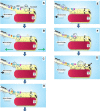
Emerging model of stretch-evoked firing in muscle spindles. Cartoon schematic showing the steps in the processes evoked by stretch in a muscle spindle afferent. The points of interest at each step are circled while changes in afferent discharge rate are indicated by the arrow. (A) Tonic secretion of glutamate from SLVs maintains stretch sensitivity of the ending via low-level activation of the PLD-mGluR, and a low, tonic firing at rest. (B) Stretch opens a mechanosensitive Na+ channel, producing the depolarising RP. (C) This, in turn, opens the voltage-gated Na+ channels in the AP initiation site, probably in the first heminode (see Cope article in this volume), greatly increasing the afferent discharge rate. (D) The depolarisation also opens voltage-gated Ca2+ channels, activating SK2 Ca2+ -activated K+ channels (E). The resulting K+ efflux repolarises the membrane, reducing the afferent discharge rate to more modest levels. (F) Meanwhile, in the terminal, stretch also opens a Ca2+ channel, which enhances SLV exocytosis (G), increasing the PLD-mGluR activation. How this maintains ending sensitivity is not clear at present, but may be through regulating mechanosensory Na+ channel insertion from the vesicle store. These may be the same, or a different, pool to the SLVs. From Bewick & Banks (2015), with permission.
Fig 12
Fig 12
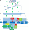
Progressive geometrical abstraction of a single terminal of a spindle primary ending, leading to a flow-chart summarising the events of mechanosensory transduction. Green block arrows in (A–C) indicate the direction and distribution of stretch applied to the terminal when the primary ending is lengthened during muscle stretch or fusimotor stimulation. (A) A single terminal in its annulospiral form, taken from a primary ending reconstructed from serial sections. Several such terminals typically enclose a single intrafusal muscle fibre. The terminal is connected to its associated heminode by a short, unmyelinated preterminal axonal branch at the point shown. (B) The terminal unrolled and turned through 90 °. Note that individual terminals may be repeatedly branched and that the direction of stress during stretch is orthogonal to the long axis of the terminal. (C) A terminal and its associated unmyelinated preterminal branch shown in abstract as cylinders whose diameters indicate the relative diameters of these structures in a spindle Ia primary afferent. The smaller preterminal branch to the right is about 1 μm diameter. The lengths, especially those of the much larger terminal to the left, are highly variable. (D) Flow chart to illustrate the main events of mechanosensory transduction, as described in this review. The principal feedforward pathway from stimulus (stretch) to output (APs) is shown by the white block arrows. We envisage that the overall gain of this pathway is controlled by several feedback pathways: negative feedback 1 is at present hypothetical and is included to account for the reversible silencing of the primary ending by PCCG-13 inhibition of the PLD-linked mGluR; the positive feedback pathway is the well-established SLV/glutamatergic loop; negative feedbacks 2 and 3 involve different kinds of K[Ca], one located in the terminal, the other in the heminode and both perhaps triggered by APs opening voltage-gated Ca channels. Green lines and arrowheads indicate enhancing/excitatory actions; red lines and circles indicate reducing/inhibitory actions. From Bewick & Banks (2015), with permission.

Similar articles

Cited by

References

    1. Aguado F, Majó G, Ruiz-Montasell B, et al. Syntaxin 1A and 1B display distinct distribution patterns in the rat peripheral nervous system. Neuroscience. 1999;88:437–446. - PubMed
    1. Akoev GN, Alekseev NP, Krylov BV. Mechanoreceptors: Their Functional Organisation. Berlin: Springer; 1988.
    1. Albani-Torregrossa S, Attucci S, Marinozzi M, et al. Antagonist pharmacology of metabotropic glutamate receptors coupled to phospholipase D activation in adult rat hippocampus: focus on (2R,1’S,2’R,3’S)-2-(2′-carboxy-3′-phenylcyclopropyl)glycine versus 3, 5-dihydroxyphenylglycine. Mol Pharmacol. 1999;55:699–707. - PubMed
    1. Arnadóttir J, Chalfie M. Eukaryotic mechanosensitive channels. Ann Rev Biophy. 2010;39:111–137. - PubMed
    1. Banks RW, Bewick GS, Reid B, et al. Evidence for activity-dependent modulation of sensory-terminal excitability in spindles by glutamate release from synaptic-like vesicles. Adv Exp Med Biol. 2002;508:13–18. - PubMed

Publication types

MeSH terms

LinkOut - more resources