. 2015 Oct;197(19):3097-109.
doi: 10.1128/JB.00276-15. Epub 2015 Jul 13.

Orientia tsutsugamushi Strain Ikeda Ankyrin Repeat-Containing Proteins Recruit SCF1 Ubiquitin Ligase Machinery via Poxvirus-Like F-Box Motifs

Affiliations

Orientia tsutsugamushi Strain Ikeda Ankyrin Repeat-Containing Proteins Recruit SCF1 Ubiquitin Ligase Machinery via Poxvirus-Like F-Box Motifs

Andrea R Beyer et al. J Bacteriol. 2015 Oct.

Abstract

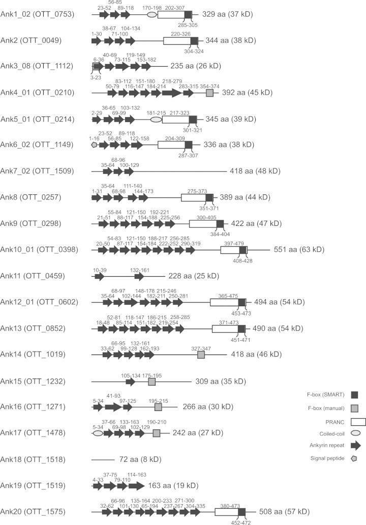
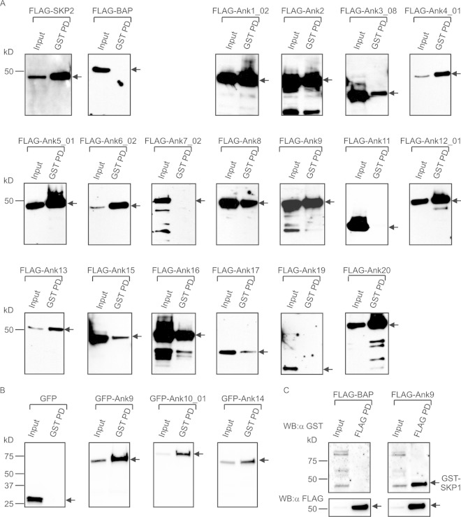
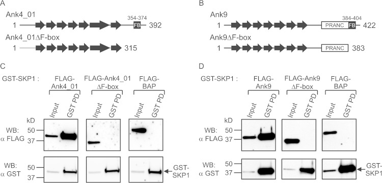
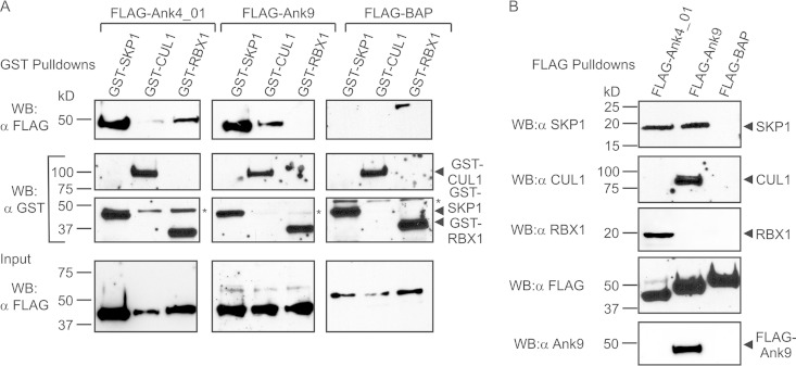
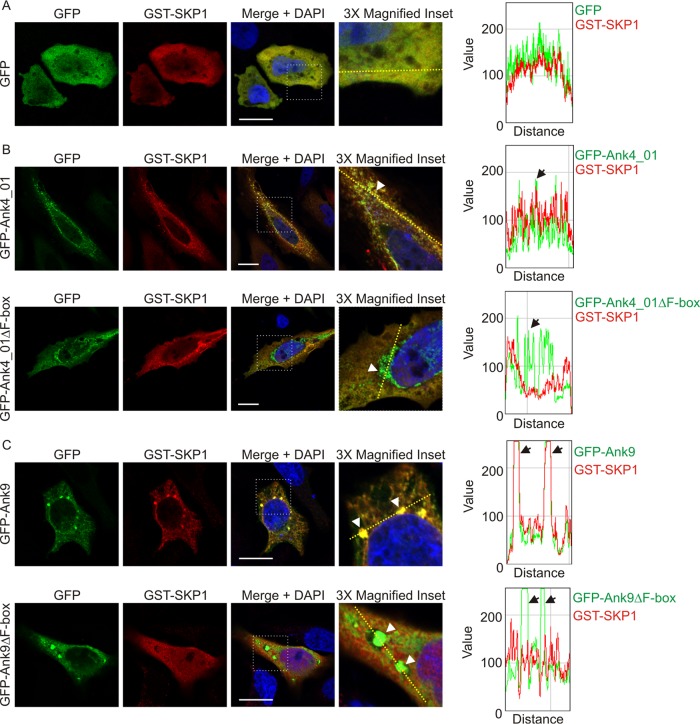
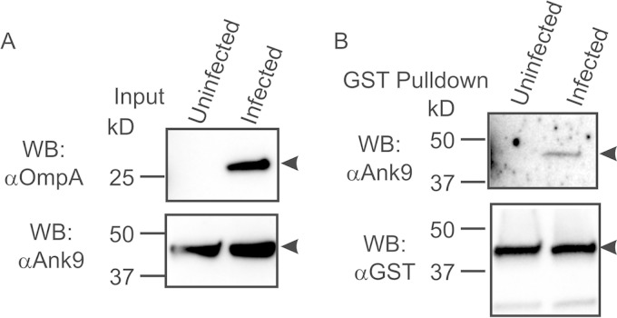
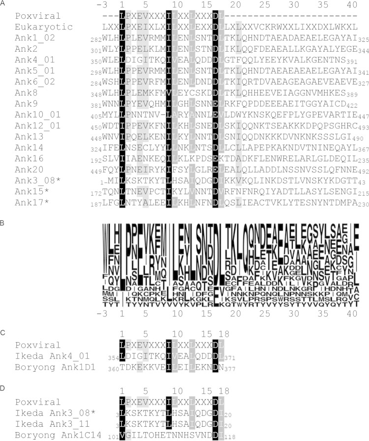
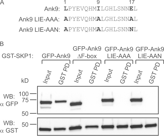
A rising theme among intracellular microbes is the delivery of ankyrin repeat-containing effectors (Anks) that interact with target proteins to co-opt host cell functions. Orientia tsutsugamushi, an obligate intracellular bacterium and the etiologic agent of scrub typhus, encodes one of the largest Ank repertoires of any sequenced microorganism. They have been previously identified as type 1 secretion system substrates. Here, in silico and manual sequence analyses revealed that a large proportion of O. tsutsugamushi strain Ikeda Anks bear a eukaryotic/poxvirus-like F-box motif, which is known to recruit host cell SCF1 ubiquitin ligase machinery. We assessed the Anks for the ability to serve as F-box proteins. Coimmunoprecipitation assays demonstrated that F-box-containing Anks interact with overexpressed and/or endogenous SCF1 components. When coexpressed with FLAG-Ank4_01 or FLAG-Ank9, a glutathione S-transferase (GST)-tagged version of the SCF1 component SKP1 localized to subcellular sites of FLAG-Ank accumulation. The abilities of recombinant Anks to interact and colocalize with SKP1 were F-box dependent. GST-SKP1 precipitated O. tsutsugamushi-derived Ank9 from infected host cells, verifying both that the pathogen expresses Ank9 during infection and the protein's capability to bind SKP1. Aligning O. tsutsugamushi, poxviral, and eukaryotic F-box sequences delineated three F-box residues that are highly conserved and likely to be functionally important. Substitution of these residues ablated the ability of GFP-Ank9 to interact with GST-SKP1. These results demonstrate that O. tsutsugamushi strain Ikeda Anks can co-opt host cell polyubiquitination machinery, provide the first evidence that an O. tsutsugamushi Ank does so during infection, and advance overall understanding of microbial F-box proteins.

Importance: Ankyrin repeat-containing proteins (Anks) are important virulence factors of intracellular bacteria that mediate protein-protein interactions with host cell targets. Orientia tsutsugamushi, which causes a debilitating infection called scrub typhus in one of the most densely populated regions of the world, encodes one of the largest Ank armamentariums of any sequenced bacterium. This study demonstrates that O. tsutsugamushi strain Ikeda Anks also bear F-box motifs that interact with host cell polyubiquitination machinery. By proving that an Orientia-derived Ank interacts with SKP1 in infected cells, this evidences the first bona fide Orientia effector and the first example of an endogenous F-box-containing Ank-mammalian-host ligand interaction for any intracellular bacterium. Also, importantly, this work identifies key residues that are essential for microbial F-box function.


Figures

FIG 1
FIG 1
Schematic of domains found within O. tsutsugamushi strain Ikeda Anks. Each Ank is listed by name, followed by its gene annotation in parentheses. The amino acid locations of ankyrin repeats, putative F-boxes, coiled-coil domains, signal peptides, and PRANC domains are noted above or below the corresponding shape. F-boxes predicted using the SMART algorithm are found within PRANC domains, while those found via manual sequence searches are shown as separate boxes. The length of each Ank is noted to the right of its respective schematic, with the estimated protein size. Though Ank18 does not contain any ankyrin repeats itself, it is annotated as an Ank because it shares homology with nonrepeat regions of O. tsutsugamushi strain Boryong Ank1u7 (OTBS_1195), which does contain ankyrin repeats. aa, amino acids.
FIG 2
FIG 2
O. tsutsugamushi Anks interact with GST-SKP1. (A) GST-SKP1 coprecipitates FLAG-tagged Anks. Lysates of transfected HeLa cells expressing GST-SKP1 together with FLAG-tagged Anks, positive-control FLAG-SKP2, or negative control FLAG-BAP were incubated with glutathione-Sepharose to precipitate GST-SKP1 and thereby coprecipitate interacting proteins. The resulting Western blots were screened with FLAG antibody. (B) GST-SKP1 coprecipitates GFP-tagged Anks. Lysates of transfected HeLa cells expressing GST-SKP1 together with GFP-tagged Ank9, Ank10, Ank14, or negative-control GFP alone were Western blotted and screened with GFP antibody. (C) FLAG-Ank9 coimmunoprecipitates GST-SKP1. Lysates of transfected HeLa cells expressing GST-SKP1 together with FLAG-Ank9 or FLAG-BAP were incubated with FLAG antibody-conjugated agarose beads to immunoprecipitate FLAG-tagged proteins and coprecipitate interacting proteins. The resulting Western blots (WB) were probed with GST antibody (α-GST). Precipitation of FLAG-tagged proteins was confirmed by probing the stripped blots with FLAG antibody (α-FLAG). (A to C) For each experiment, expression of the protein of interest was confirmed in the “Input” lane containing 3% of the sample. The pulldown (PD) lanes represent proteins that coprecipitated with GST-SKP1 (A and B) or coimmunoprecipitated with FLAG-tagged protein (C). Precipitation of GST-SKP1 was confirmed, but is not shown, for each sample in panels A and B. The arrows indicate the expected sizes of the prey proteins. All the blots are aligned at the 50-kDa marker, as indicated on the leftmost blot in each row. The data presented are representative of two to five experiments with similar results.
FIG 3
FIG 3
Ank-SKP1 interactions are F-box dependent. (A and B) Schematics of wild-type Ank4_01 and Ank9 proteins and corresponding Anks with their F-box-containing C termini deleted (Ank4_01ΔF-box and Ank9ΔF-box). Ankyrin repeats are depicted as arrows, with the PRANCs and F-boxes (FB) indicated. The length of each protein is marked with amino acid numbers at its N and C termini, and residues comprising the F-boxes are denoted above. (C and D) GST-SKP1 and interacting proteins precipitated from lysates of HeLa cells coexpressing GST-SKP1 and FLAG-tagged Ank4_01 or Ank9, Ank4_01ΔF-box or Ank9ΔF-box, or the BAP negative control were Western blotted and screened with FLAG antibody to determine which FLAG-tagged proteins coprecipitated with GST-SKP1. GST-SKP1 pulldown was confirmed by probing the stripped blots with GST antibody. The input lanes represent 3% of the total protein input. The data are representative of two or three experiments with similar results.
FIG 4
FIG 4
FLAG-tagged Ank4_01 and Ank9 interact with ectopically expressed and endogenous SCF1 ubiquitin ligase complex components. (A) GST-tagged SCF1 ubiquitin ligase components coprecipitate FLAG-Ank4_01 and FLAG-Ank9. FLAG-tagged Ank4_01, Ank9, and BAP (negative control) were each coexpressed in HeLa cells with GST-tagged SKP1, CUL1, or RBX1. (Top row) GST pulldowns were performed, and the resulting Western blots were probed with FLAG antibody to detect coprecipitated FLAG proteins. (Middle rows) Precipitation of GST-SKP1, GST-CUL1, and GST-RBX1 was confirmed by stripping and reprobing the blots with GST antibody. The asterisk denotes a nonspecific background band that was detected by GST antibody. (Bottom row) FLAG-tagged-protein expression was confirmed using FLAG antibody to screen input samples. (B) FLAG-tagged Ank4_01 and Ank9 coprecipitate endogenous SCF1 components. FLAG-tagged Ank4_01, Ank9, and BAP were precipitated from lysates of transfected HeLa cells using FLAG affinity resin. Native SCF1 ubiquitin ligase components that coprecipitated with FLAG-tagged proteins were detected with SKP1, CUL1, and RBX1 antibodies. Expression and precipitation of FLAG-tagged proteins was confirmed using FLAG antibody. The specificity of Ank9 antiserum was verified by probing the FLAG pulldown blots to detect FLAG-Ank9 but neither FLAG-Ank4_01 nor FLAG-BAP. The data presented are representative of at least two experiments with similar results.
FIG 5
FIG 5
Colocalization of GFP-tagged Anks with GST-SKP1 is F-box dependent. HeLa cells expressing GST-SKP1 together with GFP (A), GFP-Ank4_01 or GFP-Ank4_01ΔF-box (B), or GFP-Ank9 or GFP-Ank9ΔF-box (C) were screened with GFP and GST antibodies, stained with DAPI, and examined using LSCM. Scale bars, 20 μm. The areas demarcated by dotted boxes in the “Merge + DAPI” column are magnified 3-fold in the right column. The graphs represent the levels of green and red pixels at each point along the dashed line (moving left to right) in each image in the right column. The arrowheads in the inset images point to a region of interest that correlates with the color peaks denoted by arrows on each respective graph. The images are representative of multiple cells imaged over two to four experiments with similar results.
FIG 6
FIG 6
GST-SKP1 precipitates O. tsutsugamushi-derived Ank9. HeLa cells expressing GST-SKP1 were infected with O. tsutsugamushi for 48 h prior to lysis and GST pulldown analysis. Uninfected HeLa cells served as a negative control. (A) Ank9 antiserum recognizes a host cell background protein with an apparent molecular mass similar to that of Ank9. To confirm infection, lysates (Input) were subjected to Western blotting with O. tsutsugamushi OmpA antibody. Probing the lysates with Ank9 antiserum detected a background band in both uninfected and infected cell lysates that was the same size as that expected for Ank9 (∼47 kDa). (B) Ank9 antiserum detects O. tsutsugamushi-derived Ank9 coprecipitated by GST-SKP1. To verify that O. tsutsugamushi expresses Ank9 during infection and that it is capable of interacting with SKP1, Western blots of proteins coprecipitated by GST affinity resin were probed with Ank9 antiserum. Screening with GST antibody confirmed expression and precipitation of GST-SKP1. Protein sizes are indicated on the left. The arrowheads point to bands of interest in each blot. The data are representative of at least two experiments with similar results.
FIG 7
FIG 7
Identification of highly conserved O. tsutsugamushi strain Ikeda Ank F-box residues. (A) Alignment of F-boxes from the 16 O. tsutsugamushi strain Ikeda Anks that bind SKP1 with canonical poxviral and eukaryotic F-box sequences. Regions containing each Ank protein's F-box domain were aligned to canonical poxviral and eukaryotic F-box sequences using Geneious software. The shaded residues indicate amino acid similarity, with black highlighting residues that are similar among 100% of the aligned sequences, dark gray highlighting residues that are similar among 83.3 to 94.4% of the aligned sequences, and light gray highlighting residues that are similar among 72.2 to 77.8% of the aligned sequences. Residues were considered similar if they fell within the following groups: A, V, L, I, and M; D and E; K and R; P and G; or F, Y, and W (39). The three Anks marked with asterisks contain a predicted F-box sequence but interacted weakly with SKP1 in pulldown assays compared to the other 13 Anks, as shown in Fig. 2. The numbers at the N and C termini of each sequence indicate the amino acid positions of the F-box motif within the full-length Ank. (B) Sequence logo representation corresponding to the alignment in panel A, showing Ank amino acid conservation at each position. The letter size indicates the relative frequency of a given residue at its respective position in the alignment. (C) Alignment of the Ikeda Ank4_01 F-box sequence with that of its Boryong homolog, Ank1D1. (D) Alignment of the sequence of the Ikeda Ank3_08 F-box, which was shown to bind weakly to SKP1 in this study (Fig. 2), with that of its Ikeda paralog, Ank3_11, and its homolog, Boryong Ank1C14. The numbers above (A, C, and D) and below (B) the alignment serve as references for amino acid positions. The number 1 denotes the first position of the canonical poxviral F-box residue.
FIG 8
FIG 8
Mutation of conserved F-box residues abolishes the ability of GFP-Ank9 to interact with GST-SKP1. (A) Alignment of the F-box motifs of Ank9, Ank9 LIE-AAA, and Ank9 LIE-AAN. The numbers above the alignment indicate the relative positions of the O. tsutsugamushi Ank consensus F-box motif. The boldface residues of Ank9 correspond to those that are mutated to alanine or asparagine in Ank9 LIE-AAA and Ank9 LIE-AAN. (B) Lysates of transfected HeLa cells expressing GST-SKP1 together with GFP-tagged Ank9, Ank9 LIE-AAA, Ank9 LIE-AAN, or the negative-control Ank9ΔF-box were Western blotted and screened with GFP antibody. Stripping and probing the blot with GST antibody confirmed precipitation of GST-SKP1 in all samples. The results are representative of three experiments with similar results.

Similar articles

Cited by

References

    1. Paris DH, Phetsouvanh R, Tanganuchitcharnchai A, Jones M, Jenjaroen K, Vongsouvath M, Ferguson DP, Blacksell SD, Newton PN, Day NP, Turner GD. 2012. Orientia tsutsugamushi in human scrub typhus eschars shows tropism for dendritic cells and monocytes rather than endothelium. PLoS Negl Trop Dis 6:e1466. doi:10.1371/journal.pntd.0001466. - DOI - PMC - PubMed
    1. Chattopadhyay S, Richards AL. 2007. Scrub typhus vaccines: past history and recent developments. Hum Vaccin 3:73–80. doi:10.4161/hv.3.3.4009. - DOI - PubMed
    1. Ge Y, Rikihisa Y. 2011. Subversion of host cell signaling by Orientia tsutsugamushi. Microbes Infect 13:638–648. doi:10.1016/j.micinf.2011.03.003. - DOI - PubMed
    1. Moron CG, Popov VL, Feng HM, Wear D, Walker DH. 2001. Identification of the target cells of Orientia tsutsugamushi in human cases of scrub typhus. Mod Pathol 14:752–759. doi:10.1038/modpathol.3880385. - DOI - PubMed
    1. Kelly DJ, Richards AL, Temenak J, Strickman D, Dasch GA. 2002. The past and present threat of rickettsial diseases to military medicine and international public health. Clin Infect Dis 34:S145–S169. doi:10.1086/339908. - DOI - PubMed

Publication types

MeSH terms