. 2015 Jul 14:8:41.
doi: 10.1186/s13041-015-0129-7.

Arf6 controls beta-amyloid production by regulating macropinocytosis of the Amyloid Precursor Protein to lysosomes

Affiliations

Arf6 controls beta-amyloid production by regulating macropinocytosis of the Amyloid Precursor Protein to lysosomes

Weihao Tang et al. Mol Brain. .

Abstract

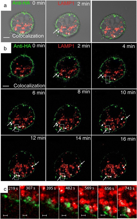
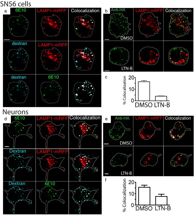
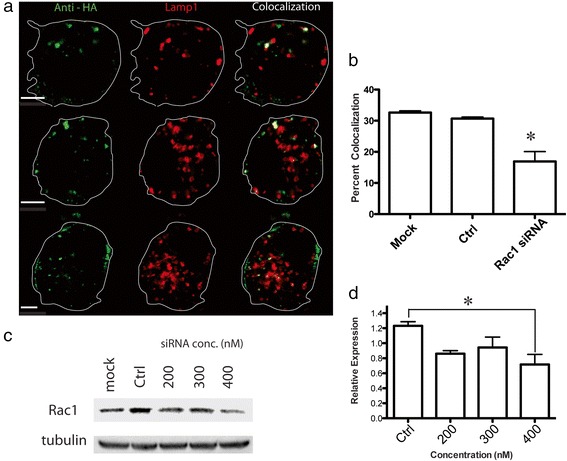
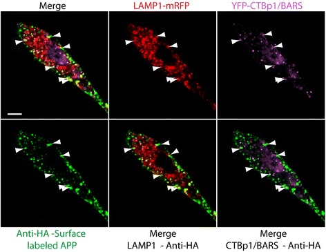
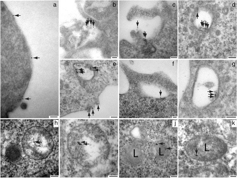
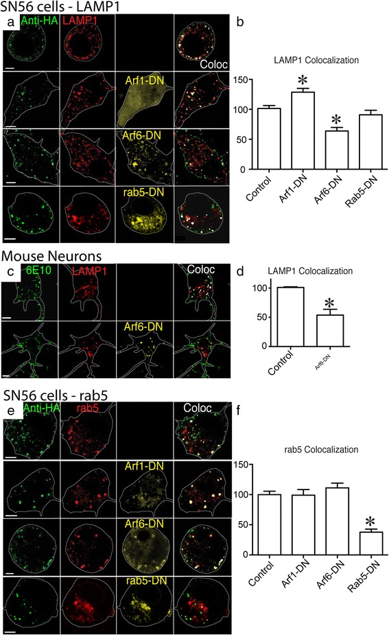
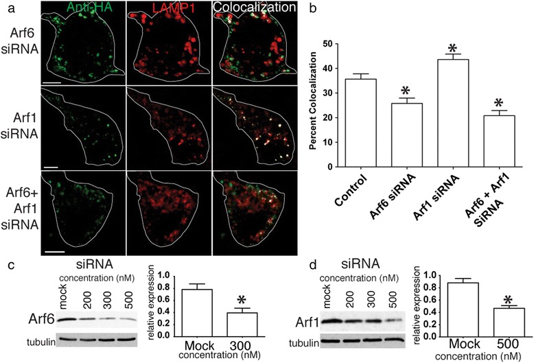
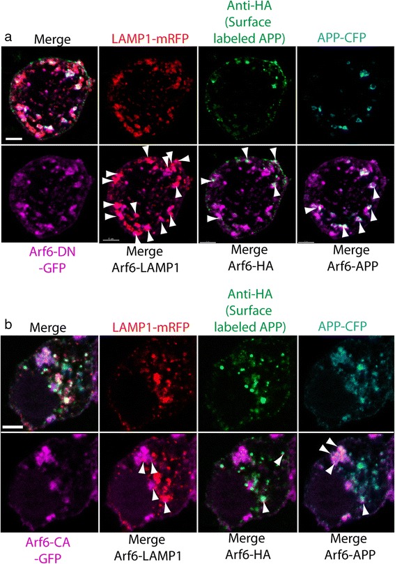
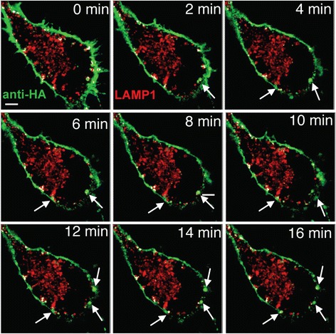
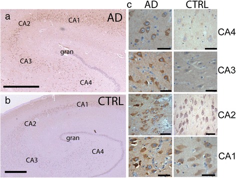
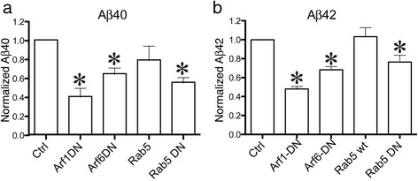
Alzheimer's disease (AD) is characterized by the deposition of Beta-Amyloid (Aβ) peptides in the brain. Aβ peptides are generated by cleavage of the Amyloid Precursor Protein (APP) by the β - and γ - secretase enzymes. Although this process is tightly linked to the internalization of cell surface APP, the compartments responsible are not well defined. We have found that APP can be rapidly internalized from the cell surface to lysosomes, bypassing early and late endosomes. Here we show by confocal microscopy and electron microscopy that this pathway is mediated by macropinocytosis. APP internalization is enhanced by antibody binding/crosslinking of APP suggesting that APP may function as a receptor. Furthermore, a dominant negative mutant of Arf6 blocks direct transport of APP to lysosomes, but does not affect classical endocytosis to endosomes. Arf6 expression increases through the hippocampus with the development of Alzheimer's disease, being expressed mostly in the CA1 and CA2 regions in normal individuals but spreading through the CA3 and CA4 regions in individuals with pathologically diagnosed AD. Disruption of lysosomal transport of APP reduces both Aβ40 and Aβ42 production by more than 30 %. Our findings suggest that the lysosome is an important site for Aβ production and that altering APP trafficking represents a viable strategy to reduce Aβ production.


Figures

Fig. 1
Fig. 1
Live cell imaging demonstrates rapid transport of APP to Lysosomes. SN56 cells transfected with LAMP1-mRFP (red) and HA-βAPP-CFP (not shown) were surface labeled with Zenon-647 anti-HA antibodies (green) for 30 min, and then placed on a microscope stage at 37 °C and imaged by confocal microscopy. Images were taken about every 30 s. a Images showing a DIC brightfield channel superimposed on confocal microscopy images. Co-localized pixels were identified by Imaris software and are overlaid in white (see Methods). b Images showing confocal channels and colocalization channels without the brightfield channel. Surface labeled APP can be seen internalizing in very large vesicles (indicated by arrows) that fuse directly with lysosomes (arrow heads). Rectangle shows region enlarged in part c. (See also Additional file 2: Video S1A and B). Scale bar = 5 microns. c Enlarged inset of a single vesicle forming and fusing with a LAMP1 positive lysosome. Scale Bar = 1 micron
Fig. 2
Fig. 2
Rapid lysosomal transport of APP occurs by Macropinocytosis. a SN56 cells were transfected with untagged full length APP695 and Lamp1-mRFP (red) and surface labeled the antibody 6E10 labeled with Alexa Fluor 647 (green) on ice. Cells were placed in HBSS containing fluorescent dextran (blue) and allowed to internalize at 37 °C for 15 min, washed in HBSS and fixed. The approximate outline of each cell is superimposed for clarity. Colocalized pixels identified by Imaris software are overlayed in white. These images show that Surface labeled APP and dextran are both rapidly co-internalized together to a LAMP1 positive compartment. Scale bar = 5 microns. b SN56 cells were transfected with HA-βAPP-CFP and Lamp1-mRFP (red), surface labeled with Alexa Fluor 647 anti-HA antibodies (green) on ice and incubated at 37 °C for 15 min with or without 2.5 μM latrunculin B. c The percentage of green pixels (internalized surface-labeled APP) colocalized with red pixels (LAMP1) was calculated by Imaris Software and from at least 38 cells drawn from 3 separate experiments is graphed. * denotes p < 0.05 (t-test). d Mouse cortical neurons were transfected with untagged full length APP695 and Lamp1-mRFP (red) and surface labeled the antibody 6E10 labeled with Alexa Fluor 647 (green) on ice and then placed in HBSS containing fluorescent dextran (blue) at 37 °C for 15 min. Colocalized pixels are overlayed in white. APP and dextran are both rapidly internalize together to a LAMP1 positive compartment. Scale bar = 5 microns. e SN56 cells were transfected with HA-βAPP-CFP and Lamp1-mRFP (red), surface labeled with Alexa Fluor 647 anti-HA antibodies (green) on ice and incubated at 37 °C for 15 min with or without 2.5 μM latrunculin B. f Quantitation of the colocalization of internalized surface-labeled APP and LAMP1 was generated using Imaris software from at least 30 cells drawn from 4 independent experiments. * denotes p < 0.05 (t-test)
Fig. 3
Fig. 3
Rac1 knockdown with siRNA blocks direct lysosomal transport of APP. a SN56 cells were transfected with HA-βAPP-CFP (not shown) and LAMP1-mRFP (red), and 400 nM siRNA against Rac1. Cells were surface labeled on ice for with 647-labeled Anti-HA antibodies for 30 min and allowed to internalize APP for 15 min at 37 °C. Dye-labeled negative control siRNA was included with these transfections to confirm that they were transfected with siRNA (not shown). The approximate outline of each cell is superimposed for clarity. Co-localized pixels are shown in white. Scale bar = 5 microns. b Co-localization of internalized APP and LAMP1 in the presence of Rac1 siRNA was quantitated in at least 38 cells from at drawn from at least 4 experiments using a one-way ANOVA with Tukey post-test * denotes p < 0.05. c SN56 cells were grown in 60 mm dishes and transfected with increasing amounts of siRNA directed against Rac1. Cells were lysed and western blotted with antibodies against Rac1, stripped and reprobed with antibodies against tubulin. d Western blots from at least 3 experiments were quantified using ImageJ, and normalized to tubulin expression in 4 experiments using a one-way ANOVA with Tukey post-test. * denotes p < 0.05
Fig. 4
Fig. 4
CTBp1/BARS is colocalized with Anti-HA and LAMP1-mRFP in macropinosomes at the cell surface. SN56 cells transfected with LAMP1-mRFP (red) and HA-βAPP-CFP (not shown), and CTBp1/BARS-YFP (purple) were surface labeled with Zenon-647 anti-HA antibodies (green) for 30 min, incubated at 37 °C for 15 min, fixed and imaged. Colocalization of surface labled APP, CTBp1/BARS, and LAMP1 appears in structures near the cell surface. Scale bar = 5 microns
Fig. 5
Fig. 5
Electron Microscopy of APP Endocytosed into Macropinosomes and Lysosomes. SN56 cells were transfected with HA-βAPP-CFP and surface-labeled with Anti-HA antibody, followed by an anti-mouse antibody labeled with 10 nm gold and Alexa Fluor 488 on ice. When cells were fixed and sectioned immediately (a) gold particles are seen only on the cell surface. After incubation for 15 min at 37 °C, gold particles are seen collected at the cell surface in regions of membrane ruffling (b, c). Gold particles were seen in structures surrounded by vesicles and tubules suggestive of sorting endosomes (d) as well as in large clear structures with the appearance of macropinosomes (e, f, g). Gold particles were also seen in structures containing a mixture of clear areas and electron dense regions, suggestive of macropinosomes fused with primary lysosomes (e, h, i) and in electron dense structures typical of lysosomes (j, k). Scale Bar = 100 nm
Fig. 6
Fig. 6
Cell surface antibody binding enhances rapid transport of APP to the lysosome but not to the early endosome. a SN56 cells were transfected with βAPP-CFP (not shown) and either LAMP1-mRFP or Rab5-mRFP or (red). APP was surface-labeled with FlAsH reagent (green) on ice for 3 min. No antibody labeling was used. Cells were either directly fixed (0 min) or incubated at 37 °C for 15 min and then fixed. Co-localized green/red pixels are overlayed in white. Each image shows a single cell, with approximate outline superimposed for clarity. In the absence of antibody, FlAsH labeled APP moves mostly to the early endosome. Approximate outlines of each cell is overlaid. Scale bar = 5 microns. b SN56 cells were transfected with HA-βAPP-CFP (not shown) and LAMP1-mRFP (red). APP was surface-labeled with FlAsH reagent (yellow) on ice for 3 min and then with 647-labeled anti-HA antibody (green) for 30 min on ice. After 15 min at 37 °C, both labels show extensive co-localization with lysosome. c Co-localized pixels were quantitated from the antibody crosslinking experiments in A and B in at least 10 cells from at least 3 experiments using an ANOVA with Tukey post-test. * denotes p < 0.05. Scale bar = 5 microns
Fig. 7
Fig. 7
An Arf6-DN mutant blocks APP transport to the lysosome but not the early endosome. a SN56 cells were transfected with HA-βAPP-CFP (not shown), LAMP1-mRFP (red) and a dominant negative mutant of a GTPase fused to fluorescent protein (yellow or cyan) as indicated. Cells were then surface-labeled with 647-labeled anti-HA antibody (green) for 30 min on ice and then allowed to internalize APP for 15 min at 37 °C and fixed. Each panel shows a single cell with approximate its outline superimposed for clarity. Merged red and green channels are shown, with co-localized pixels overlayed in white. Cell outlines are overlaid in white. Scale bar = 5 microns. c Quantitation of APP internalization to the lysosome with values normalized to HA-βAPP-CFP/LAMP1-mRFP cells examined at the time of the experiment. Colocalization was determined from more than 37 cells drawn from at least 4 transfections, and analyzed by a one way ANOVA with Tukey Post-test. * denotes p < 0.05. b Mouse cortical neurons were transfected with APP695, and LAMP1-mRFP (red), with or without Arf6-DN. Cells were then surface-labeled with 647-labeled 6E10 (green) and then allowed to internalize. Merged red and green channels are shown. Co-localized pixels overlayed in white. Scale bar = 5 microns. d Quantitation of APP internalization to the lysosome normalized to lysosome with values normalized to APP695/LAMP1-mRFP transfected cells at the time of the experiment. Data is from at least 58 cells in each group drawn from 5 experiments using a one-way ANOVA with Tukey post-test. * denotes p < 0.05. e SN56 cells were transfected with HA-βAPP-CFP (not shown), rab5-mRFP (red) and a dominant negative mutant of a GTPase as indicated. Merged red and green channels are shown, with co-localized pixels overlayed in white. Scale bar = 5 microns. f Quantitation of APP internalization to the early endosome, with values normalized to HA-βAPP-CFP/rab5-mRFP cells examined at the time of the experiment. Data is from more the 30 cells drawn from at least 4 transfections using one-way ANOVA with Tukey post-test. * denotes p < 0.05
Fig. 8
Fig. 8
Arf6 and Arf1 knockdown with siRNA also blocks direct lysosomal transport of APP. a SN56 cells were transfected with HA-βAPP-CFP (not shown) and LAMP1-mRFP (red), and siRNAs against Arf6 and Arf1. Cells were surface labeled on ice for with 647-labeled Anti-HA antibodies for 30 min and allowed to internalize APP for 15 min at 37 °C. Co-localized pixels are shown in white. Scale bar = 5 microns. The approximate outline of each cell is superimposed for clarity. b Co-localization of internalized APP and LAMP1 in the presence of ARF6 and Arf1 siRNAs was quantitated in at least 30 cells from at drawn from at least 3 experiments. * denotes p < 0.05. c Western blots demonstrating Arf6 and Arf1 are knocked down with increasing amounts of siRNA. d) were quantitated in from at least 3 experiments. These are normalized to tubulin expression and quantified
Fig. 9
Fig. 9
An Arf6-DN mutant blocks is localized at the cell surface and on lysosomes. SN56 cells were transfected with HA-βAPP-CFP, LAMP1-mRFP, and Arf6 bearing either a dominant negative GFP-Arf6-T27N-DN or constitutively active GFP-Arf6-Q67L-CA mutation. Cells were surface labeled Alexa Fluor 647 labeled anti-HA antibodies on ice and then allowed to internalize at 37 °C and imaged by confocal microscopy. a Images show the individual color channels of LAMP1, HA (surface-labeled APP), and CFP (APP) along the top row. On the bottom row is the distribution of GFP-Arf6-DN by itself, and merged each of the above channeled. Arf6-DN protein appears on the cell surface and in large intracellular organelles, colocalized with LAMP1 (arrowheads), surface-labeled HA-APP (arrowheads) and with the CFP tag on APP (arrowheads). b The constitutively active GFP-Arf6-Q67L-CA, is also present at the cell surface, the cytosol, and on many intracellular organelles. It demonstrates on a much less localization with LAMP1 (arrowheads), surface-labeled HA-APP (arrowheads) or the CFP tag on APP (arrowheads)
Fig. 10
Fig. 10
Arf6-DN arrests APP internalization at the cell surface. SN56 cells transfected with HA-βAPP-CFP (not shown) and LAMp1-mRFP (red) and were surface labeled with Zenon-647 anti-HA antibodies (green) for 30 min. Cells were placed on a microscope stage at 37 °C and imaged by confocal microscopy every 30 s. Co-localized red and green pixels are overlayed in white. Surface labeled APP can be seen entering large vesicles that fail to fuse with lysosomes. (See also Additional file 7: Video S4). Scale bar = 5 microns
Fig. 11
Fig. 11
Arf6 expression spreads through the Hippocampus with progression of Alzheimer’s disease. Hippocampal sections from Alzheimer’s disease and Control subjects were stained with an antibody to Arf6. In low power images images (Aperio Slide scanner), Arf6 can be seen darkly staining CA1-CA4 pyramidal cells in AD brain (a) but only CA1 and CA2 in Control sample (b) Hippocampal Granule cells (gran) are only lightly stained. Scale Bar 1000 μm. Panel c shows high power (40X) images of representative pyramidal cells from each hippocampal region (scale bar 50 μm)
Fig. 12
Fig. 12
Arf6 and Arf1 Dominant negative mutations reduce Aβ production. SN56 cells were transfected with HA- βAPP-CFP, a construct bearing rab5, Arf1 or Arf6 as indicated. After 3 days, media was assayed using an Aβ42 or Aβ40 ELISA. Results are from 4-8 independent experiments with assays performed in duplicate or triplicate, and normalized to control cells. Analysis was performed using a one-way ANOVA with Tukey post-test. a Concentrations of Aβ40 normalized to control. b Concentrations of Aβ42 normalized to control. * denotes p < 0.05

Similar articles

Cited by

References

    1. Hussain I, Powell DJ, Howlett DR, Chapman GA, Gilmour L, Murdock PR, et al. ASP1 (BACE2) cleaves the amyloid precursor protein at the beta-secretase site. Mol Cell Neurosci. 2000;16(5):609–19. doi: 10.1006/mcne.2000.0884. - DOI - PubMed
    1. Kimberly WT, Xia W, Rahmati T, Wolfe MS, Selkoe DJ. The transmembrane aspartates in presenilin 1 and 2 are obligatory for gamma-secretase activity and amyloid beta-protein generation. J Biol Chem. 2000;275(5):3173–8. doi: 10.1074/jbc.275.5.3173. - DOI - PubMed
    1. Selkoe DJ, Wolfe MS. Presenilin: running with scissors in the membrane. Cell. 2007;131(2):215–21. doi: 10.1016/j.cell.2007.10.012. - DOI - PubMed
    1. Pasternak SH, Callahan JW, Mahuran DJ. The role of the endosomal/lysosomal system in amyloid-beta production and the pathophysiology of Alzheimer’s disease: Reexamining the spatial paradox from a lysosomal perspective. J Alzheimers Dis. 2004;6(1):53–65. - PubMed
    1. Grbovic OM, Mathews PM, Jiang Y, Schmidt SD, Dinakar R, Summers-Terio NB, et al. Rab5-stimulated up-regulation of the endocytic pathway increases intracellular beta-cleaved amyloid precursor protein carboxyl-terminal fragment levels and Abeta production. J Biol Chem. 2003;278(33):31261–8. doi: 10.1074/jbc.M304122200. - DOI - PubMed

Publication types

MeSH terms

LinkOut - more resources