Tacrolimus Protects Podocytes from Injury in Lupus Nephritis Partly by Stabilizing the Cytoskeleton and Inhibiting Podocyte Apoptosis
- PMID: 26161538
- PMCID: PMC4498640
- DOI: 10.1371/journal.pone.0132724
Tacrolimus Protects Podocytes from Injury in Lupus Nephritis Partly by Stabilizing the Cytoskeleton and Inhibiting Podocyte Apoptosis
Abstract
Objective: Several studies have reported that tacrolimus (TAC) significantly reduced proteinuria in lupus nephritis (LN) patients and mouse models. However, the mechanism for this effect remains undetermined. This study explored the mechanism of how TAC protects podocytes from injury to identify new targets for protecting renal function.
Methods: MRL/lpr mice were given TAC at a dosage of 0.1 mg/kg per day by intragastric administration for 8 weeks. Urine and blood samples were collected. Kidney sections (2 μm) were stained with hematoxylin-eosin (HE), periodic acid-Schiff base (PAS) and Masson's trichrome stain. Mouse podocyte cells (MPC5) were treated with TAC and/or TGF-β1 for 48 h. The mRNA levels and protein expression of synaptopodin and Wilms' tumor 1 (WT1) were determined by real-time PCR, Western blotting and/or immunofluorescence, respectively. Flow cytometry was used to detect cell apoptosis with annexin V. Podocyte foot processes were observed under transmission electron microscopy. IgG and C3 deposition were assessed with immunofluorescence assays and confocal microscopy.
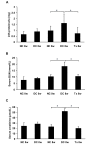
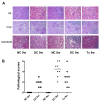
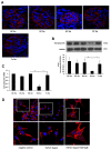
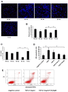
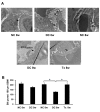
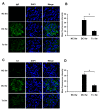
Results: Synaptopodin expression significantly decreased in MRL/lpr disease control mice, accompanied by increases in 24-h proteinuria, blood urea nitrogen, and serum creatinine. TAC, however, reduced proteinuria, improved renal function, attenuated renal pathology, restored synaptopodin expression and preserved podocyte numbers. In MPC5 cells, TGF-β1 enhanced F-actin damage in podocytes and TAC stabilized it. TAC also decreased TGF-β1-induced podocyte apoptosis in vitro and inhibited foot process fusion in MRL/lpr mice. In addition, our results also showed TAC inhibited glomerular deposition of IgG and C3.
Conclusion: This study demonstrated that TAC reduced proteinuria and preserved renal function in LN through protecting podocytes from injury partly by stabilizing podocyte actin cytoskeleton and inhibiting podocyte apoptosis.
Conflict of interest statement
Figures
Similar articles
-
Effects of complement factor D deficiency on the renal disease of MRL/lpr mice.Kidney Int. 2004 Jan;65(1):129-38. doi: 10.1111/j.1523-1755.2004.00371.x. Kidney Int. 2004. PMID: 14675043
-
Mesenchymal stem cells prevent podocyte injury in lupus-prone B6.MRL-Faslpr mice via polarizing macrophage into an anti-inflammatory phenotype.Nephrol Dial Transplant. 2019 Apr 1;34(4):597-605. doi: 10.1093/ndt/gfy195. Nephrol Dial Transplant. 2019. PMID: 29982691
-
Nestin protects podocyte from injury in lupus nephritis by mitophagy and oxidative stress.Cell Death Dis. 2020 May 5;11(5):319. doi: 10.1038/s41419-020-2547-4. Cell Death Dis. 2020. PMID: 32371936 Free PMC article.
-
Unraveling the podocyte injury in lupus nephritis: Clinical and experimental approaches.Semin Arthritis Rheum. 2017 Apr;46(5):632-641. doi: 10.1016/j.semarthrit.2016.10.005. Epub 2016 Oct 17. Semin Arthritis Rheum. 2017. PMID: 27839739 Review.
-
The safety of isotretinoin in patients with lupus nephritis: a comprehensive review.Cutan Ocul Toxicol. 2017 Mar;36(1):77-84. doi: 10.3109/15569527.2016.1169284. Epub 2016 May 10. Cutan Ocul Toxicol. 2017. PMID: 27160965 Review.
Cited by
-
[Lupus nephritis: from diagnosis to treatment].Inn Med (Heidelb). 2023 Mar;64(3):225-233. doi: 10.1007/s00108-023-01489-y. Epub 2023 Feb 10. Inn Med (Heidelb). 2023. PMID: 36763102 Review. German.
-
Effect of Tacrolimus vs Intravenous Cyclophosphamide on Complete or Partial Response in Patients With Lupus Nephritis: A Randomized Clinical Trial.JAMA Netw Open. 2022 Mar 1;5(3):e224492. doi: 10.1001/jamanetworkopen.2022.4492. JAMA Netw Open. 2022. PMID: 35353167 Free PMC article. Clinical Trial.
-
A Comprehensive Literature Review on Managing Systemic Lupus Erythematosus: Addressing Cardiovascular Disease Risk in Females and Its Autoimmune Disease Associations.Cureus. 2023 Aug 18;15(8):e43725. doi: 10.7759/cureus.43725. eCollection 2023 Aug. Cureus. 2023. PMID: 37727166 Free PMC article. Review.
-
A network meta-analysis of randomized controlled trials comparing the effectiveness and safety of voclosporin or tacrolimus plus mycophenolate mofetil as induction treatment for lupus nephritis.Z Rheumatol. 2023 Sep;82(7):580-586. doi: 10.1007/s00393-021-01087-z. Epub 2021 Sep 20. Z Rheumatol. 2023. PMID: 34545430 English.
-
Cytoskeleton Rearrangement in Podocytopathies: An Update.Int J Mol Sci. 2024 Jan 4;25(1):647. doi: 10.3390/ijms25010647. Int J Mol Sci. 2024. PMID: 38203817 Free PMC article. Review.
References
-
- Kitamura N, Matsukawa Y, Takei M, Sawada S. Antiproteinuric effect of angiotensin-converting enzyme inhibitors and an angiotensin II receptor blocker in patients with lupus nephritis. J Int Med Res. 2009;37: 892–898. - PubMed
Publication types
MeSH terms
Substances
Grants and funding
LinkOut - more resources
Full Text Sources
Other Literature Sources
Miscellaneous

