. 2015 Jul 7:6:7285.
doi: 10.1038/ncomms8285.

Phosphoproteomics reveals malaria parasite Protein Kinase G as a signalling hub regulating egress and invasion

Affiliations

Phosphoproteomics reveals malaria parasite Protein Kinase G as a signalling hub regulating egress and invasion

Mahmood M Alam et al. Nat Commun. .

Abstract

Our understanding of the key phosphorylation-dependent signalling pathways in the human malaria parasite, Plasmodium falciparum, remains rudimentary. Here we address this issue for the essential cGMP-dependent protein kinase, PfPKG. By employing chemical and genetic tools in combination with quantitative global phosphoproteomics, we identify the phosphorylation sites on 69 proteins that are direct or indirect cellular targets for PfPKG. These PfPKG targets include proteins involved in cell signalling, proteolysis, gene regulation, protein export and ion and protein transport, indicating that cGMP/PfPKG acts as a signalling hub that plays a central role in a number of core parasite processes. We also show that PfPKG activity is required for parasite invasion. This correlates with the finding that the calcium-dependent protein kinase, PfCDPK1, is phosphorylated by PfPKG, as are components of the actomyosin complex, providing mechanistic insight into the essential role of PfPKG in parasite egress and invasion.


Figures
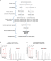
Figure 1
Figure 1. A schematic representation of the quantitative global phosphoproteomic study to determine the cellular targets for PfPKG.
(a) Late schizont stage parasites were purified on MACS column and incubated with or without Compound 2 (2 μM, 60 min) after which parasite lysates were prepared and digested with trypsin. Peptide samples from individual experiments were labelled with isobaric mass tags and the peptides from three independent experiments were pooled, then phosphopeptides were enriched by sequential fractionation using anion-exchange, PHOS-select (IMAC) and TiO2 beads before being analysed by LC-MS/MS. (b,c) Illustrative mass spectra showing an example of a phosphopeptide that was less abundant following Compound 2 treatment (b) and one that was unchanged by Compound 2 treatment (c).
Figure 2
Figure 2. Cellular targets for PfPKG in schizonts.
By comparing the impact on the phosphoproteome of the PfPKG inhibitor, Compound 2, in wild-type 3D7 parasites and a mutant parasite strain expressing the Compound 2 insensitive PfPKGT618Q, we identified 107 phosphorylation sites on 69 parasite proteins as cellular targets of PfPKG. The proteins were divided into Gene Ontology groups. Those marked with an * are proteins that share an association with the motor complex, suggesting that they might function as motor-associated proteins.
Figure 3
Figure 3. PfCDPK1 is a PfPKG cellular target.
(a,b) Representative spectra from the global phosphoproteomic data demonstrating that phosphorylation of serine-64 in PfCDPK1 is downregulated following Compound 2 treatment (2 μM, 60 min; Comp 2) of wild-type schizonts (a) but not in PfPKGT618Q mutant parasites (b). The inset on the left is a zoom of each spectrum to show the relative abundance of reporter ions from three experiments (126 and 127 from Expt. 1, 128 and 129 from Expt. 2, and 130 and 131 from Expt. 3). The inset on the right hand side is the fragmentation table showing the observed b-ions in orange and y-ions in blue. (c) Box plot representation of the quantitative changes in abundance of a non-phosphorylated PfCDPK1 peptide derived from wild-type and PfPKGT618Q mutant parasites and (d) of peptides spanning the serine-64 phosphorylation site in PfCDPK1. (e,f) Representative western blots and quantification of n=5 experiments ±s.e.m. showing changes in phosphorylation at serine-64 of PfCDPK1 in wild-type parasites (e) and PfPKGT618Q parasites (f) using an in-house antibody to phosphoserine-64 on CDPK1 (CDPK1-pS64). Blots were stripped and re-blotted using a structural PfCDPK1 antibody as a loading control. Student's paired t-test was applied to test statistical significance ***P<0.001.
Figure 4
Figure 4. Parasite myosin A (PfMyoA) is a cellular target for PfPKG.
(a,b) Box plot display of the quantitative changes in abundance of (a) a non-phosphorylated PfMyoA peptide derived from wild-type and PfPKGT618Q parasites treated with vehicle or Compound 2 (2 μM; Comp 2) and (b) of a peptide containing the serine-19 phosphorylation of PfMyoA. (c,d) Western blot of parasite lysates prepared from (c) wild-type parasites and (d) PfPKGT618Q parasites, treated with vehicle or Comp 2 (2 μM). The blots were probed either with a phospho-specific antibody to phosphorylated serine-19 on PfMyoA (MyoA-pS19) or with an antibody against PfMyoA as a loading control. The western blot data shown are representative of three independent experiments and the quantification of n=3 experiments ±s.e.m. Student's paired t-test was applied to test statistical significance ***P<0.001.
Figure 5
Figure 5. PfCDPK1 is phosphorylated by PfPKG in vitro.
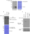
(a) An in vitro kinase reaction with [32P]-ATP was carried out using a recombinant HIS-tagged PfPKG (1 μg) with histone (2 μg) as a substrate in the presence and absence of cGMP (10 μM). (b) An in vitro kinase reaction with GST-tagged ‘kinase dead' mutant of PfCDPK1 where asparate-191 was substituted for an asparagine (CDPK1-KD, 1 μg). This construct was used as a substrate for purified recombinant HIS-tagged PfPKG (1 μg) in the presence and absence of cGMP (10 μM). Coomassie-stained gel is shown as a loading control. (c) In vitro PfPKG kinase assay performed with recombinant GST-tagged CDPK1-KD (50 ng) as a substrate or a mutant version of CDPK1-KD where serine-64 was substituted by alanine (CDPK1-KD(S-A); 50 ng). The kinase assay was conducted either in the presence or absence of PfPKG (50 ng) and the resulting reaction probed with a phospho-specific antibody to phosphoserine-64 on CDPK1 (CDPK1-pS64). Blots were stripped and probed with a structural PfCDPK1 antibody as a loading control. The results shown are representative of three independent experiments.
Figure 6
Figure 6. Phosphorylation of PfCDPK1 at S64 occurs during the schizont (Schiz) stage of parasite development.
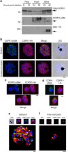
(a) Parasite lysates (15 μg) prepared from ring, trophozoite (Troph) or Schiz stage and were probed with the structural PfCDPK1 antibody, the phospho-specific CDPK1-pS64 antibody or the Schiz-specific marker PfEBA-175. (b) A late Schiz culture treated with vehicle or Compound 2 (2μM; 60min) was fixed and probed with DAPI stain to reveal the nuclei (blue), the anti-HA antibody to reveal PfCDPK1-HA expressed in transgenic parasites (red) or the phospho-specific CDPK1-pS64 antibody (green). A merge of images from all three stains is shown as well as a differential interference contrast (DIC) image. (c,d) Free merozoites were fixed and probed with DAPI stain to reveal the nuclei (blue), the anti-HA antibody to reveal PfCDPK1-HA (red) or the phospho-specific CDPK1-pS64 antibody (green). A merge of images from all three stains is shown. (e) A rendered image where deconvoluted z stacks (see; Supplementary Movie 1) were reconstructed in 3D, with interpolation, of wild-type parasites stained with DAPI (blue), the structural CDPK1 antibody to reveal the total pool of PfCDPK1 (red) and the CDPK1-pS64 antibody (green). (f) Same as e but in this case the images (see; Supplementary Movie 2—video for z stacks) are of a free merozoite. These results are representative of at least three experiments. Scale bars, b=1 μm, c=0.5 μm, d=0.5 μm, e=1 μm, f=0.5 μm
Figure 7
Figure 7. PfCDPK1 phosphorylated at serine-64 is associated with apical parasite structures.
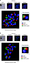
(a) A schizont stage parasite stained with DAPI to reveal the nuclei (blue), an antibody to PfEBA-175 to reveal the micronemes (red) and the phospho-specific CDPK1-pS64 antibody (green). A merge of images from all three stains is shown on the far right; this is a representative image from at least three experiments. Also shown is a rendered image where deconvoluted z stacks (see: Supplementary Movie 3) were reconstructed in 3D, with interpolation. The inset shows a rendered image of a free merozoite from the same preparation (see: Supplementary Movie 4). (b) The same as a, but instead of probing with an anti-EBA-175 antibody the preparation was probed with an antibody to the rhoptry marker PfTRAMP (red;see: Supplementary Movie 5 (schizonts) and Supplementary Movie 6 (merozoites) for z stacks). Scale bars, a=1 μm (insert=0.5 μm), b=1 μm (insert=0.5 μm),
Figure 8
Figure 8. PfCDPK1 exists as a high-molecular-weight (Mwt) complex that is regulated by PfPKG.
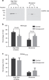
(a) A parasite lysate was prepared from parasites expressing a HA-tagged PfCDPK1. The lysate was fractionated over a Superdex 200 gel filtration column and the fractions probed by western blot with an anti-HA antibody. Shown is a representative of three independent experiments. (b) Quantification of the relative proportion of CDPK1-HA staining that appears in the high-Mwt complex and in the monomeric form—from parasites treated with vehicle and from parasites treated with Compound 2. (c) The experiment shown in b was repeated using the PfPKGT618Q mutant parasites. In this instance, the fractions from the Superdex column were probed with the structural CDPK1 antibody to detect the high-Mwt complex and the monomeric form of PfCDPK1. The results shown are from three experiments ±s.e.m. Student's paired t-test was applied to test statistical significance.
Figure 9
Figure 9. Parasite red blood cell invasion is dependent on PfPKG activity.
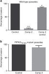
(a) Invasion of wild-type free merozoites into red blood cells was monitored in the absence or presence of Compound 2 (2 μM; Comp 2) by the determination of the number of infected red blood cells 24 h post invasion. Comp 2 was also added 1 h post infection and the number of infected red blood cells 24 h later was determined (Comp 2 Control); this was to control for the possibility that Comp 2 treatment might affect the survival of the parasites post invasion. (b) The invasion of PfPKGT618Q merozoites into red blood cells was monitored. The results are the mean of 3–5 experiments ±s.e.m. Student's paired t-test was applied to test statistical significance. ***P<0.001, *P<0.05.
Figure 10
Figure 10. Schematic representation of the role of cGMP/PfPKG signalling in egress/invasion.
The present study has identified a number of cellular targets for PfPKG, which appear to map to the reported roles for cGMP and calcium signalling in egress and invasion. We describe here the PfPKG-dependent phosphorylation of a subpopulation of PfCDPK1 localized at the apical pole, which together with other calcium-dependent kinases, most notably PfCDPK5, represents an interplay between calcium and cGMP/PfPKG signalling necessary to mediate egress. Interestingly, this may also involve phosphorylation of additional proteins involved in egress, such as the recently described PfSEA1, which we demonstrate here to be a target (either direct or indirect) for PfPKG. Furthermore, the identification of a number of proteins involved in parasite invasion, particularly those associated with the actomyosin motor complex, provides a mechanistic explanation for the essential role played by PfPKG in invasion that is described in our study.

Similar articles

Cited by

References

    1. Hunter T. & Pawson T. The evolution of protein phosphorylation. Preface. Philos. Trans. R. Soc. Lond. B Biol. Sci. 367, 2512 (2012). - PMC - PubMed
    1. Newman R. H. et al.. Construction of human activity-based phosphorylation networks. Mol. Syst. Biol. 9, 655 (2013). - PMC - PubMed
    1. Jin J. & Pawson T. Modular evolution of phosphorylation-based signalling systems. Philos. Trans. R. Soc. Lon. B Biol. Sci. 367, 2540–2555 (2012). - PMC - PubMed
    1. Johnson L. N. Protein kinase inhibitors: contributions from structure to clinical compounds. Q. Rev. Biophys. 42, 1–40 (2009). - PubMed
    1. Lucet I. S., Tobin A., Drewry D., Wilks A. F. & Doerig C. Plasmodium kinases as targets for new-generation antimalarials. Future Med. Chem. 4, 2295–2310 (2012). - PubMed

Publication types

MeSH terms

Substances