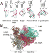
RNA Structures as Mediators of Neurological Diseases and as Drug Targets
- PMID: 26139368
- PMCID: PMC4508199
- DOI: 10.1016/j.neuron.2015.06.012
RNA Structures as Mediators of Neurological Diseases and as Drug Targets
Abstract
RNAs adopt diverse folded structures that are essential for function and thus play critical roles in cellular biology. A striking example of this is the ribosome, a complex, three-dimensionally folded macromolecular machine that orchestrates protein synthesis. Advances in RNA biochemistry, structural and molecular biology, and bioinformatics have revealed other non-coding RNAs whose functions are dictated by their structure. It is not surprising that aberrantly folded RNA structures contribute to disease. In this Review, we provide a brief introduction into RNA structural biology and then describe how RNA structures function in cells and cause or contribute to neurological disease. Finally, we highlight successful applications of rational design principles to provide chemical probes and lead compounds targeting structured RNAs. Based on several examples of well-characterized RNA-driven neurological disorders, we demonstrate how designed small molecules can facilitate the study of RNA dysfunction, elucidating previously unknown roles for RNA in disease, and provide lead therapeutics.
Copyright © 2015 Elsevier Inc. All rights reserved.
Figures


Similar articles
-
Repeat RNA expansion disorders of the nervous system: post-transcriptional mechanisms and therapeutic strategies.Crit Rev Biochem Mol Biol. 2021 Feb;56(1):31-53. doi: 10.1080/10409238.2020.1841726. Epub 2020 Nov 10. Crit Rev Biochem Mol Biol. 2021. PMID: 33172304 Free PMC article. Review.
-
RNA-protein interactions in unstable microsatellite diseases.Brain Res. 2014 Oct 10;1584:3-14. doi: 10.1016/j.brainres.2014.03.039. Epub 2014 Apr 4. Brain Res. 2014. PMID: 24709120 Free PMC article. Review.
-
2D and 3D FISH of expanded repeat RNAs in human lymphoblasts.Methods. 2017 May 1;120:49-57. doi: 10.1016/j.ymeth.2017.04.002. Epub 2017 Apr 9. Methods. 2017. PMID: 28404480
-
RNA-mediated neurodegeneration in repeat expansion disorders.Ann Neurol. 2010 Mar;67(3):291-300. doi: 10.1002/ana.21948. Ann Neurol. 2010. PMID: 20373340 Free PMC article. Review.
-
Small molecule targeting of RNA structures in neurological disorders.Ann N Y Acad Sci. 2020 Jul;1471(1):57-71. doi: 10.1111/nyas.14051. Epub 2019 Apr 9. Ann N Y Acad Sci. 2020. PMID: 30964958 Free PMC article. Review.
Cited by
-
Critical Design Factors for Electrochemical Aptasensors Based on Target-Induced Conformational Changes: The Case of Small-Molecule Targets.Biosensors (Basel). 2022 Oct 1;12(10):816. doi: 10.3390/bios12100816. Biosensors (Basel). 2022. PMID: 36290952 Free PMC article. Review.
-
Diagnostic and therapeutic potential of microRNAs in neuropsychiatric disorders: Past, present, and future.Prog Neuropsychopharmacol Biol Psychiatry. 2017 Feb 6;73:87-103. doi: 10.1016/j.pnpbp.2016.03.010. Epub 2016 Apr 9. Prog Neuropsychopharmacol Biol Psychiatry. 2017. PMID: 27072377 Free PMC article. Review.
-
Identifying and validating small molecules interacting with RNA (SMIRNAs).Methods Enzymol. 2019;623:45-66. doi: 10.1016/bs.mie.2019.04.027. Epub 2019 May 15. Methods Enzymol. 2019. PMID: 31239057 Free PMC article.
-
Huntingtin and Its Role in Mechanisms of RNA-Mediated Toxicity.Toxins (Basel). 2021 Jul 14;13(7):487. doi: 10.3390/toxins13070487. Toxins (Basel). 2021. PMID: 34357961 Free PMC article. Review.
-
RNA structure determination: From 2D to 3D.Fundam Res. 2023 Jun 12;3(5):727-737. doi: 10.1016/j.fmre.2023.06.001. eCollection 2023 Sep. Fundam Res. 2023. PMID: 38933295 Free PMC article. Review.
References
-
- Akimoto C, Volk AE, van Blitterswijk MM, Van den Broeck M, Leblond CS, Lumbroso S, Camu W, Neitzel B, Onodera O, van Rheenen W, et al. A blinded international study on the reliability of genetic testing for GGGGCC-repeat expansions in C9orf72 reveals marked differences in results among 14 laboratories. J Med Genet. 2014;51:419–424. - PMC - PubMed
-
- Antar LN, Li C, Zhang H, Carroll RC, Bassell GJ. Local functions for FMRP in axon growth cone motility and activity-dependent regulation of filopodia and spine synapses. Mol Cell Neurosci. 2006;32:37–48. - PubMed
-
- Arocena DG, Iwahashi CK, Won N, Beilina A, Ludwig AL, Tassone F, Schwartz PH, Hagerman PJ. Induction of inclusion formation and disruption of lamin A/C structure by premutation CGG-repeat RNA in human cultured neural cells. Hum Mol Genet. 2005;14:3661–3671. - PubMed
Publication types
MeSH terms
Substances
Grants and funding
LinkOut - more resources
Full Text Sources
Other Literature Sources
Medical

