Development of a recombinant human ovarian (BG1) cell line containing estrogen receptor α and β for improved detection of estrogenic/antiestrogenic chemicals
- PMID: 26139245
- PMCID: PMC4772679
- DOI: 10.1002/etc.3146
Development of a recombinant human ovarian (BG1) cell line containing estrogen receptor α and β for improved detection of estrogenic/antiestrogenic chemicals
Erratum in
-
Corrigendum.Environ Toxicol Chem. 2017 May;36(5):1405. doi: 10.1002/etc.3528. Epub 2016 Jul 7. Environ Toxicol Chem. 2017. PMID: 28423203 No abstract available.
Abstract
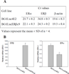
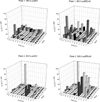
Estrogenic endocrine-disrupting chemicals are found in environmental and biological samples, commercial and consumer products, food, and numerous other sources. Given their ubiquitous nature and potential for adverse effects, a critical need exists for rapidly detecting these chemicals. The authors developed an estrogen-responsive recombinant human ovarian (BG1Luc4E2) cell line recently accepted by the US Environmental Protection Agency (USEPA) and Organisation for Economic Co-operation and Development (OECD) as a bioanalytical method to detect estrogen receptor (ER) agonists/antagonists. Unfortunately, these cells appear to contain only 1 of the 2 known ER isoforms, ERα but not ERβ, and the differential ligand selectivity of these ERs indicates that the currently accepted screening method only detects a subset of total estrogenic chemicals. To improve the estrogen screening bioassay, BG1Luc4E2 cells were stably transfected with an ERβ expression plasmid and positive clones identified using ERβ-selective ligands (genistein and Br-ERβ-041). A highly responsive clone (BG1LucERβc9) was identified that exhibited greater sensitivity and responsiveness to ERβ-selective ligands than BG1Luc4E2 cells, and quantitative reverse-transcription polymerase chain reaction confirmed the presence of ERβ expression in these cells. Screening of pesticides and industrial chemicals identified chemicals that preferentially stimulated ERβ-dependent reporter gene expression. Together, these results not only demonstrate the utility of this dual-ER recombinant cell line for detecting a broader range of estrogenic chemicals than the current BG1Luc4E2 cell line, but screening with both cell lines allows identification of ERα- and ERβ-selective chemicals.
Keywords: BG1Luc ER TA; Environmental screening; Estrogen receptor bioassay; Estrogenic chemicals.
© 2015 SETAC.
Conflict of interest statement
The authors declare that they have no conflict or competing interests with regards to this manuscript.
Figures


Similar articles
-
Identification of putative estrogen receptor-mediated endocrine disrupting chemicals using QSAR- and structure-based virtual screening approaches.Toxicol Appl Pharmacol. 2013 Oct 1;272(1):67-76. doi: 10.1016/j.taap.2013.04.032. Epub 2013 May 23. Toxicol Appl Pharmacol. 2013. PMID: 23707773 Free PMC article.
-
Integrated Model of Chemical Perturbations of a Biological Pathway Using 18 In Vitro High-Throughput Screening Assays for the Estrogen Receptor.Toxicol Sci. 2015 Nov;148(1):137-54. doi: 10.1093/toxsci/kfv168. Epub 2015 Aug 13. Toxicol Sci. 2015. PMID: 26272952 Free PMC article.
-
Endocrine-Disrupting Chemicals (EDCs): In Vitro Mechanism of Estrogenic Activation and Differential Effects on ER Target Genes.Environ Health Perspect. 2013 Apr;121(4):459-66. doi: 10.1289/ehp.1205951. Epub 2013 Feb 5. Environ Health Perspect. 2013. PMID: 23384675 Free PMC article.
-
Estrogen Receptor β as a Possible Double-Edged Sword Molecule in Breast Cancer: A Mechanism of Alteration of Its Role by Exposure to Endocrine-Disrupting Chemicals.Biol Pharm Bull. 2021;44(11):1594-1597. doi: 10.1248/bpb.b21-00468. Biol Pharm Bull. 2021. PMID: 34719637 Review.
-
Regulation of specific target genes and biological responses by estrogen receptor subtype agonists.Curr Opin Pharmacol. 2010 Dec;10(6):629-36. doi: 10.1016/j.coph.2010.09.009. Epub 2010 Oct 14. Curr Opin Pharmacol. 2010. PMID: 20951642 Free PMC article. Review.
Cited by
-
Screening of Estrogenic-Disrupting Compounds in Dairy Products Based on the Estrogen Receptor Cocktail.Foods. 2022 Apr 19;11(9):1178. doi: 10.3390/foods11091178. Foods. 2022. PMID: 35563901 Free PMC article.
-
The small heat shock protein B8 (HSPB8) modulates proliferation and migration of breast cancer cells.Oncotarget. 2017 Feb 7;8(6):10400-10415. doi: 10.18632/oncotarget.14422. Oncotarget. 2017. PMID: 28060751 Free PMC article.
-
Using Estrogenic Activity and Nontargeted Chemical Analysis to Identify Contaminants in Sewage Sludge.Environ Sci Technol. 2021 May 18;55(10):6729-6739. doi: 10.1021/acs.est.0c07846. Epub 2021 Apr 28. Environ Sci Technol. 2021. PMID: 33909413 Free PMC article.
-
Estrogenic activity of capsule coffee using the VM7Luc4E2 assay.Curr Res Toxicol. 2021 May 18;2:210-216. doi: 10.1016/j.crtox.2021.05.003. eCollection 2021. Curr Res Toxicol. 2021. PMID: 34345863 Free PMC article.
-
Identification of nonmonotonic concentration-responses in Tox21 high-throughput screening estrogen receptor assays.Toxicol Appl Pharmacol. 2022 Oct 1;452:116206. doi: 10.1016/j.taap.2022.116206. Epub 2022 Aug 19. Toxicol Appl Pharmacol. 2022. PMID: 35988584 Free PMC article.
References
-
- Bergman k, Heindel JJ, Jobling S, Kidd KA, Zoeller RT World Health Organization., United Nations Environment Programme. State of the science of endocrine disrupting chemicals - 2012: an assessment of the state of the science of endocrine disruptors prepared by a group of experts for the United Nations Environment Programme (UNEP) and WHO. Geneva, Switzerland: UNEP: WHO; 2013.
-
- Tyler CR, Jobling S, Sumpter JP. Endocrine disruption in wildlife: a critical review of the evidence. Crit Rev Toxicol. 1998;28:319–361. - PubMed
-
- Rogers JM, Denison MS. Recombinant cell bioassays for endocrine disruptors: Development of a stably transfected human ovarian cell line for the detection of estrogenic and anti-estrogenic chemicals. In Vitro Mol Toxicol. 2000;13:67–82. - PubMed
-
- Klein KO. Is there a role for estrogen activity assays? Recombinant cell bioassay for estrogen: Development and applications. Steroids. 2014;9:108–112. - PubMed
-
- Van der Linden SC, Heringa MB, Man HY, Sonneveld E, Puijker LM, Brouwer A, Van der Burg B. Detection of multiple hormonal activities in wastewater effluents and surface water, using a panel of steroid receptor CALUX bioassays. Environ Sci Technol. 2008;42:5814–5820. - PubMed
Publication types
MeSH terms
Substances
Grants and funding
LinkOut - more resources
Full Text Sources
Other Literature Sources
Research Materials
Miscellaneous
