. 2015 Jul 1;35(26):9676-88.
doi: 10.1523/JNEUROSCI.0513-15.2015.

α6-Containing GABAA Receptors Are the Principal Mediators of Inhibitory Synapse Strengthening by Insulin in Cerebellar Granule Cells

Affiliations

α6-Containing GABAA Receptors Are the Principal Mediators of Inhibitory Synapse Strengthening by Insulin in Cerebellar Granule Cells

Michael V Accardi et al. J Neurosci. .

Abstract

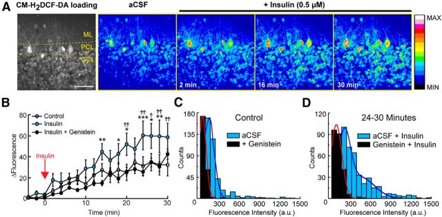
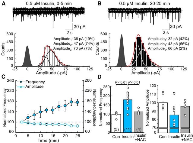
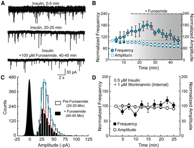
Activity-dependent strengthening of central synapses is a key factor driving neuronal circuit behavior in the vertebrate CNS. At fast inhibitory synapses, strengthening is thought to occur by increasing the number of GABAA receptors (GABARs) of the same subunit composition to preexisting synapses. Here, we show that strengthening of mouse cerebellar granule cell GABAergic synapses occurs by a different mechanism. Specifically, we show that the neuropeptide hormone, insulin, strengthens inhibitory synapses by recruiting α6-containing GABARs rather than accumulating more α1-containing receptors that are resident to the synapse. Because α6-receptors are targeted to functionally distinct postsynaptic sites from α1-receptors, we conclude that only a subset of all inhibitory synapses are strengthened. Together with our recent findings on stellate cells, we propose a general mechanism by which mature inhibitory synapses are strengthened. In this scenario, α1-GABARs resident to inhibitory synapses form the hardwiring of neuronal circuits with receptors of a different composition fulfilling a fundamental, but unappreciated, role in synapse strengthening.

Keywords: inhibition; insulin; metabolism; mitochondria; plasticity mechanism; reactive oxygen species.


Figures

Figure 1.
Figure 1.
mROS selectively increase the occurrence of inhibitory events of small amplitude. A, B, Top, mIPSCs from the same granule cell (cell #140307000) at the beginning (0–5 min) and during (20–25 min) pipette perfusion with antimycin-A (2 μm). Bottom, Amplitude histograms comparing the data obtained at two time periods (i.e., 0–5 min, left; 20–25 min, right, n = 4). Each was fit with the sum of three Gaussian functions (red line), with individual functions shown in either black (left panel) or white (right panel). In this figure and elsewhere, the closed point distribution (filled; dark gray) was scaled to the fitted peak of the lowest event amplitude and represents the average baseline noise observed during the recordings. C, Summary plot of the time course of mIPSC frequency (filled circle) and amplitude (open circle) during pipette perfusion with antimycin-A. Data are mean ± SEM. D, Bar graphs of maximal normalized frequency (left) and amplitude (right) obtained in the different recording conditions. Anti, antimycin-A; NAC, N-acetylcysteine (1 mm). Statistical test was a one-way ANOVA with Tukey's post hoc test. The frequency comparison of “Control” versus “Anti + NAC” (D; left) was not significant and, for clarity, was not indicated in the figure. Amplitude comparisons were not significant (p = 0.58; one-way ANOVA).
Figure 2.
Figure 2.
Furosemide block distinguishes between α1- and α6-containing GABAA receptors. A, Overlay of typical membrane currents elicited by short 1 ms applications of 10 mm GABA on recombinant α1β2γ2 (left, patch #141219p8; n = 4) and α6β2γ2 (middle, patch #141219p3; n = 5) or averaged mIPSC from a granule cell (right, cell #140605000; n = 5) before (black line) and after (red line) bath application of 100 μm furosemide. B, Bar graph comparing the degree of furosemide block on charge transfer between recombinant and native GABARs. Data are mean ± SEM, with paired Student's t tests and Bonferroni correction. C, Plot comparing mIPSC amplitude versus decay kinetics observed in the first 5 min (0–5 min, open circles) and last 5 min (20–25 min, filled circles) of recording in antimycin-A (2 μm).
Figure 3.
Figure 3.
Pharmacological block of α6-containing GABAA receptors prevents synaptic strengthening by mROS. A, mIPSCs from two time points (cell #140329013) from the same cell during bath application of furosemide (100 μm) and internal perfusion with antimycin-A (2 μm). B, Plot summarizing the effect of antimycin-A on event frequency (filled circle) and amplitude (open circle) (n = 5). C, Amplitude histograms of data from the same cells at two time points fit with the sum of 2 Gaussian functions (purple represents 0–5 min; red represents 20–25 min). D, Bar graphs comparing the effect of furosemide on mIPSC amplitude (left) and frequency (right) during internal patch perfusion with antimycin-A. In all cases, data are mean ± SEM.
Figure 4.
Figure 4.
Synaptic strengthening induced by mROS is reversed by pharmacological block of α6-containing GABAA receptors. A, Representative electrophysiological traces of a control (left, cell #140526000) and antimycin-A (2 μm, right, cell #140619000) treated granule cell at three separate time points. B, Amplitude distributions of mIPSCs from two time points (n = 5 in each case). For clarity, only the summed Gaussian functions are shown (purple represents Pre-Furosemide; red represents + Furosemide). C, Summary plot showing the time course of mIPSC frequency (red circle) and amplitude (open circle). Furosemide (100 μm) was added 15 min after the start of the experiment. Gray gradient represents the gradual equilibration of furosemide in the bath. D, Summary plots of furosemide's effect on mIPSC frequency and amplitude. Data are mean ± SEM.
Figure 5.
Figure 5.
mROS fail to strengthen inhibitory synapses of δ-KO mice. A, Representative recordings from a granule cell lacking the δ-subunit before (top) and after (bottom) applying furosemide (100 μm, cell #140621010) to the bath 15 min after the start of the experiment. B, Amplitude distributions comparing mIPSCs at the two time points (n = 5). For clarity, summed Gaussian functions are shown (purple represents Control, 0–5 min; red represents + Furosemide, 40–45 min). C, Averaged mIPSCs (from 15 random events) comparing the response profile of wild-type (cell #140605000) and δ-KO (cell #1406210010) cells before (black line) and after (red line) furosemide application. D, Summary plot showing mIPSC frequency (left) and amplitude (right) before (“control,” open circles, 0–5 min) and after (“+ furo,” filled circle, 40–45 min) furosemide (100 μm) application. Statistical test is a paired Student's t test. n.s, Not significant. E, Summary plots showing the time course of mIPSC frequency (left) and amplitude (right) of granule cells (n = 5) lacking the δ-subunit in the presence (filled circle) and absence (open circle) of antimycin-A (2 μm). Data are mean ± SEM.
Figure 6.
Figure 6.
Insulin increases cytosolic ROS in cerebellar granule cells. A, Example DCF images from a typical insulin-treated experiment showing elevations in cytosolic ROS. The images are displayed using a color-coded map of fluorescence intensity (16 colors) ranging from black (no fluorescence), to white (maximum signal for any pixel over the course of an individual experiment). A representative two-photon single focal plane image (far left) of a cerebellar parasagittal slice with the cellular layers denoted. Ml, Molecular layer; PCL, Purkinje cell layer; GCL, granule cell layer. Scale bar, 5 μm. Right, The 16 color version of this image. B, Summary plot of the time course of fluorescence intensity changes in control (white circle), insulin (cyan circles), or insulin + genistein (black circles) treated conditions. Data are mean ± SEM. Two-way repeated-measures ANOVA with Holm-Sidak's multiple-comparison test: *Comparisons between “insulin” and “insulin + genistein.” †Comparisons between “insulin” and “control.” *p < 0.05. †p < 0.05. **p < 0.01. ††p < 0.01. ***p < 0.001. C, Histograms comparing the level of fluorescence intensity obtained in the presence (black) or absence (cyan) of 50 μm genistein. Each histogram was fit with a single Gaussian function (red represents genistein; dark blue represents aCSF). D, Histograms comparing the level of fluorescence intensity obtained after bath application of insulin (0.5 μm) in the presence (black) or absence (cyan) of 50 μm genistein at the end of a typical imaging recording. Each histogram was fit with either a single (red represents genistein + insulin) or the sum of two (dark blue represents aCSF + insulin), Gaussian functions.
Figure 7.
Figure 7.
Insulin selectively increases the occurrence of inhibitory events of small amplitude. A, B, Top, mIPSCs from the same granule cell (cell #1503150007) at the beginning (0–5 min) and during (20–25 min) bath application with insulin (0.5 μm). Bottom, Amplitude histograms comparing the data obtained at two time periods (i.e., 0–5 min, left; 20–25 min, right; n = 11). Each was fit with the sum of three Gaussian functions (red line) with individual functions shown in either black (left) or white (right). C, Summary plot of the time course of mIPSC frequency (filled circle) and amplitude (open circle) during constant bath application with insulin. Data are mean ± SEM. D, Bar graphs of maximal normalized frequency (left) and amplitude (right) obtained in the different recording conditions. Control, Con (n = 5); N-acetylcysteine (1 mm), NAC (n = 4). Statistical test was a one-way ANOVA with Tukey's post hoc test. The frequency comparison of “Con” versus “Insulin + NAC” (D; left) was not significant and, for clarity, was not indicated in the figure. Amplitude comparisons were not significant (p = 0. 26; one-way ANOVA).
Figure 8.
Figure 8.
Synaptic strengthening induced by insulin is attenuated by pharmacological block of α6-containing GABAA receptors. A, Representative electrophysiological traces of a cerebellar granule cell in the presence of insulin (0.5 μm, cell #150319000) at three separate time points. B, Summary plot showing the time course of mIPSC frequency (filled cyan circle) and amplitude (open circle). Furosemide (100 μm) was added 15 min after the start of the experiment. Insulin was present within the bath throughout the duration of the experiment. Gray gradient represents the gradual equilibration of furosemide in the bath. C, Amplitude distributions of mIPSCs from two time points (n = 6). For clarity, only the summed Gaussian functions are shown (cyan represents Pre-Furosemide: red represents + Furosemide). D, Summary plot showing the time course of mIPSC frequency (filled circle) and amplitude (open circle) during internal perfusion of wortmannin (1 μm) during continuous bath application of insulin (n = 6). Data are mean ± SEM.

Similar articles

Cited by

References

    1. Accardi MV, Daniels BA, Brown PM, Fritschy JM, Tyagarajan SK, Bowie D. Mitochondrial reactive oxygen species regulate the strength of inhibitory GABA-mediated synaptic transmission. Nat Commun. 2014;5:3168. doi: 10.1038/ncomms4168. - DOI - PMC - PubMed
    1. Attwell D, Buchan AM, Charpak S, Lauritzen M, Macvicar BA, Newman EA. Glial and neuronal control of brain blood flow. Nature. 2010;468:232–243. doi: 10.1038/nature09613. - DOI - PMC - PubMed
    1. Barnard EA, Skolnick P, Olsen RW, Möhler H, Sieghart W, Biggio G, Braestrup C, Bateson AN, Langer SZ. International Union of Pharmacology: XV. Subtypes of gamma-aminobutyric acid A receptors: classification on the basis of subunit structure and receptor function. Pharmacol Rev. 1998;50:291–313. - PubMed
    1. Belelli D, Harrison NL, Maguire J, Macdonald RL, Walker MC, Cope DW. Extrasynaptic GABAA receptors: form, pharmacology, and function. J Neurosci. 2009;29:12757–12763. doi: 10.1523/JNEUROSCI.3340-09.2009. - DOI - PMC - PubMed
    1. Bogdanov Y, Michels G, Armstrong-Gold C, Haydon PG, Lindstrom J, Pangalos M, Moss SJ. Synaptic GABAA receptors are directly recruited from their extrasynaptic counterparts. EMBO J. 2006;25:4381–4389. doi: 10.1038/sj.emboj.7601309. - DOI - PMC - PubMed

Publication types

MeSH terms