. 2015 Jul 14;112(28):8768-73.
doi: 10.1073/pnas.1501831112. Epub 2015 Jun 29.

Neural activity and CaMKII protect mitochondria from fragmentation in aging Caenorhabditis elegans neurons

Affiliations

Neural activity and CaMKII protect mitochondria from fragmentation in aging Caenorhabditis elegans neurons

Hao-Ching Jiang et al. Proc Natl Acad Sci U S A. .

Abstract

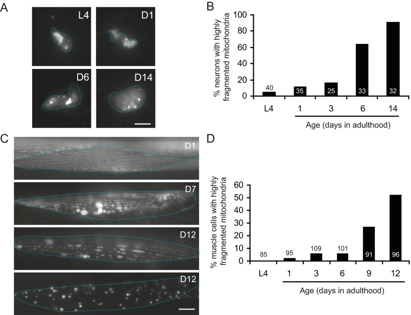
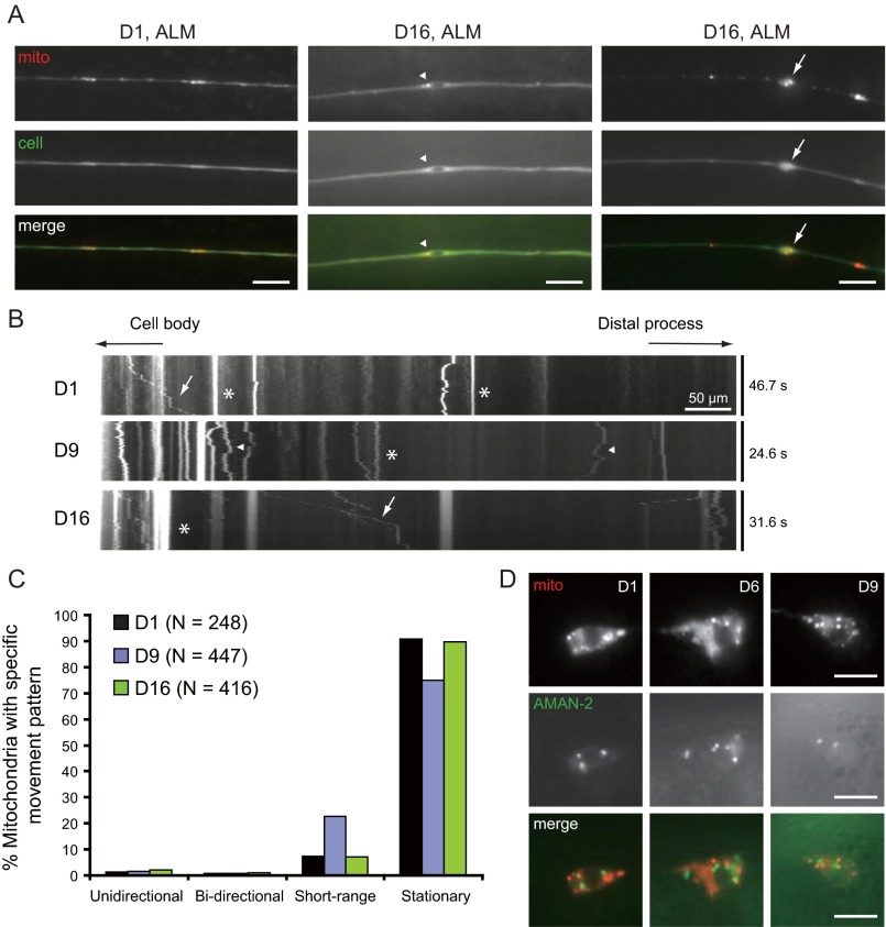
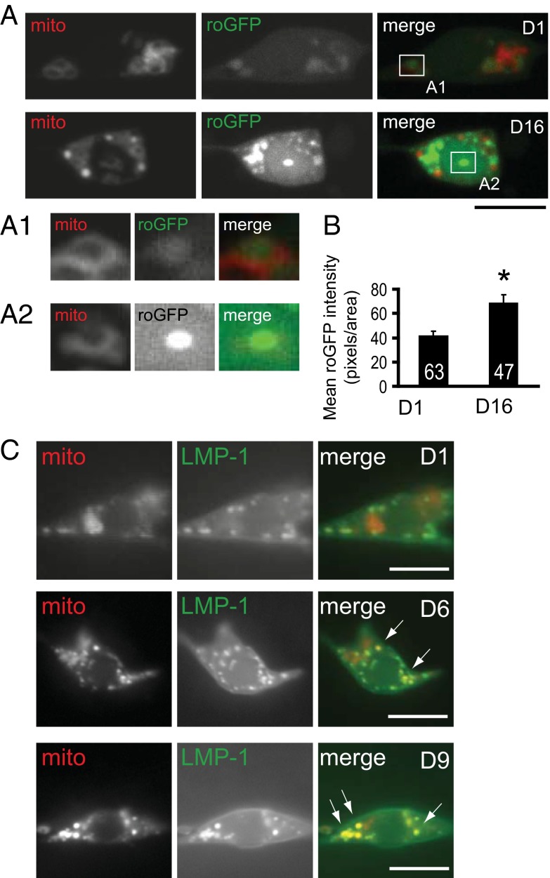
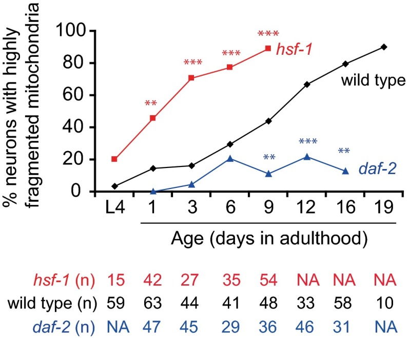
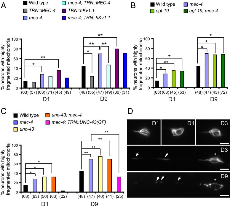
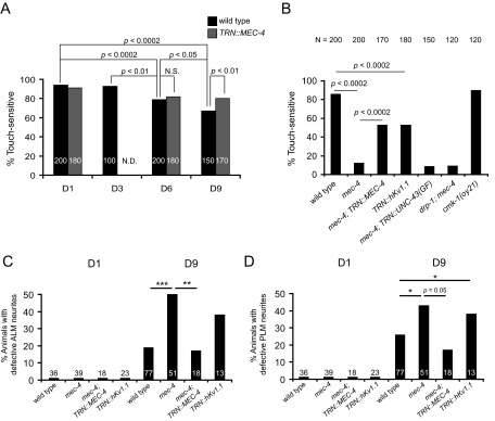
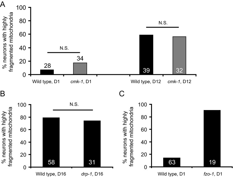
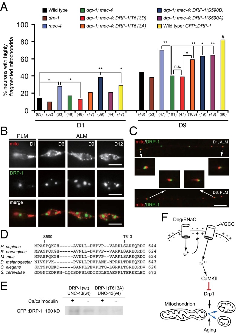
Decline in mitochondrial morphology and function is a hallmark of neuronal aging. Here we report that progressive mitochondrial fragmentation is a common manifestation of aging Caenorhabditis elegans neurons and body wall muscles. We show that sensory-evoked activity was essential for maintaining neuronal mitochondrial morphology, and this activity-dependent mechanism required the Degenerin/ENaC sodium channel MEC-4, the L-type voltage-gated calcium channel EGL-19, and the Ca/calmodulin-dependent kinase II (CaMKII) UNC-43. Importantly, UNC-43 phosphorylated and inhibited the dynamin-related protein (DRP)-1, which was responsible for excessive mitochondrial fragmentation in neurons that lacked sensory-evoked activity. Moreover, enhanced activity in the aged neurons ameliorated mitochondrial fragmentation. These findings provide a detailed description of mitochondrial behavior in aging neurons and identify activity-dependent DRP-1 phosphorylation by CaMKII as a key mechanism in neuronal mitochondrial maintenance.

Keywords: C. elegans; CaMKII; mitochondria; neural activity; neuronal aging.


Conflict of interest statement

The authors declare no conflict of interest.

Figures

Fig. 1.
Fig. 1.
Age-dependent mitochondrial fragmentation in C. elegans touch neurons. (A) Projected confocal z-stack images of mitochondria in the ALM touch neurons visualized with mito::mCherry. (B) Quantification of neurons with highly fragmented (>50%) mitochondria in the touch neuron soma. n = numbers of neurons scored. See SI Materials and Methods for detail of data analysis. (C) Representative images of FRAP from D4 ALM neurons. Regions subjected to photobleaching were boxed. (D) Quantification of FRAP from D4 ALM neurons. n = 10 and 12 regions of interest (ROIs) on mitochondria for the networked or fragmented categories, respectively. *P < 0.05, **P < 0.01, Mann–Whitney u test. (Scale bars: A, 5 μm; C, 1 μm.)
Fig. S1.
Fig. S1.
Age-dependent mitochondrial fragmentation in C. elegans thermosensory neurons and muscles. (A) Mitochondria in the AFD thermosensory neurons were visualized with Pgcy-8::mito::mCherry, and cells were visualized with Pgcy-8::GFP. Ages of the neurons were indicated. Blue dotted lines outline AFD cell boundaries. (B) Quantification of neurons with highly fragmented mitochondria in the AFD soma. n = numbers of neurons scored. (C) Mitochondria in the body wall muscles were visualized with Pmyo-3::mito::GFP. Ages of the muscle cells were indicated. Blue dotted lines outline single muscle cells. (D) Quantification of muscle cells with highly fragmented mitochondria. n = numbers of body wall muscle cells scored. (Scale bars: A, 5 μm; C, 10 μm.)
Fig. S2.
Fig. S2.
Mitochondria in the neurite of aging C. elegans touch neurons. (A) Mitochondria frequently accumulated at neurite beading sites (arrows) or near the ends of the large bubble-like lesions (arrowheads) in the aged touch neurons. (B) Mitochondrial movements in the PLM neurite during aging. In the representative kymographs, examples of movement patterns include unidirectional anterograde movement (arrows), short-range movement (arrowheads), and stationary (asterisks). (C) Quantification of mitochondrial movement patterns in the PLM neurite. n = total numbers of mitochondrial puncta analyzed for movement patterns. (D) Fluorescence images of touch neurons expressing mito::mCherry and AMAN-2::GFP that marks the Golgi apparatus. (Scale bars: 5 μm.)
Fig. 2.
Fig. 2.
Mitochondrial oxidation, lysosomal recruitment, and disrupted ER-Mitochondrial contact during neuronal aging. (A) Projected z-stack confocal fluorescence images of touch neurons expressing roGFP and mito::mCherry. Select mitochondria (A1 and A2) were highlighted to demonstrate roGFP (matrix) and mito::mCherry (outer mitochondrial membrane). (B) Quantification of roGFP intensity. *P < 0.01, Student’s t test. n = numbers of mitochondria quantified for roGFP signal intensity. Error bars are SE of means. (C) Fluorescence images of touch neurons expressing mito::mCherry and LMP-1::GFP (lysosomes). Arrows are foci of colocalization between mitochondria and lysosomes. (Scale bars: 5 μm.)
Fig. 3.
Fig. 3.
Mitochondrial morphology in longevity mutants. Quantification of highly fragmented mitochondria in the touch neurons of the wild type, hsf-1, or daf-2. Data for the wild type were replotted from those in Fig. 1B. n = numbers of neurons scored. **P < 0.005; ***P < 0.0001, Fisher’s exact test or two-proportion z test. NA, not assessed.
Fig. 4.
Fig. 4.
Neural activity maintains mitochondrial morphology through VGCCs and CaMKII. (AC) Quantification of highly fragmented mitochondria in the touch neurons of the wild type as well as mec-4 (A), egl-19 (B), or unc-43 mutants (C). TRN is the mec-7 promoter. n = numbers of neurons scored. *P < 0.05; **P < 0.01, two-proportion z test. (D) Elongated mitochondria in the ALM neurons of the mec-4 mutants overexpressing UNC-43(GF). Epifluorescence images were shown, with arrows and asterisks indicating mitochondria and the ALM soma, respectively. (Scale bars: 5 μm.)
Fig. S3.
Fig. S3.
Sensory-evoked activity acts cell-autonomously for touch sensitivity, neurite integrity, and mitochondrial maintenance. (A) Sensitivity to gentle touch in animals during aging or with altered touch neuron excitability. Responsiveness to gentle touch in the anterior body region (ALM sensory field) of wild-type aging animals. n = number of trials of responses to gentle touch, with 10 trials tested for an individual animal. (B) Responsiveness to gentle touch in the anterior body region of animals in which touch neuron excitability was altered by mutations in mec-4 or expression of a hyperpolarizing hKv1.1 potassium channel. D1 animals were tested. TRN is the mec-7 promoter. N is as described in A. (C and D) Age-dependent defects of touch neuron processes. Quantification of age-dependent defects in the ALM (C) or the PLM (D) neurites. TRN is the mec-7 promoter. n = numbers of neurons scored. *P < 0.05; **P < 0.01; ***P < 0.001, two-proportion z-test or Fisher’s exact test.
Fig. S4.
Fig. S4.
Mitochondrial fragmentation in the cmk-1, drp-1, and fzo-1 mutants. (A) Mitochondria in the touch neurons of D1 and D12 wild-type or cmk-1 mutant animals. n = numbers of neurons scored. (B) Mitochondria in the touch neurons of D16 wild-type or drp-1 mutant animals. n = numbers of neurons scored. (C) Mitochondria in the touch neurons of D1 wild-type or fzo-1 mutant animals. n = number of neurons scored. N.S., not significant, two-proportion z-test or Fisher’s exact test.
Fig. 5.
Fig. 5.
CaMKII maintains mitochondrial morphology by phosphorylating and inhibiting DRP-1. (A) Quantification of highly fragmented mitochondria in the touch neurons. *P < 0.05; *P < 0.01, two-proportion z test; #P < 0.05, Fisher’s exact test, wild type vs. wild type with GFP::DRP-1. n = numbers of neurons scored. (B) Epifluorescence images of touch neurons expressing mito::mCherry and GFP::DRP-1. (C) GFP::DRP-1 in the PLM neurite. Select DRP-1 puncta were enlarged to show the close association of GFP::DRP-1 with mitochondria. (D) DRP-1 sequence alignment of various species. For C. elegans, DRP-1b is shown. (E) In vitro kinase assay of DRP-1 by UNC-43. (F) Schematic model of mitochondrial maintenance by activity-dependent Drp1/DRP-1 inhibition. Unknown, age-dependent factors contribute to mitochondrial fragmentation independently of Drp1 in wild-type neurons. (Scale bars: B, 5 μm; C, 10 μm.)
Fig. S5.
Fig. S5.
In vitro kinase assay of UNC-43 for DRP-1 phosphorylation. (A) Results of three independent experiments were shown. Asterisks were artifacts due to bubble formation during transfer. (B) Two independent Coomassie blue staining experiments as controls for protein quantity loaded for in vitro kinase assays.
Fig. S6.
Fig. S6.
Additional assessment of mitochondrial defects and touch sensitivity in the wild type and mutants. Mitochondrial fragmentation (AD) and touch sensitivity (EH) were assayed in additional independent populations of wild-type, mec-4 mutant, and transgenic animals expressing hKv1.1 (TRN::hKv1.1) or MEC-4 (TRN::MEC-4) in the touch neurons. Numbers are the number of animals (AD) or numbers of trial of touch assay (EH) scored in each indicated population. Asterisks indicate the samples presented in the text and figures.

Similar articles

Cited by

References

    1. Wallace DC. A mitochondrial paradigm of metabolic and degenerative diseases, aging, and cancer: A dawn for evolutionary medicine. Annu Rev Genet. 2005;39:359–407. - PMC - PubMed
    1. Chan DC. Fusion and fission: Interlinked processes critical for mitochondrial health. Annu Rev Genet. 2012;46:265–287. - PubMed
    1. Scheckhuber CQ, et al. Reducing mitochondrial fission results in increased life span and fitness of two fungal ageing models. Nat Cell Biol. 2007;9(1):99–105. - PubMed
    1. Hughes AL, Gottschling DE. An early age increase in vacuolar pH limits mitochondrial function and lifespan in yeast. Nature. 2012;492(7428):261–265. - PMC - PubMed
    1. Wilson PD, Franks LM. The effect of age on mitochondrial ultrastructure. Gerontologia. 1975;21(2):81–94. - PubMed

Publication types

MeSH terms

Substances

LinkOut - more resources