

Opposing Roles of Dectin-1 Expressed on Human Plasmacytoid Dendritic Cells and Myeloid Dendritic Cells in Th2 Polarization
- PMID: 26123355
- PMCID: PMC4530104
- DOI: 10.4049/jimmunol.1402276
Opposing Roles of Dectin-1 Expressed on Human Plasmacytoid Dendritic Cells and Myeloid Dendritic Cells in Th2 Polarization
Abstract
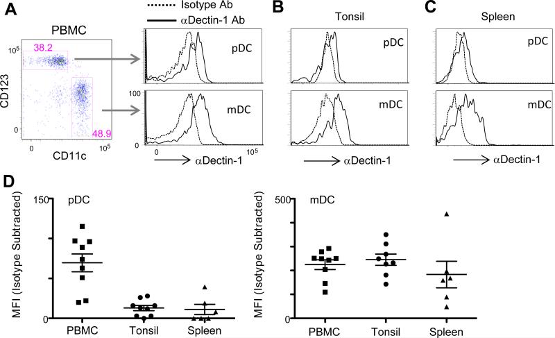
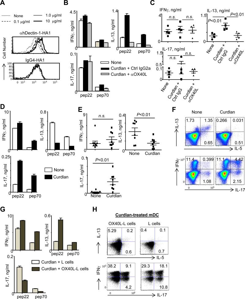
Dendritic cells (DCs) can induce and control host immune responses. DC subset-dependent functional specialties and their ability to display functional plasticity, which is mainly driven by signals via pattern recognition receptors, identify DCs as immune orchestrators. A pattern recognition receptor, Dectin-1, is expressed on myeloid DCs and known to play important roles in Th17 induction and activation during fungal and certain bacterial infections. In this study, we first demonstrate that human plasmacytoid DCs express Dectin-1 in both mRNA and protein levels. More interestingly, Dectin-1-activated plasmacytoid DCs promote Th2-type T cell responses, whereas Dectin-1-activated myeloid DCs decrease Th2-type T cell responses. Such contrasting outcomes of Th2-type T cell responses by the two DC subsets are mainly due to their distinct abilities to control surface OX40L expression in response to β-glucan. This study provides new insights for the regulation of host immune responses by Dectin-1 expressed on DCs.
Copyright © 2015 by The American Association of Immunologists, Inc.
Figures
Similar articles
-
Syk-dependent cytokine induction by Dectin-1 reveals a novel pattern recognition pathway for C type lectins.Immunity. 2005 Apr;22(4):507-17. doi: 10.1016/j.immuni.2005.03.004. Immunity. 2005. PMID: 15845454
-
Dectin-1/2-induced autocrine PGE2 signaling licenses dendritic cells to prime Th2 responses.PLoS Biol. 2018 Apr 18;16(4):e2005504. doi: 10.1371/journal.pbio.2005504. eCollection 2018 Apr. PLoS Biol. 2018. PMID: 29668708 Free PMC article.
-
Dectin-2 promotes house dust mite-induced T helper type 2 and type 17 cell differentiation and allergic airway inflammation in mice.Am J Respir Cell Mol Biol. 2014 Aug;51(2):201-9. doi: 10.1165/rcmb.2013-0522OC. Am J Respir Cell Mol Biol. 2014. PMID: 24588637
-
The role of Dectin-1 in the host defence against fungal infections.Curr Opin Microbiol. 2011 Aug;14(4):392-9. doi: 10.1016/j.mib.2011.07.001. Epub 2011 Jul 29. Curr Opin Microbiol. 2011. PMID: 21803640 Review.
-
Dendritic cells: a conductor of T cell differentiation.Allergol Int. 2007 Sep;56(3):193-9. doi: 10.2332/allergolint.R-07-146. Epub 2007 Aug 1. Allergol Int. 2007. PMID: 17646736 Review.
Cited by
-
Functional Specialty of CD40 and Dendritic Cell Surface Lectins for Exogenous Antigen Presentation to CD8(+) and CD4(+) T Cells.EBioMedicine. 2016 Jan 28;5:46-58. doi: 10.1016/j.ebiom.2016.01.029. eCollection 2016 Mar. EBioMedicine. 2016. PMID: 27077111 Free PMC article.
-
Immune Response in Bacterial and Candida Sepsis.Eur J Microbiol Immunol (Bp). 2019 Oct 4;9(4):105-113. doi: 10.1556/1886.2019.00011. eCollection 2019 Dec 25. Eur J Microbiol Immunol (Bp). 2019. PMID: 31934361 Free PMC article. Review.
-
Dectin-1 Positive Dendritic Cells Expand after Infection with Leishmania major Parasites and Represent Promising Targets for Vaccine Development.Front Immunol. 2018 Feb 26;9:263. doi: 10.3389/fimmu.2018.00263. eCollection 2018. Front Immunol. 2018. PMID: 29535708 Free PMC article.
-
Dectin-1 Signaling Update: New Perspectives for Trained Immunity.Front Immunol. 2022 Feb 14;13:812148. doi: 10.3389/fimmu.2022.812148. eCollection 2022. Front Immunol. 2022. PMID: 35237264 Free PMC article. Review.
-
C-Type Lectin Receptors in Asthma.Front Immunol. 2018 Apr 11;9:733. doi: 10.3389/fimmu.2018.00733. eCollection 2018. Front Immunol. 2018. PMID: 29696023 Free PMC article. Review.
References
-
- Steinman RM, Hawiger D, Nussenzweig MC. Tolerogenic dendritic cells. Annu Rev Immunol. 2003;21:685–711. - PubMed
-
- Figdor CG, van Kooyk Y, Adema GJ. C-type lectin receptors on dendritic cells and Langerhans cells. Nat Rev Immunol. 2002;2:77–84. - PubMed
-
- Brown GD. Dectin-1: a signalling non-TLR pattern-recognition receptor. Nat Rev Immunol. 2006;6:33–43. - PubMed
-
- Geijtenbeek TB, van Vliet SJ, Engering A, t Hart BA, van Kooyk Y. Self- and nonself-recognition by C-type lectins on dendritic cells. Annu Rev Immunol. 2004;22:33–54. - PubMed
-
- Caparros E, Munoz P, Sierra-Filardi E, Serrano-Gomez D, Puig-Kroger A, Rodriguez-Fernandez JL, Mellado M, Sancho J, Zubiaur M, Corbi AL. DC-SIGN ligation on dendritic cells results in ERK and PI3K activation and modulates cytokine production. Blood. 2006;107:3950–3958. - PubMed
Publication types
MeSH terms
Substances
Associated data
- Actions
- Actions
- Actions
- Actions
- Actions
- Actions
- Actions
- Actions
Grants and funding
LinkOut - more resources
Full Text Sources
Other Literature Sources
Miscellaneous