. 2015 Jun 25;10(6):e0130826.
doi: 10.1371/journal.pone.0130826. eCollection 2015.

MED30 Regulates the Proliferation and Motility of Gastric Cancer Cells

Affiliations

MED30 Regulates the Proliferation and Motility of Gastric Cancer Cells

Yong Joo Lee et al. PLoS One. .

Abstract

MED30 is an essential member of the mediator complex that forms a hub between transcriptional activators and RNA polymerase II. However, the expressions and roles of MED30 have been poorly characterized in cancer. In this study, we examined the functional roles of MED30 during gastric cancer progression. It was found that MED30 was overexpressed in gastric cancer tissues and cell lines. Moreover, MED30 overexpression increased the proliferation, migration, and invasion of gastric cancer cells, whereas MED30 knockdown inhibited these effects. Furthermore the knockdown significantly inhibited tumorigenicity in SCID mice. MED30 also promoted the expressions of genes related to epithelial-mesenchymal transition and induced a fibroblast-like morphology. This study shows MED30 has pathophysiological roles in the proliferation, migration, and invasion of gastric cancer cells and suggests that MED30 should be viewed as a potent therapeutic target for malignant gastric carcinoma.


Conflict of interest statement

Competing Interests: The authors have declared that no competing interests exist.

Figures

Fig 1
Fig 1. MED30 was overexpressed in gastric cancer tissues.
(A-D) Immunohistochemical staining showed the overexpression of MED30 in gastric cancer tissues (B-D) versus normal gastric mucosae (A). Its overexpression was observed in invading cancer cells (C) and metastatic cancer cells nearby lymph nodes (D) in addition to the primary cancer site (B). (E) The overexpression of MED30 in gastric cancer cells was determined by real-time PCR using specific primers (NT; normal tissue). GAPDH was used to normalize all data. Values are the means ± SDs of the three independent experiments performed in triplicate. *, p< 0.01 (Student’s t test, versus NT). (F) The overexpression of MED30 in gastric cancer tissues was examined by real-time PCR using specific primers. GAPDH was used to normalize all data. Values are the means ± SDs of the three independent experiments performed in triplicate. *, p< 0.01; **, p< 0.05 (Student’s t test, versus normal gastric tissues). (G) MED30 protein expression in higher N stages (N stages 2 and 3) was significantly greater than in lower N stages (N stage 0, 1) (N = 41, p< 0.05, Mann-Whitney U test).
Fig 2
Fig 2. MED30 regulated the proliferation of gastric cancer cells.
Gastric cancer cells (SNU216 and SNU638) were transfected with MED30 siRNA (100 nM) or scrambled (SCR) siRNA. MED30 knockdown efficiency was determined by western blot (A) and real-time PCR (C) at 48 hr post-transfection. MED30 expression was also examined in cells stably overexpressing MED30 (MED30-over) and in empty control vector-transfected (Mock) cells. (B) MED30 protein levels relative to β-actin were quantified using Multi Gauge V3.1 software. Values indicate MED30 to β-actin band intensity ratios and are the means ± SDs of three independent experiments performed in triplicate. *, p < 0.01 (Student’s t test, versus SCR or Mock). (D) Effect of MED30 knockdown or overexpression on the proliferation of gastric cancer cells (SNU16, SNU216, SNU620, and SNU638). To check the effect of MED30 knockdown, five days after the transfection with 100 nM MED30 siRNA or SCR siRNA, cell viability assays were performed, and to examine the effect of MED30 overexpression, cell viability assays were performed after 3 days of incubation. Values are the means ± SDs of three independent experiments performed in quadruplicate. *, p< 0.01 (Student’s t test, versus SCR or Mock).
Fig 3
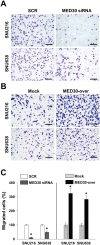
Fig 3. MED30 accelerated the migration of gastric cancer cells.
Cell migration was evaluated using a Boyden chamber assay as described in ‘Materials and Methods’. (A) MED30 knockdown with MED30 siRNA significantly inhibited the FBS-induced migration of SNU216 and SNU638 cells compared with SCR siRNA. (B) MED30 overexpression (MED30-over) significantly increased migration compared to the mock control. (C) Migrated cells were counted and results are presented as a bar graph. Values are the means ± SDs of three independent experiments performed in triplicate. *, p< 0.01 (Student’s t test, versus SCR or Mock). Bar; 100 μm.
Fig 4
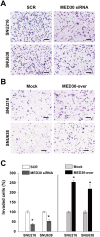
Fig 4. MED30 induced the invasion of gastric cancer cells.
Cell invasion was examined using a Matrigel invasion assay. (A) Knockdown of MED30 significantly inhibited the FBS-induced invasions of SNU216 and SNU638 cells as compared with SCR siRNA. (B) MED30 overexpression (MED30-over) enhanced cell invasion compared with the mock control. (C) Invaded cells were counted and results are presented as a bar graph. Values are the means ± SDs of three independent experiments performed in triplicate. *, p< 0.01 (Student’s t test, versus SCR or Mock). Bar; 100 μm.
Fig 5
Fig 5. Knockdown of MED30 suppressed tumor growth in SCID mice.
(A) SNU638 cells were transfected with MED30 or SCR siRNA and then injected subcutaneously into two sites per mouse. Tumor volumes were measured weekly from week three to seven post-injection. At week seven, mice were sacrificed and tumor volumes (B) and weights (C) were measured. Values are the means ± SDs of three independent experiments performed in triplicate. *, p< 0.01, **p< 0.05 (Student’s t test or one way ANOVA, versus SCR or Mock cells).
Fig 6
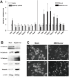
Fig 6. MED30 induced EMT in gastric cancer cells.
Real-time PCR (A) and western blot analysis (B) of E-cadherin (CDH1), N-cadherin (CDH2), P-cadherin (CDH3), twist family bHLH transcription factor 1 (TWIST1/2), vimentin (VIM), and snail family zinc finger 1 and 2 (SNAI1/2) were performed after the knockdown or overexpression of MED30 in SNU638 cells. GAPDH and β-actin were used as internal control for real-time PCR and western blot analysis respectively. Values are the means ± SDs of three independent experiments performed in quadruplicate. *, p< 0.01, **p< 0.05 (Student’s t test, versus Mock). (C) SNU638 cells (MED30-over and mock) were grown in medium containing 10% FBS. After 2 days of culture, the morphologies of mock and MED30-overexpressing cells were observed by bright field microscopy.

Similar articles

Cited by

References

    1. Smith MG, Hold GL, Tahara E, El-Omar EM. Cellular and molecular aspects of gastric cancer. World journal of gastroenterology: WJG. 2006;12(19):2979–90. Epub 2006/05/24. . - PMC - PubMed
    1. Jemal A, Bray F, Center MM, Ferlay J, Ward E, Forman D. Global cancer statistics. CA: a cancer journal for clinicians. 2011;61(2):69–90. Epub 2011/02/08. 10.3322/caac.20107 . - DOI - PubMed
    1. Crew KD, Neugut AI. Epidemiology of gastric cancer. World journal of gastroenterology: WJG. 2006;12(3):354–62. Epub 2006/02/21. - PMC - PubMed
    1. Lauren P. The Two Histological Main Types of Gastric Carcinoma: Diffuse and So-Called Intestinal-Type Carcinoma. An Attempt at a Histo-Clinical Classification. Acta pathologica et microbiologica Scandinavica. 1965;64:31–49. Epub 1965/01/01. . - PubMed
    1. Pacelli F, Papa V, Caprino P, Sgadari A, Bossola M, Doglietto GB. Proximal compared with distal gastric cancer: multivariate analysis of prognostic factors. The American surgeon. 2001;67(7):697–703. Epub 2001/07/14. . - PubMed

Publication types

Grants and funding

This work was supported by the Bio & Medical Technology Development Program (2012M3A9C6050213) and Basic Science Research Foundation (2012R1A1A3010521) of the National Research Foundation (NRF) funded by the Korean government (MEST) and a grant from the National R&D Program for Cancer Control, Ministry for Health, Welfare and Family Affairs, Republic of Korea (0920050). The funders had no role in study design, data collection and analysis, decision to publish, or preparation of the manuscript.