Oleanolic acid induces mitochondrial-dependent apoptosis and G0/G1 phase arrest in gallbladder cancer cells
- PMID: 26109845
- PMCID: PMC4472077
- DOI: 10.2147/DDDT.S84448
Oleanolic acid induces mitochondrial-dependent apoptosis and G0/G1 phase arrest in gallbladder cancer cells
Abstract
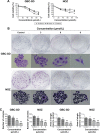
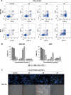
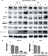
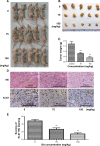
Oleanolic acid (OA), a naturally occurring triterpenoid, exhibits potential antitumor activity in many tumor cell lines. Gallbladder carcinoma is the most common malignancy of the biliary tract, and is a highly aggressive tumor with an extremely poor prognosis. Unfortunately, the effects of OA on gallbladder carcinoma are unknown. In this study, we investigated the effects of OA on gallbladder cancer cells and the underlying mechanism. The results showed that OA inhibits proliferation of gallbladder cancer cells in a dose-dependent and time-dependent manner on MTT and colony formation assay. A flow cytometry assay revealed apoptosis and G0/G1 phase arrest in GBC-SD and NOZ cells. Western blot analysis and a mitochondrial membrane potential assay demonstrated that OA functions through the mitochondrial apoptosis pathway. Moreover, this drug inhibited tumor growth in nude mice carrying subcutaneous NOZ tumor xenografts. These data suggest that OA inhibits proliferation of gallbladder cancer cells by regulating apoptosis and the cell cycle process. Thus, OA may be a promising drug for adjuvant chemotherapy in gallbladder carcinoma.
Keywords: apoptosis; cell cycle arrest; gallbladder carcinoma; mitochondrial pathway; oleanolic acid.
Figures


Similar articles
-
Oleanolic acid inhibits cell survival and proliferation of prostate cancer cells in vitro and in vivo through the PI3K/Akt pathway.Tumour Biol. 2016 Jun;37(6):7599-613. doi: 10.1007/s13277-015-4655-9. Epub 2015 Dec 18. Tumour Biol. 2016. PMID: 26687646
-
Shikonin induces apoptosis and G0/G1 phase arrest of gallbladder cancer cells via the JNK signaling pathway.Oncol Rep. 2017 Dec;38(6):3473-3480. doi: 10.3892/or.2017.6038. Epub 2017 Oct 17. Oncol Rep. 2017. PMID: 29039581
-
20(S)-ginsenoside Rg3 promotes senescence and apoptosis in gallbladder cancer cells via the p53 pathway.Drug Des Devel Ther. 2015 Aug 10;9:3969-87. doi: 10.2147/DDDT.S84527. eCollection 2015. Drug Des Devel Ther. 2015. PMID: 26309394 Free PMC article.
-
Oridonin induces apoptosis and cell cycle arrest of gallbladder cancer cells via the mitochondrial pathway.BMC Cancer. 2014 Mar 21;14:217. doi: 10.1186/1471-2407-14-217. BMC Cancer. 2014. PMID: 24655726 Free PMC article.
-
Anticancer activity of oleanolic acid and its derivatives modified at A-ring and C-28 position.J Asian Nat Prod Res. 2023 Jun;25(6):581-594. doi: 10.1080/10286020.2022.2120863. Epub 2022 Sep 24. J Asian Nat Prod Res. 2023. PMID: 36151896 Review.
Cited by
-
Preparation and antitumor evaluation of self-assembling oleanolic acid-loaded Pluronic P105/d-α-tocopheryl polyethylene glycol succinate mixed micelles for non-small-cell lung cancer treatment.Int J Nanomedicine. 2016 Nov 28;11:6337-6352. doi: 10.2147/IJN.S119839. eCollection 2016. Int J Nanomedicine. 2016. PMID: 27932881 Free PMC article.
-
Oleanolic acid inhibits cell survival and proliferation of prostate cancer cells in vitro and in vivo through the PI3K/Akt pathway.Tumour Biol. 2016 Jun;37(6):7599-613. doi: 10.1007/s13277-015-4655-9. Epub 2015 Dec 18. Tumour Biol. 2016. PMID: 26687646
-
Self-Nanoemulsifying Drug Delivery System (SNEDDS) Using Lipophilic Extract of Viscum album subsp. austriacum (Wiesb.) Vollm.Int J Nanomedicine. 2024 Jun 14;19:5953-5972. doi: 10.2147/IJN.S464508. eCollection 2024. Int J Nanomedicine. 2024. PMID: 38895147 Free PMC article.
-
Oleanolic acid induces p53-dependent apoptosis via the ERK/JNK/AKT pathway in cancer cell lines in prostatic cancer xenografts in mice.Oncotarget. 2018 May 29;9(41):26370-26386. doi: 10.18632/oncotarget.25316. eCollection 2018 May 29. Oncotarget. 2018. PMID: 29899865 Free PMC article.
-
Potential Effects of Nutraceuticals in Retinopathy of Prematurity.Life (Basel). 2021 Jan 22;11(2):79. doi: 10.3390/life11020079. Life (Basel). 2021. PMID: 33499180 Free PMC article. Review.
References
-
- Cao Y, Liu X, Lu W, et al. Fibronectin promotes cell proliferation and invasion through mTOR signaling pathway activation in gallbladder cancer. Cancer Lett. 2015;360:141–150. - PubMed
-
- Li M, Zhang Z, Li X, et al. Whole-exome and targeted gene sequencing of gallbladder carcinoma identifies recurrent mutations in the ErbB pathway. Nat Genet. 2014;46:872–876. - PubMed
Publication types
MeSH terms
Substances
LinkOut - more resources
Full Text Sources
Medical

