. 2015 Jul 7;112(27):E3466-75.
doi: 10.1073/pnas.1501557112. Epub 2015 Jun 22.

An eIF2α-binding motif in protein phosphatase 1 subunit GADD34 and its viral orthologs is required to promote dephosphorylation of eIF2α

Affiliations

An eIF2α-binding motif in protein phosphatase 1 subunit GADD34 and its viral orthologs is required to promote dephosphorylation of eIF2α

Margarito Rojas et al. Proc Natl Acad Sci U S A. .

Abstract

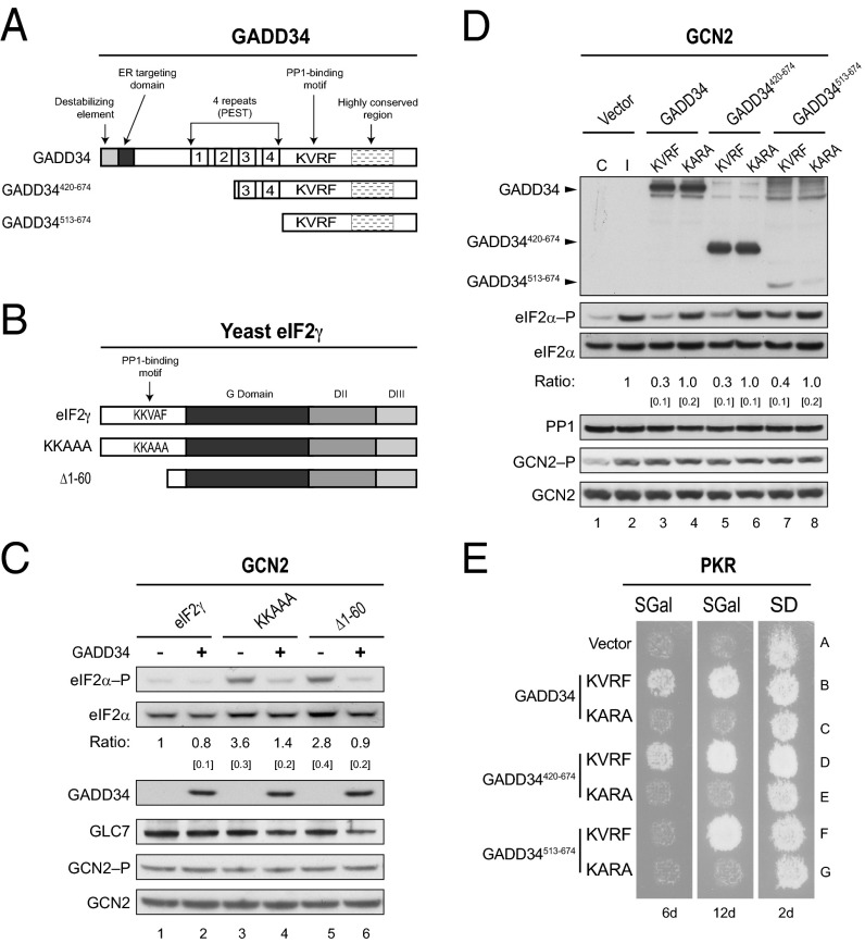
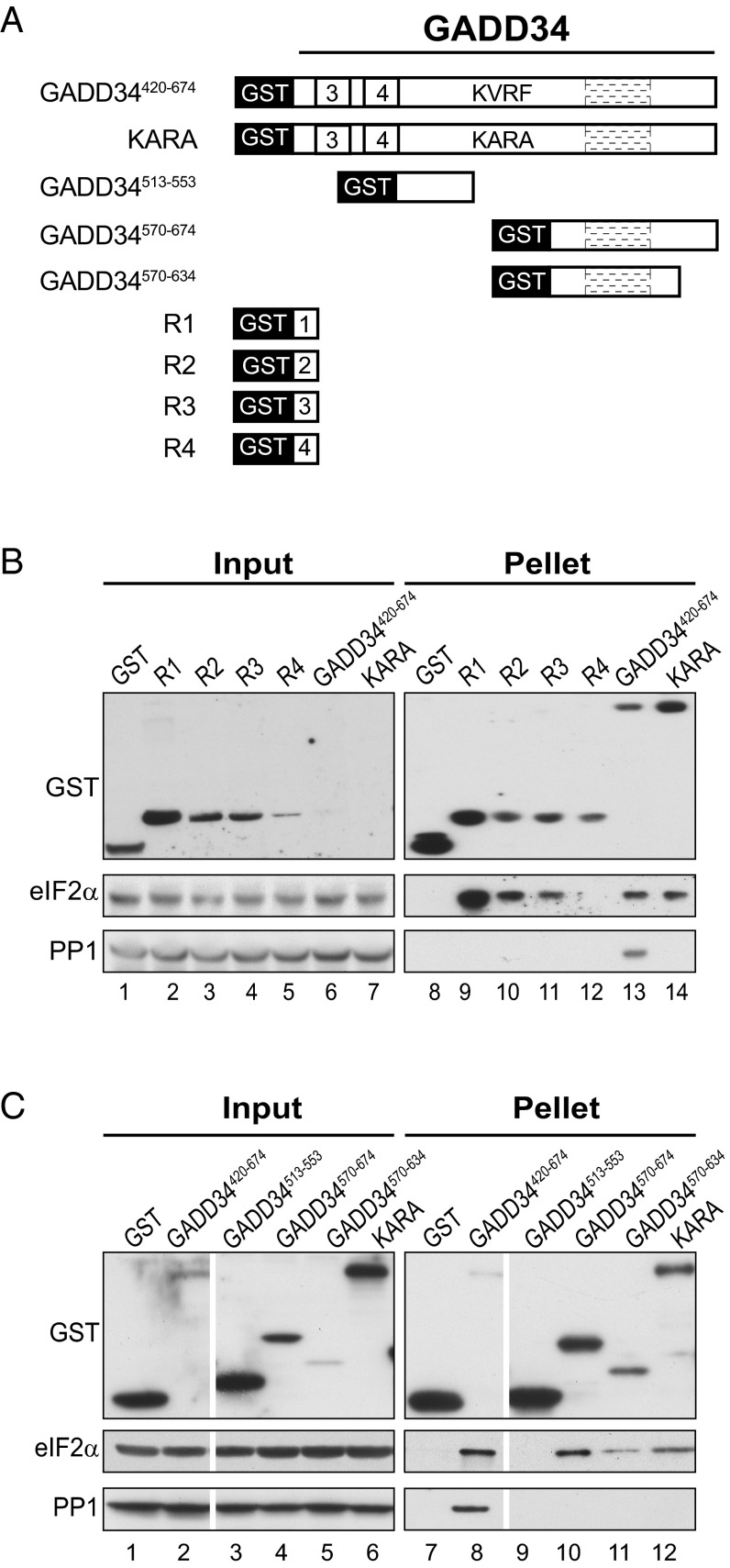
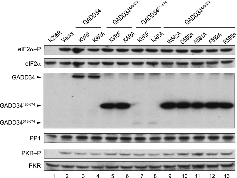
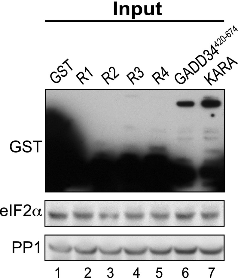
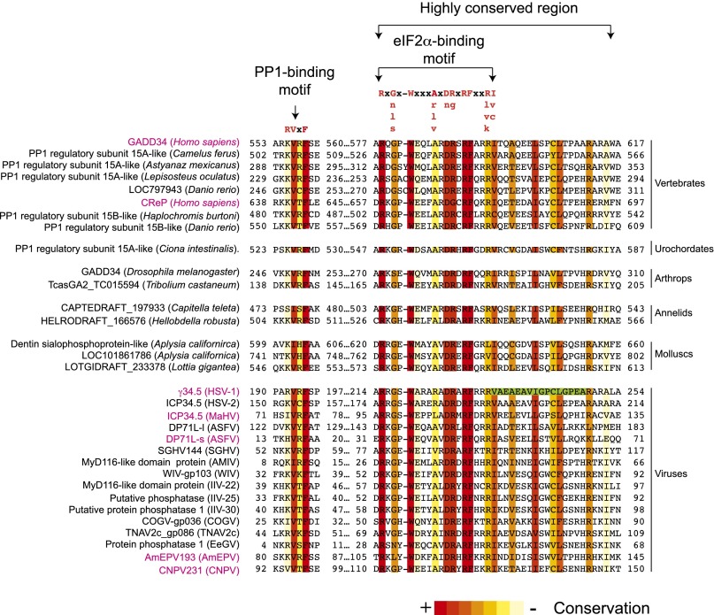
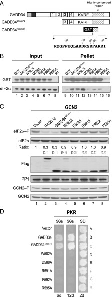
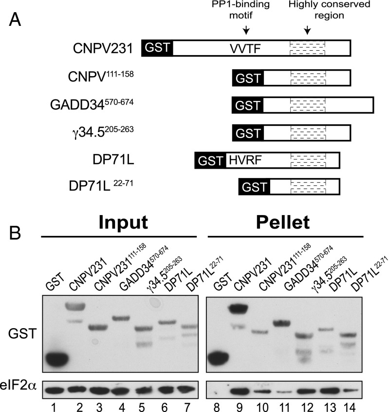
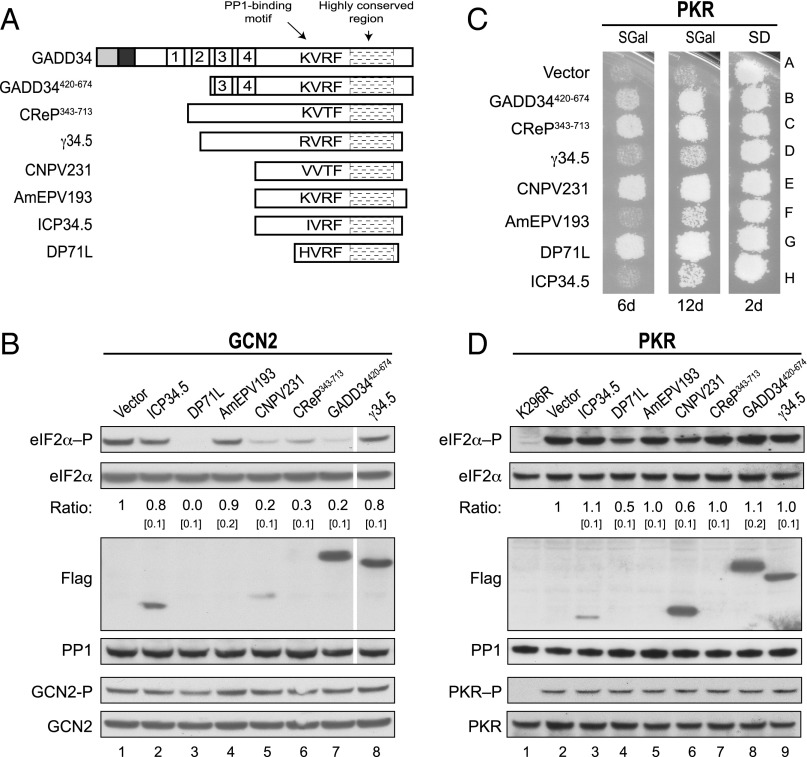
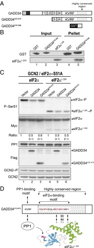
Transient protein synthesis inhibition, mediated by phosphorylation of the α subunit of eukaryotic translation initiation factor 2 (eIF2α), is an important protective mechanism cells use during stress conditions. Following relief of the stress, the growth arrest and DNA damage-inducible protein GADD34 associates with the broadly acting serine/threonine protein phosphatase 1 (PP1) to dephosphorylate eIF2α. Whereas the PP1-binding motif on GADD34 has been defined, it remains to be determined how GADD34 directs PP1 to specifically dephosphorylate eIF2α. In this report, we map a novel eIF2α-binding motif to the C terminus of GADD34 in a region distinct from where PP1 binds to GADD34. This motif is characterized by the consensus sequence Rx[Gnl]x(1-2)Wxxx[Arlv]x[Dn][Rg]xRFxx[Rlvk][Ivc], where capital letters are preferred and x is any residue. Point mutations altering the eIF2α-binding motif impair the ability of GADD34 to interact with eIF2α, promote eIF2α dephosphorylation, and suppress PKR toxicity in yeast. Interestingly, this eIF2α-docking motif is conserved among viral orthologs of GADD34, and is necessary for the proteins produced by African swine fever virus, Canarypox virus, and Herpes simplex virus to promote eIF2α dephosphorylation. Taken together, these data indicate that GADD34 and its viral orthologs direct specific dephosphorylation of eIF2α by interacting with both PP1 and eIF2α through independent binding motifs.

Keywords: CReP; DP71L; PKR; PP1; canarypox.


Conflict of interest statement

The authors declare no conflict of interest.

Figures

Fig. 1.
Fig. 1.
GADD34 promotes eIF2α dephosphorylation in yeast. (A) Schematics of GADD34 and two N-terminally truncated derivatives. Positions of the ER targeting domain, destabilizing element, four repeated sequence elements, PP1-binding motif (KVRF sequence), and the highly conserved region are indicated. (B) Schematic diagram showing S. cerevisiae eIF2γ. The PP1-binding motif (KKVAF sequence) in the N-terminal extension of eIF2γ, the locations of the GTP-binding (G) domain and domains II (DII) and III (DIII), and eIF2γ mutations designed to alter or eliminate the KKVAF motif are indicated. (C) Derivatives of yeast strain YM103 expressing eIF2γ, or its KKAAA or Δ1–60 derivative, and carrying either an empty vector (-) or a plasmid expressing GADD34 (+) under the control of a galactose-inducible promoter were grown in synthetic galactose (SGal) medium to log phase under nonstarvation conditions, and then equivalent amounts of WCEs were subjected to SDS/PAGE, followed by immunoblot analysis with antibodies to detect phosphorylated eIF2α–P. The membrane was then sequentially stripped and probed with antibodies against total yeast eIF2α (SUI2), the Myc-tag on GLC7, and the Flag-tag on GADD34. The relative level of phosphorylated to total eIF2α was determined by quantitative densitometry by using ImageJ software and normalized to the ratio obtained in lane 1, mean and SEs (in brackets) were calculated from at least three independent experiments. GCN2 was immunoprecipitated from yeast and subjected sequentially to immunoblot analysis by using antibodies against phosphorylated Thr882–P or total GCN2. (D) The humanized yeast strain YM100 expressing human eIF2α and human PP1 in place of yeast eIF2α (SUI2) and GLC7 was transformed with empty vector or plasmids expressing the indicated variant of GADD34. Cells were grown in SGal medium and then incubated for 1 h under nonstress control (C) conditions (lane 1) or in the presence of 1 μg/mL SM to induce (I) activation of GCN2 (lanes 2–8); equivalent amounts of WCEs were subjected to sequential immunoblot analysis by using phosphospecific antibodies against phosphorylated Ser51 of eIF2α (eIF2α–P), monoclonal antibodies against the Myc-tag on human eIF2α, monoclonal antibodies against the Flag-tag on GADD34, and monoclonal antibodies against human PP1. GCN2 was immunoprecipitated and analyzed as described for C. The relative levels of phosphorylated to total eIF2α were determined as described above. (E) Transformants of yeast strain YM77 (+PKR) bearing an empty vector or a plasmid that expresses the indicated version of GADD34 were grown to confluence on synthetic dextrose (SD) plates and then replica-plated to SD plates or SGal plates to induce PKR and GADD34 expression. To limit the appearance of revertants, plates were incubated at 18 °C for 6 or 12 d.
Fig. 2.
Fig. 2.
Multiple eIF2α binding sites in GADD34. (A) Schematic diagram illustrating serial deletions of GADD34 that were fused in-frame with GST. (B and C) GST or the indicated GST-GADD34 fusion protein was overexpressed in the yeast strain YM100 that expresses human eIF2α and human PP1. WCEs were mixed with glutathione-Sepharose beads, and after washing, bound proteins were eluted with SDS-loading buffer, separated by SDS/PAGE, and detected by immunoblotting with antibodies against GST, PP1, or the Myc-epitope on eIF2α; 5% (vol/vol) of input and 20% (vol/vol) of pellet fractions were analyzed; white lines indicate splicing of lanes from the same original blot to generate the final figure. A longer exposure of the input blot from B is presented in Fig. S2.
Fig. S1.
Fig. S1.
GADD34 does not reduce PKR activation. Transformants of yeast strain YM77 (+PKR) carrying an empty vector or expressing the indicated version of GADD34 were grown in SD medium and then incubated for 1 h in SGal medium to induce expression of PKR and GADD34. Lanes 2–8 correspond to the strains described in Fig. 1E, and lanes 9–13 correspond to the strains described in Fig. 4D. Lane 1 is from a yeast strain carrying a galactose-inducible PKR-K296R plasmid. Equivalent amounts of WCEs were subjected to SDS/PAGE followed by immunoblot analysis to detect eIF2α–P, eIF2α-Myc, the Flag tag on GADD34, and PP1. In parallel, PKR was immunoprecipitated from the WCEs and subjected sequentially to immunoblot analysis with antibodies against phosphorylated Thr446 or total PKR.
Fig. S2.
Fig. S2.
Longer exposure of GST blot from Input image of Fig. 2B. GST or the indicated GST-GADD34 fusion protein was overexpressed in the yeast strain YM100 that expresses human eIF2α and human PP1. WCEs [5% (wt/vol) of the input samples used for GST pull-down assays] were subjected to SDS/PAGE and immunoblot analysis by using antibodies against GST, the Myc-epitope on eIF2α, or PP1.
Fig. 3.
Fig. 3.
Identification of a conserved eIF2α-binding motif in GADD34. Multiple sequence alignment of the C-terminal portion of human GADD34 and 31 related proteins was built by using the T-coffee web server (48, 49). Numbers correspond to residue positions in the full-length proteins. Residues are colored according to conservation. The consensus PP1-binding RVxF motif and the eIF2α-binding motif are indicated; uppercase letters indicate the most preferred residue in the eIF2α-binding motif. The green box highlights residues that have been shown to be important for the interaction between γ34.5 and eIF2α (29).
Fig. 4.
Fig. 4.
The C-terminal region of GADD34 contains an eIF2α-binding motif that is essential to promote dephosphorylation of eIF2α. (A) Schematic of GADD34. The eIF2α-binding motif (residues 578–596 of GADD34) is indicated; numbers indicate positions of mutated residues. (B) E. coli cells expressing GST or the indicated GST-GADD34578-596 fusion protein were mixed with E. coli cells expressing human eIF2α. WCEs were prepared and mixed with glutathione-Sepharose beads, and after washing, bound proteins were eluted with SDS-loading buffer and subjected to immunoblot analysis by using monoclonal antibodies against the His-tag on eIF2α and polyclonal antibodies against the GST tag on GADD34. (C) Derivatives of the yeast strain YM100 expressing full-length GADD34, GADD34420-674, or the indicated GADD34420-674 mutant were grown in SGal medium, and then shifted to SGal medium supplemented with 1 μg/mL SM for 1 h to trigger activation of GCN2. Equivalent amounts of WCEs were subjected to immunoblot analysis to detect phosphorylated eIF2α–P, total eIF2α-Myc, human PP1, and Flag-GADD34. GCN2 was immunoprecipitated from yeast and subjected sequentially to immunoblot analysis by using antibodies against Thr882–P or total GCN2. eIF2α phosphorylation ratios were calculated from three independent experiments as described for Fig. 1C. (D) Transformants of yeast strain YM77 (+PKR) expressing the indicated form of GADD34 as described in C were grown to confluence on SD plates, and then replica-plated to SD or SGal plates and incubated for 2, 6, or 12 d at 18 °C.
Fig. 5.
Fig. 5.
Viral GADD34-related proteins directly bind eIF2α. (A) Schematic diagram of GST fusion proteins containing the indicated full-length or truncated versions of GADD34 or related viral proteins. The RVxF motifs in CNPV231 (sequence VVTF) and DP71L (sequence HVRF) are indicated. (B) E. coli cells expressing the indicated GST fusion protein were mixed with E. coli cells expressing human eIF2α. WCEs were prepared and mixed with glutathione-Sepharose beads, and after washing, bound proteins were eluted with SDS-loading. Five percent (vol/vol) of input and 20% (vol/vol) of pellet fractions were subjected to immunoblot analysis by using monoclonal antibodies against the His-tag on eIF2α and polyclonal antibodies against GST.
Fig. 6.
Fig. 6.
Viral GADD34-related proteins promote eIF2α dephosphorylation. (A) Schematic diagram showing the relation among GADD34, GADD34420-674, CReP343-713, and full-length γ34.5, CNPV231, AmEPV193, ICP34.5, and DP71L; their respective PP1-binding motifs and highly conserved regions are indicated. (B) Derivatives of yeast strain YM100 containing an empty vector or expressing Flag-tagged versions of GADD34 or the indicated viral protein were grown in SGal medium and then incubated for 1 h in the presence of 1 μg/mL SM. Equivalent amounts of WCEs were subjected to SDS/PAGE followed by immunoblot analysis to detect eIF2α–P, eIF2α-Myc, and the indicated Flag-tagged protein. The relative level of phosphorylated to total eIF2α was determined by quantitative densitometry as described for Fig. 1C. White lines indicate splicing of lanes from the same original blot to generate the final figure. GCN2 was immunoprecipitated from yeast cells and subjected sequentially to immunoblot analysis with antibodies against Thr882–P or total GCN2. (C) Transformants of yeast strain YM77 (+PKR) carrying an empty vector or expressing GADD34, CReP, or the indicated viral protein were grown to confluence on SD plates, and then replica-plated to SD or SGal plates and incubated for 2, 6, or 12 d at 18 °C. (D) Yeast strains described in C were grown in SD medium and then incubated for 1 h in SGal medium to induce expression of PKR and the indicated viral protein. Equivalent amounts of WCEs were subjected to SDS/PAGE followed by immunoblot analysis to detect eIF2α–P, eIF2α-Myc, PP1, and the indicated Flag-tagged protein. The relative level of phosphorylated to total eIF2α was determined as described for Fig. 1C. In parallel, PKR was immunoprecipitated from the WCEs and subjected sequentially to immunoblot analysis with antibodies against phosphorylated Thr446-P or total PKR.
Fig. 7.
Fig. 7.
GADD34 recognizes the N-terminal 200 residues of human eIF2α to promote Ser51 dephosphorylation. (A) Schematic representation of GADD34 derivatives. (B) GST or a GST-GADD34578-596 fusion protein were immobilized on glutathione-Sepharose beads, incubated for 1 h with purified human eIF2α1-200, and after washing, bound proteins were eluted with SDS-loading buffer. Five percent (vol/vol) of input and 20% (vol/vol) of pellet fractions were subjected to immunoblot analysis by using monoclonal antibodies against the His-tag on eIF2α1-200 and polyclonal antibodies against GST. (C) Plasmids encoding WT human eIF2α, eIF2α1-200, and the indicated form of GADD34 were introduced into the strain YM107, in which human PP1 and the phosphorylation site mutant eIF2α-S51A are expressed in place of yeast GLC7 and eIF2α (SUI2). WCEs were prepared from cells grown in SGal medium and treated with 1 μg/mL SM to induce activation of GCN2. Proteins were separated by SDS/PAGE and analyzed by immunoblotting to detect phospho-Ser51, the Myc-tag on eIF2α, human PP1, or the Flag-tag on GADD34. The relative level of phosphorylated eIF2α was determined as described for Fig. 1C. GCN2 was immunoprecipitated from yeast cells grown in the presence of SM and subjected sequentially to immunoblot analysis by using antibodies against Thr882–P or total GCN2. (D) Model depicting the PP1–GADD34–eIF2α complex. The PP1-binding motif in GADD34 (KVRF) is indicated. Residues in red are conserved among multiple orthologs of GADD34; arrows indicate residues that are critical for GADD34 binding and Ser51 dephosphorylation. The ribbons image presents the N-terminal β-barrel (cyan) and the helical (green) domains of eIF2α1-200; Ser51 in eIF2α is shown in magenta.

Similar articles

Cited by

References

    1. Cohen P. The structure and regulation of protein phosphatases. Annu Rev Biochem. 1989;58:453–508. - PubMed
    1. Fernandez A, Brautigan DL, Lamb NJ. Protein phosphatase type 1 in mammalian cell mitosis: Chromosomal localization and involvement in mitotic exit. J Cell Biol. 1992;116(6):1421–1430. - PMC - PubMed
    1. Dever TE, et al. Phosphorylation of initiation factor 2 α by protein kinase GCN2 mediates gene-specific translational control of GCN4 in yeast. Cell. 1992;68(3):585–596. - PubMed
    1. Proud CG. eIF2 and the control of cell physiology. Semin Cell Dev Biol. 2005;16(1):3–12. - PubMed
    1. Hinnebusch AG. Mechanism and regulation of initiator methionyl-tRNA binding to ribosomes. In: Sonenberg N, Hershey JWB, Mathews MB, editors. Translational Control of Gene Expression. Cold Spring Harbor Lab Press; Plainview, NY: 2000. pp. 185–243.

Publication types

Substances