

Tumor-Specific Effector CD8+ T Cells That Can Establish Immunological Memory in Humans after Adoptive Transfer Are Marked by Expression of IL7 Receptor and c-myc
- PMID: 26100671
- PMCID: PMC4537826
- DOI: 10.1158/0008-5472.CAN-15-0584
Tumor-Specific Effector CD8+ T Cells That Can Establish Immunological Memory in Humans after Adoptive Transfer Are Marked by Expression of IL7 Receptor and c-myc
Abstract
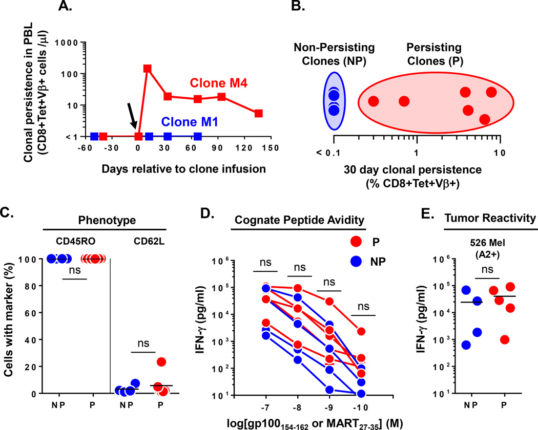
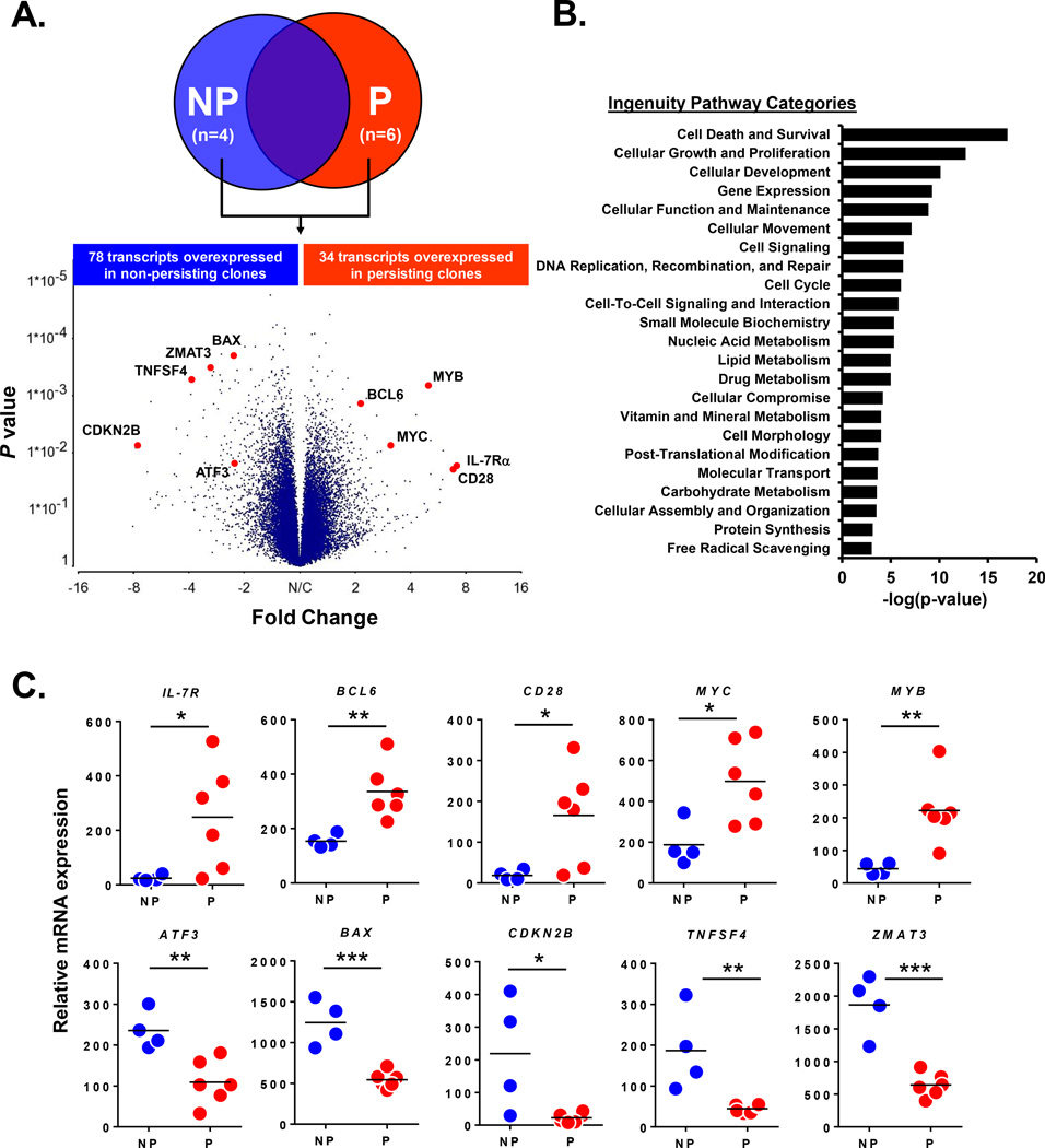
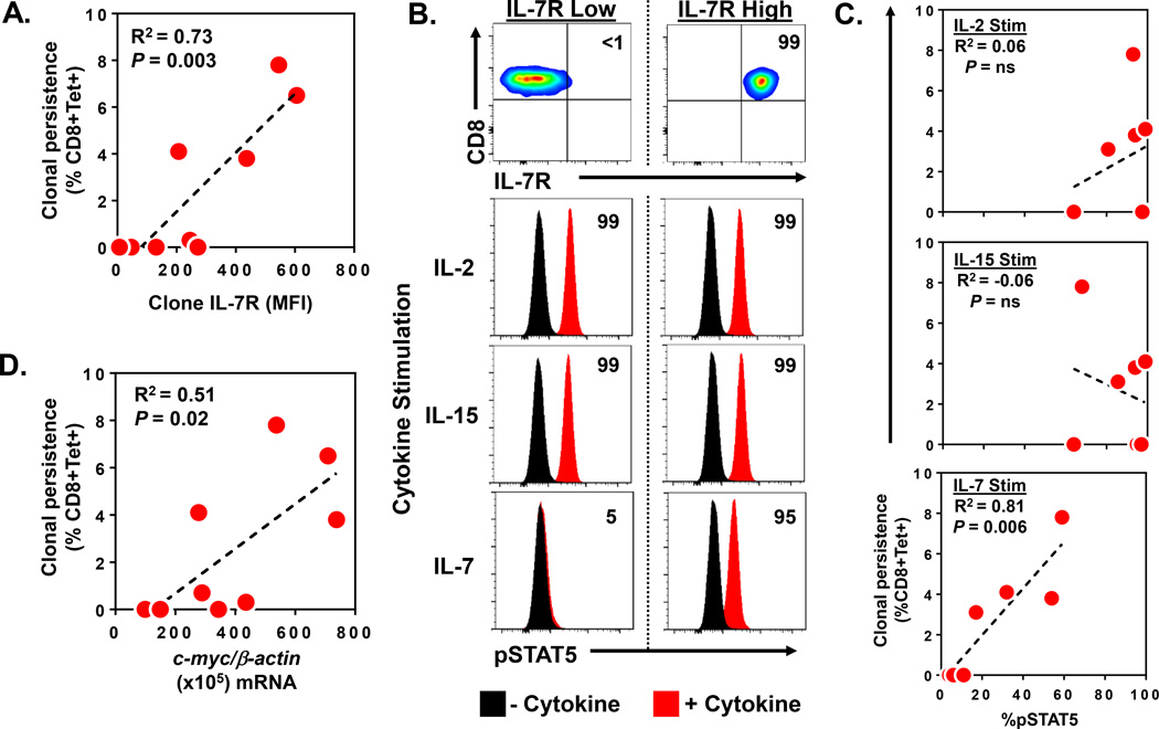
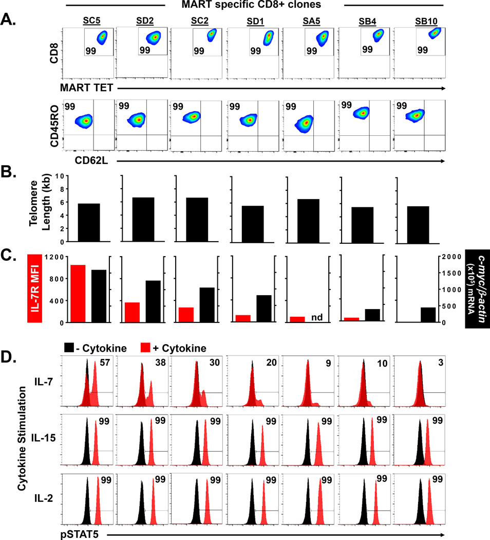
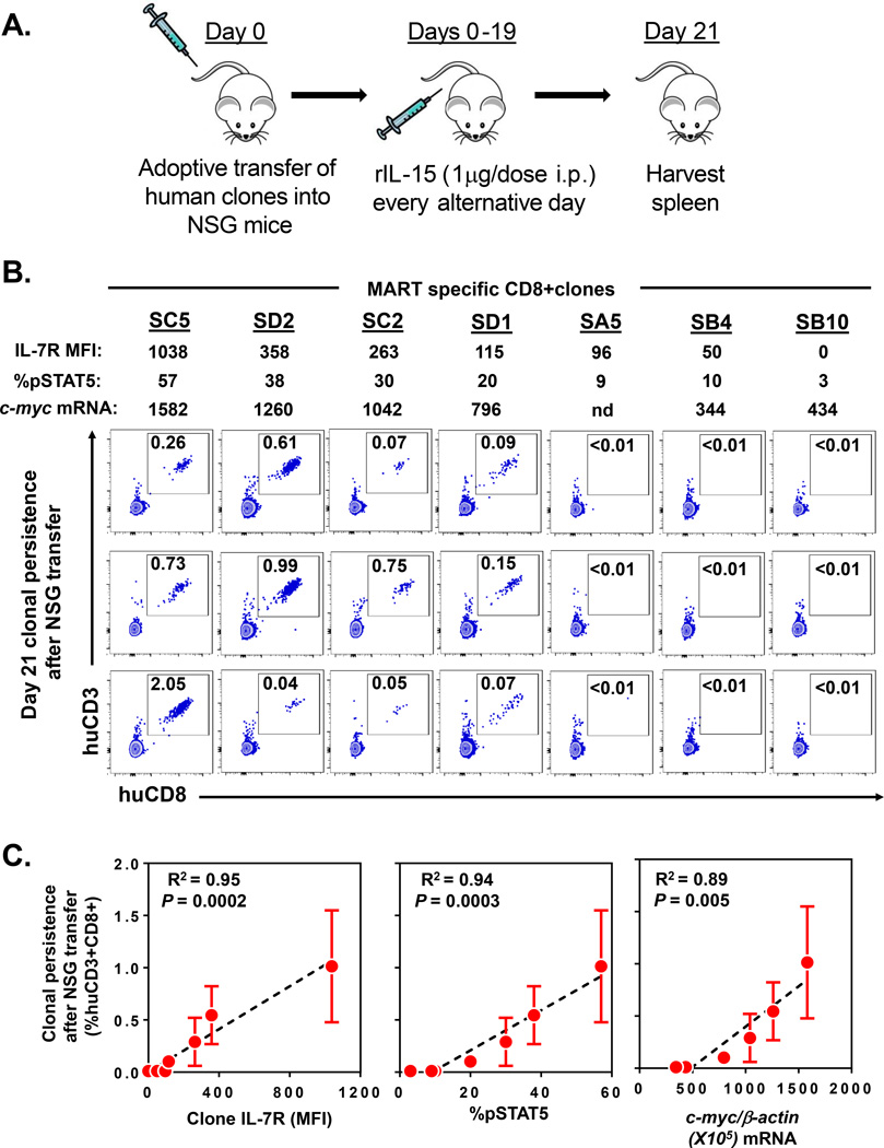
The optimal T-cell attributes for adoptive cancer immunotherapy are unclear. Recent clinical trials of ex vivo-expanded tumor-infiltrating lymphocytes indicated that differentiated T effector cells can elicit durable antitumor responses in some patients with cancer, with their antitumor activity tightly correlated with their persistence in the host. Thus, there is great interest in the definition of intrinsic biomarkers that can predict the conversion of short-lived tumor antigen-specific T effector cells into long-lived T memory cells. Long-term persistence of ex vivo-expanded tumor-specific CD8+ T effector clones has been reported in refractory metastatic melanoma patients after adoptive T-cell transfer. By using highly homogeneous clone populations from these preparations, we performed a comparative transcriptional profiling to define preinfusion molecular attributes that can be ascribed to an effector-to-memory transition. Through this route, we discovered that preinfusion T-cell clones that expressed the IL7 receptor (IL7R) and c-myc were more likely to persist longer after adoptive transfer to patients. The predictive value of these two biomarkers was strengthened by using IL7R protein, IL7-induced pSTAT5, and c-myc mRNA expression to prospectively identify human tumor-specific T effector clones capable of engraftment into immunodeficient mice. Overall, our findings reveal IL7R and c-myc expression as intrinsic biomarkers that can predict the fate of CD8+ T effector cells after adoptive transfer.
©2015 American Association for Cancer Research.
Conflict of interest statement
Conflicts of Interest: None
Figures
Similar articles
-
Chimeric γc cytokine receptors confer cytokine independent engraftment of human T lymphocytes.Mol Immunol. 2013 Nov;56(1-2):1-11. doi: 10.1016/j.molimm.2013.03.021. Epub 2013 Apr 27. Mol Immunol. 2013. PMID: 23628622
-
In Vitro Priming of Adoptively Transferred T Cells with a RORγ Agonist Confers Durable Memory and Stemness In Vivo.Cancer Res. 2018 Jul 15;78(14):3888-3898. doi: 10.1158/0008-5472.CAN-17-3973. Epub 2018 May 16. Cancer Res. 2018. PMID: 29769201 Free PMC article.
-
The stoichiometric production of IL-2 and IFN-γ mRNA defines memory T cells that can self-renew after adoptive transfer in humans.Sci Transl Med. 2012 Aug 29;4(149):149ra120. doi: 10.1126/scitranslmed.3004306. Sci Transl Med. 2012. PMID: 22932225 Free PMC article.
-
Adoptive T-cell transfer in melanoma.Immunotherapy. 2013 Jan;5(1):79-90. doi: 10.2217/imt.12.143. Immunotherapy. 2013. PMID: 23256800 Review.
-
Understanding memory CD8+ T cells.Immunol Lett. 2017 May;185:32-39. doi: 10.1016/j.imlet.2017.02.012. Epub 2017 Mar 6. Immunol Lett. 2017. PMID: 28274794 Free PMC article. Review.
Cited by
-
Cell-Intrinsic Barriers of T Cell-Based Immunotherapy.Trends Mol Med. 2016 Dec;22(12):1000-1011. doi: 10.1016/j.molmed.2016.10.002. Epub 2016 Nov 4. Trends Mol Med. 2016. PMID: 27825667 Free PMC article. Review.
-
T cell receptor therapeutics: immunological targeting of the intracellular cancer proteome.Nat Rev Drug Discov. 2023 Dec;22(12):996-1017. doi: 10.1038/s41573-023-00809-z. Epub 2023 Oct 27. Nat Rev Drug Discov. 2023. PMID: 37891435 Free PMC article. Review.
-
Immune Analysis of Radium-223 in Patients With Metastatic Prostate Cancer.Clin Genitourin Cancer. 2018 Apr;16(2):e469-e476. doi: 10.1016/j.clgc.2017.10.010. Epub 2017 Oct 24. Clin Genitourin Cancer. 2018. PMID: 29137877 Free PMC article.
-
Analyzing Prognostic Hub Genes in the Microenvironment of Cutaneous Melanoma by Computer Integrated Bioinformatics.Comput Intell Neurosci. 2022 Mar 8;2022:4493347. doi: 10.1155/2022/4493347. eCollection 2022. Comput Intell Neurosci. 2022. PMID: 35300397 Free PMC article.
-
Self-Assembled, Adjuvant/Antigen-Based Nanovaccine Mediates Anti-Tumor Immune Response against Melanoma Tumor.Polymers (Basel). 2018 Sep 25;10(10):1063. doi: 10.3390/polym10101063. Polymers (Basel). 2018. PMID: 30960988 Free PMC article.
References
Publication types
MeSH terms
Substances
Associated data
- Actions
Grants and funding
LinkOut - more resources
Full Text Sources
Other Literature Sources
Medical
Molecular Biology Databases
Research Materials
