. 2015 Sep;83(9):3479-89.
doi: 10.1128/IAI.00507-15. Epub 2015 Jun 22.

Endoplasmic Reticulum Tubule Protein Reticulon 4 Associates with the Legionella pneumophila Vacuole and with Translocated Substrate Ceg9

Affiliations

Endoplasmic Reticulum Tubule Protein Reticulon 4 Associates with the Legionella pneumophila Vacuole and with Translocated Substrate Ceg9

Eva Haenssler et al. Infect Immun. 2015 Sep.

Abstract

Intracellular growth of Legionella pneumophila occurs in a replication vacuole constructed by host proteins that regulate vesicular traffic from the host endoplasmic reticulum (ER). This process is promoted by a combination of approximately 300 Icm/Dot translocated substrates (IDTS). One of these proteins, Ceg9, was previously identified in a screen for L. pneumophila IDTS that manipulate secretory traffic when overexpressed in yeast. Using ectopic expression of Ceg9 in mammalian cells, we demonstrate that Ceg9 interacts with isoforms of host reticulon 4 (Rtn4), a protein that regulates ER tubule formation. Binding occurs under conditions that prevent association with other known reticulon binding proteins, arguing that Ceg9 binding is stable. A tripartite complex was demonstrated among Rtn4, Ceg9, and atlastin 1, a previously characterized reticulon interacting partner. The binding of Ceg9 to Rtn4 was not due to bridging by atlastin 1 but resulted from the two interacting partners binding independently to reticulon. When Ceg9 is ectopically expressed in mammalian cells, it shows a localization pattern that is indistinguishable from that of Rtn4, perhaps due to interactions between and similar structural features of the two proteins. Consistent with Rtn4 playing a role in the formation of the Legionella-containing vacuole, it was recruited to almost 50% of the vacuoles within 20 min postinfection. Our studies suggest that L. pneumophila proteins interact with ER tubules at an early stage of replication vacuole formation.


Figures

FIG 1
FIG 1
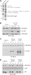
Ceg9 (lpg0246) binds Rtn4b/d when expressed in human cells. (A) Identification of proteins that coimmunoprecipitate with 3×FLAG-Ceg9. HEK293T cells transfected with 3×FLAG-Ceg9 were extracted in Triton X-100 and immunoprecipitation was performed using anti-FLAG resin (see Materials and Methods). After separation by SDS-PAGE and visualization by silver staining, candidate proteins were excised and analyzed by matrix-assisted laser desorption ionization–time of flight mass spectrometry. Vector, elution fraction of immunoprecipitates from extracts of cells harboring the 3×FLAG empty vector only; Ceg9, elution fraction of immunoprecipitates from extracts of cells harboring the 3×FLAG-Ceg9 vector. (B) Efficient coimmunoprecipitation of Rtn4 isoform with 3×FLAG-Ceg9. HEK293T cells were transfected with the 3×FLAG vector control and 3×FLAG-tagged Ceg9, extracted, and subjected to anti-FLAG immunoprecipitation, followed by Western blotting with the denoted antibodies. Lanes: Input, 0.87% of the starting material; Un., 0.91% of the unbound supernatant; IP, 12.5% of the immunoprecipitation fractions eluted by boiling in 2× SDS sample buffer (Materials and Methods). (C) Identification of Ceg9 as an Rtn4-associated protein. Cell lysates from HEK293T cells harboring the 3×FLAG vector, as well as 3×FLAG-Ceg19 and 3×FLAG-Ceg9, were subjected to anti-Rtn4 immunoprecipitation, and associated proteins were identified by Western blot analysis with anti-FLAG and anti-Rtn4 antibodies. Lanes: Input, 2% of the starting material; Un., the remaining material after the immunoprecipitation; IP, about 25% of the total immunoprecipitates. (D) Ceg9 does not pull down p97. HEK293T cells were transfected with the 3×FLAG vector control, 3×FLAG-tagged Ceg9, and 3×FLAG-Ceg19, extracted, and subjected to anti-FLAG immunoprecipitation, followed by Western blotting with the denoted antibodies. Although the 3×FLAG-Ceg9 and Rtn4 isoforms interact, binding of 3×FLAG-Ceg9 to p97 could not be confirmed.
FIG 2
FIG 2
Association of endogenous Rtn4b and 3×FLAG-tagged Ceg9 depends on the predicted hydrophobic region of Ceg9. (A) Schematic of Ceg9 and Rtn4b/d. Ceg9 is a 241-aa protein with two predicted transmembrane helices spanning approximately aa 57 to 79 and aa 94 to 116 (TMHMM Server v2.0). Truncated EGFP-tagged derivatives of Ceg9 used in immunoprecipitations and localization studies are displayed below. The reticulon 4 isoforms Rtn4b and Rtn4d have two long hydrophobic regions (gray boxes) hypothesized to form bent helical structures in membranes (65). Rtn4d splice variant has a 19-aa insertion displayed (white box). (B) Lysates from HEK293T cells transfected with the EGFP fusion vectors displayed were subjected to immunoprecipitations with anti-GFP resin (see Materials and Methods). This was followed by immunoblot analysis with anti-GFP, anti-Rtn4, anti-ATP5A, and anti-RhoGDI antibodies. Lanes: Input, 1.6% of the starting material was loaded; Un., 1.6% of the unbound supernatant was loaded; IP, 12.5% of the elution fraction obtained by boiling in SDS sample buffer.
FIG 3
FIG 3
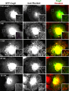
Localization of ectopically expressed EGFP-Ceg9 fusions to an ER-like compartment in Cos7 cells correlates with the presence of the transmembrane helices. The EGFP truncation derivatives described in Fig. 2 were transfected into Cos7 cells, incubated for 24 h, fixed, and probed with anti-Rtn4 to determine the subcellular localization of EGFP-Ceg9. Insets show higher-magnification regions of interest from each image. Panels: left, EGFP-Ceg9 fusion proteins; center, Rtn4a visualized by anti-Rtn4; right, merged images with anti-Rtn4 staining in red and EGFP in green. Scale bar, 20 μm.
FIG 4
FIG 4
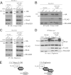
Ceg9 binding to Rtn4 does not require an atlastin bridge. (A) HA-tagged ATL1 does not interact with 3×FLAG-tagged Ceg9 in 1% Triton X-100 extracts. HEK293T cells were cotransfected with plasmids encoding HA-tagged ATL1 and 3×FLAG-tagged Ceg9, as well as with the 3×FLAG vector. 1% Triton X-100 lysates were subjected to immunoprecipitation with anti-HA resin and analyzed by Western blotting with anti-HA, anti-FLAG, and anti-RhoGDI antibodies. Lanes: Input, 0.91% of the starting material; Un., 0.91% of the unbound remaining material; IP, 11.1% of the precipitation was loaded. (B) Immunoprecipitation of 3×FLAG-Ceg9 does not result in association with ATL1 in the presence of Triton X-100. Lysates of HEK293T cells transfected with plasmids encoding HA-tagged ATL1 and 3×FLAG-tagged Ceg9, as well as the 3×FLAG vector, were used in immunoprecipitations with anti-FLAG resin. Subsequent Western blots were carried out with anti-HA, anti-FLAG, and anti-RhoGDI antibodies. In the “Input” lanes, 0.9% of the starting material was loaded, and in the “Un.” lanes, 0.9% of the unbound supernatant was loaded. The “IP” lanes contained 11% of the total elution fraction after boiling in 2× SDS sample buffer. (C) HA-tagged ATL1 and 3×FLAG-tagged Ceg9 associate in 1% digitonin lysates. HEK293T cells were cotransfected with plasmids expressing HA-tagged ATL1 and 3×FLAG-tagged Ceg9, as well as the 3×FLAG vector control. One percent digitonin cell lysates were subjected to immunoprecipitation with anti-HA resin and analyzed by Western blotting with anti-HA, anti-FLAG, and anti-RhoGDI antibodies. Lanes: Input, loaded with 0.91% of the starting material; Un., loaded with 0.91% of the material remaining from the immunoprecipitation. The “IP” lanes contained 11% of the total immunoprecipitation. (D). Rtn4 self-associates in extracts that allow complex formation with Ceg9 and ATL1. HEK293T cells were cotransfected with plasmids expressing myc-tagged Rtn4a and 3×FLAG-tagged Ceg9, as well as the 3×FLAG vector control. 1% digitonin extracts were made. The “Input” and “Un.” lanes were loaded with ca. 0.7% of the starting material and the material remaining from the immunoprecipitation, respectively. “IP” lanes contained 11% of the total immunoprecipitation. Immunoprecipitation with anti-myc (Rtn4a) and described Western blotting followed. (E) Schematic of 3×FLAG-tagged Ceg9 interactions in the presence of 1% Triton X-100 compared to 1% digitonin. Although in 1% Triton X-100 lysates 3×FLAG-tagged Ceg9 interaction with endogenous Rtn4b is maintained, HA-tagged ATL1 does not bind (62). However, in 1% digitonin lysates (62), complex formation of HA-tagged ATL1 and 3×FLAG-tagged Ceg9 can be observed.
FIG 5
FIG 5
Rtn4 localizes to the LCV during infection of A/J mouse bone marrow-derived macrophages independently of Ceg9. (A) Representative examples for visualization of Rtn4 during infection of A/J mouse bone marrow-derived macrophages with denoted L. pneumophila strains by immunofluorescence microscopy (see Materials and Methods). Panels: left, L. pneumophila; center, Rtn4; right, merge images with L. pneumophila strains in red and Rtn4 in green. (B) Host cells were challenged with the wild-type strain Lp02, the dotA-deficient strain Lp03, and a ceg9 deletion mutant at an MOI of 1. At the denoted time points, the cells were fixed and probed for Legionella and Rtn4 as described previously (see Materials and Methods). The phagosomes surrounded by aggregates of Rtn4 with a signal above background were enumerated for each time point. The data represented are the averages and standard errors of three coverslips per data point.

Similar articles

Cited by

References

    1. Asrat S, de Jesus DA, Hempstead AD, Ramabhadran V, Isberg RR. 2014. Bacterial pathogen manipulation of host membrane trafficking. Annu Rev Cell Dev Biol 30:79–109. doi:10.1146/annurev-cellbio-100913-013439. - DOI - PubMed
    1. Hackstadt T, Rockey DD, Heinzen RA, Scidmore MA. 1996. Chlamydia trachomatis interrupts an exocytic pathway to acquire endogenously synthesized sphingomyelin in transit from the Golgi apparatus to the plasma membrane. EMBO J 15:964–977. - PMC - PubMed
    1. Derre I, Swiss R, Agaisse H. 2011. The lipid transfer protein CERT interacts with the Chlamydia inclusion protein IncD and participates to ER-Chlamydia inclusion membrane contact sites. PLoS Pathog 7:e1002092. doi:10.1371/journal.ppat.1002092. - DOI - PMC - PubMed
    1. Elwell CA, Engel JN. 2012. Lipid acquisition by intracellular chlamydiae. Cell Microbiol 14:1010–1018. doi:10.1111/j.1462-5822.2012.01794.x. - DOI - PMC - PubMed
    1. Elwell CA, Jiang S, Kim JH, Lee A, Wittmann T, Hanada K, Melancon P, Engel JN. 2011. Chlamydia trachomatis co-opts GBF1 and CERT to acquire host sphingomyelin for distinct roles during intracellular development. PLoS Pathog 7:e1002198. doi:10.1371/journal.ppat.1002198. - DOI - PMC - PubMed

Publication types

MeSH terms