. 2015 Sep;89(17):9029-43.
doi: 10.1128/JVI.01331-15. Epub 2015 Jun 17.

The Nucleocapsid Protein of Coronaviruses Acts as a Viral Suppressor of RNA Silencing in Mammalian Cells

The Nucleocapsid Protein of Coronaviruses Acts as a Viral Suppressor of RNA Silencing in Mammalian Cells

Lei Cui et al. J Virol. 2015 Sep.

Abstract

RNA interference (RNAi) is a process of eukaryotic posttranscriptional gene silencing that functions in antiviral immunity in plants, nematodes, and insects. However, recent studies provided strong supports that RNAi also plays a role in antiviral mechanism in mammalian cells. To combat RNAi-mediated antiviral responses, many viruses encode viral suppressors of RNA silencing (VSR) to facilitate their replication. VSRs have been widely studied for plant and insect viruses, but only a few have been defined for mammalian viruses currently. We identified a novel VSR from coronaviruses, a group of medically important mammalian viruses including Severe acute respiratory syndrome coronavirus (SARS-CoV), and showed that the nucleocapsid protein (N protein) of coronaviruses suppresses RNAi triggered by either short hairpin RNAs or small interfering RNAs in mammalian cells. Mouse hepatitis virus (MHV) is closely related to SARS-CoV in the family Coronaviridae and was used as a coronavirus replication model. The replication of MHV increased when the N proteins were expressed in trans, while knockdown of Dicer1 or Ago2 transcripts facilitated the MHV replication in mammalian cells. These results support the hypothesis that RNAi is a part of the antiviral immunity responses in mammalian cells. IMPORTANCE RNAi has been well known to play important antiviral roles from plants to invertebrates. However, recent studies provided strong supports that RNAi is also involved in antiviral response in mammalian cells. An important indication for RNAi-mediated antiviral activity in mammals is the fact that a number of mammalian viruses encode potent suppressors of RNA silencing. Our results demonstrate that coronavirus N protein could function as a VSR through its double-stranded RNA binding activity. Mutational analysis of N protein allowed us to find out the critical residues for the VSR activity. Using the MHV-A59 as the coronavirus replication model, we showed that ectopic expression of SARS-CoV N protein could promote MHV replication in RNAi-active cells but not in RNAi-depleted cells. These results indicate that coronaviruses encode a VSR that functions in the replication cycle and provide further evidence to support that RNAi-mediated antiviral response exists in mammalian cells.


Figures

FIG 1
FIG 1
Screening of potential VSR of SARS-CoV by reversal-of-silencing assays. 293T cells were cotransfected with plasmids encoding eGFP reporter (125 ng), eGFP-specific shRNA (shGFP) (1 μg), or luciferase-specific shRNA (shLuc) (1 μg) and viral protein of SARS-CoV (500 ng), respectively. (A to C) The expression level of eGFP was determined by Western blotting at 72 h posttransfection. Empty vector (Vec) and NoV B2 (B2) were used as a mock control and a positive control. The shLuc was used as an irrelevant silencing control. β-Actin was used as a loading control. (D) The expression of SARS-CoV-encoded proteins as indicated was detected by Western blotting. (E) The relative eGFP reversion activity of different viral proteins in panels A to C was normalized by that of typical VSR NoV B2 control and is shown in a bar diagram.
FIG 2
FIG 2
N protein of coronaviruses represses shRNA-induced RNAi in mammalian cells. (A to C) HEK293T cells were cotransfected with plasmids encoding eGFP reporter (125 ng), shGFP, or shLuc (1 µg) and plasmids encoding viral proteins as indicated (300 ng). The expression of eGFP reporter was analyzed 72 h after cotransfection. (A) The intensity of eGFP was observed under fluorescence microscopy. (B) Cell lysates were harvested and analyzed by Western blotting. (C) Cellular total mRNAs were harvested and analyzed by Northern blotting. (D and E) HEK293T cells were cotransfected with plasmids encoding eGFP and SARS-CoV N protein or NoV B2, respectively. eGFP fluorescence, protein, and mRNA levels were determined by fluorescence microscopy (D) and Western and Northern blotting (E), respectively. (F) HEK293T cells were cotransfected with plasmids of mock vector pLko.1 (1 μg) or shRNA targeting endogenous gene VHL (shVHL) in the presence or absence of SARS-CoV N protein (300 ng). At 72 h posttransfection, cells were harvested and subjected to Western blotting to determine the endogenous VHL expression. (G) S2 Cells were transfected with 0.03 μg of pFR1 or 0.6 μg of pFRNA1-ΔB2 and with SARS-CoV N or FHV B2 as indicated above. At 48 h posttransfection, FHV RNA transcription was induced by incubation with CuSO4 at 0.5 mM. At 24 h after induction, the cellular total mRNA was harvested for Northern blot analysis by a probe recognizing FHV RNA1 and RNA3. The band between RNA1 and RNA3 represents the mRNA transcribed from B2 expression plasmid. (H) HEK293T cells were cotransfected with plasmids encoding eGFP, shLuc, or shGFP and N proteins of different coronaviruses (Flag-tagged N proteins of SARS-CoV and MERS-CoV and HA-tagged N proteins of MHV, PEDV, and TGEV), respectively. The eGFP expression level and mRNA level were determined by Western blotting and Northern blotting. Empty vector (Vec, Vec1, or Vec2), nsp14, and ORF6 were used as negative controls, while NoV B2 was used as a positive control. The shLuc was irrelevant control shRNA. β-Actin and rRNAs were used as loading controls for Western and Northern blotting, respectively.
FIG 3
FIG 3
N protein represses shRNA-induced RNAi in a dose-dependent and time-dependent manner in mammalian cells. Increasing amounts of the plasmid expressing SARS-CoV N protein were transfected into HEK293T cells, as indicated in the upper panel in the reversal-of-silencing assay. At 72 h after transfection, Western blotting (A) and Northern blotting (B) were performed to determine the eGFP protein and mRNA levels, respectively. (C) Northern blotting (upper panel) and Western blotting (lower panel) were performed 24, 48, and 72 h posttransfection, respectively.
FIG 4
FIG 4
N protein inhibits the production of siRNA and RNAi in both mammalian and insect cells. (A) HEK293T cells were cotransfected with plasmid as indicated above, 72 h after transfection, and small RNAs were harvested from the cells and probed with DIG-labeled oligonucleotides that correspond to the target sites of siRNA produced from shGFP. The locations of bands corresponding to shRNA and siRNA are indicated with diagrams on the right side. Short exposures and long exposures are shown on the left and right, respectively. An ethidium bromide-stained gel of low-molecular-weight RNA is shown as a loading control. Neg., mock control transfected with shLuc. (B) The ratios of shRNA to siRNA in panel A were quantified based on the corresponding exposure signals and are shown as a bar diagram. (C) eGFP expression plasmid (200 ng) was cotransfected with multiple concentrations (5 to 40 nM) of synthetic eGFP-specific siRNA (siGFP) to confirm the effective siGFP concentration in HEK293T cells. At 72 h after transfection, cell lysates were harvested to determine the reduction in eGFP expression by Western blotting. (D and E) SARS-CoV N protein inhibits siRNA-induced RNAi. HEK293T cells were transfected with plasmids as indicated above. At 72 h after transfection, eGFP fluorescence (D), protein (E), and mRNA (F) were detected as described in Fig. 2. (F) SARS-CoV N protein inhibits RNAi in Drosophila S2 cells. eGFP-specific dsRNA (dsRNA-GFP) and siGFP were used to induce RNAi in S2 cells. The mRNA of eGFP was detected by Northern blotting. Empty vector (Vec), nsp14, and ORF6 were used as negative controls. NoV B2 and FHV B2 were used as positive controls. rp49 was used as a loading control.
FIG 5
FIG 5
N protein binds to RNAs and inhibits the Dicer-like RNase III cleavage reaction in vitro. Increasing amounts of purified GST-tagged SARS-CoV N protein (GST-N) from 0 to 15 μM were incubated with 0.2 pmol of 500-nt DIG-labeled ssRNA (A) or 244-bp DIG-labeled dsRNA (B) at 25°C for 30 min. Complexes were separated on 1.2% TBE–agarose gel and subjected to Northern blotting. The free ssRNA and dsRNA are indicated on the left side. (C) GST-N up to 15 μM was incubated with 0.2 pmol of 5′-HEX-labeled 21-nt siRNA as described in panels A and B. Complexes were applied to 4% native polyacrylamide gel, and the fluorescent signal was visualized by using a Typhoon 9200. The free siRNA is indicated on the left side. (D) DIG-labeled 500-bp dsRNA was incubated with purified proteins, as indicated above, at 25°C for 30 min before the processing of Dicer-like RNase III at 37°C for 30 min. The reaction products were separated on 1.2% TBE–agarose gel and subjected to Northern blotting. The free dsRNA is indicated on the left side, and the cleaved dsRNA is indicated on the right side. The shifted protein-RNA complexes are indicated by black arrows on the right side. The protein GST and GST-tagged WhNV B2 (GST-B2) were used as a mock control and a positive control.
FIG 6
FIG 6
Conserved residues LysK258 and LysK262 of SARS-CoV N protein are critical for RNAi activity. (A) Schematic diagram of the domain architecture of the SARS-CoV N protein and multiple-sequence alignment of CTD spanning residues 248 to 281 of coronavirus N proteins. The conserved residues are indicated by solid black boxes. NTD, N-terminal domain; CTD, C-terminal domain; SR-rich, rich in serine and arginine; Linker, linkage region. (B and C) Mapping of critical residues of SARS-CoV N protein for VSR activity. The RNAi repression activity of Flag-tagged truncations of SARS-CoV N protein was analyzed by a reversal-of-silencing assay as described for Fig. 2B. The expression of truncated SARS-CoV N proteins was detected by Western blotting. (D to F) The RNAi repression activity of Flag-tagged mutants of SARS-CoV N protein was analyzed by a reversal-of-silencing assay, as described for Fig. 2. (F) The K257A, K258A, K262A, and R263A mutants were further analyzed based on the results from panel E.
FIG 7
FIG 7
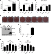
SARS-CoV N protein promotes MHV replication when provided in trans. (A and B) Mouse Neuro-2a cells were transfected with plasmids as indicated and infected with MHV strain A59 at an MOI of 0.1 at 24 h posttransfection. At 16 h after infection, culture supernatants were collected and subjected to plaque assay on L2 cells to determine the MHV titers. The relative titer fold of MHV was quantified, as shown in bar diagrams. The actual virus titers are in indicated in panel A to show MHV replication efficiency. (C) Total RNAs were extracted from the transfected cells and subjected to RT-PCR using primers targeting the subgenomic RNA7 of MHV. The data were normalized to the abundance of endogenous mouse GAPDH mRNA. (D) Virus plaque formation was analyzed on L2 cells at a dilution of 10−6. (E) Protein expression levels in transfected cell lysates were detected by Western blotting with the indicated antibodies. β-Actin was used as a loading control. (F) Neuro-2a cells were infected with MHV strain A59 at an MOI of 0.1 or transfected with 2 μg of poly(I·C). At 16 h postinfection or transfection, IFN-β production was determined by enzyme-linked immunosorbent assay (ELISA). (G and H) Total RNAs were extracted from the transfected cells as described in panel B and subjected to RT-PCR to detect Ifn-β and Isg56 mRNA. Error bars indicate the means and standard deviations for triplicate experiments. *, P < 0.05; **, P < 0.01; ns, not significant (unpaired Student t test).
FIG 8
FIG 8
Screen for siRNAs targeting mouse Dicer1 and Ago2. Mouse Neuro-2a cells were transfected with 40 nM siDicer1 (A and B) or siAgo2 (C and D). At 48 h posttransfection, total cellular mRNAs were extracted and subjected to RT-PCR to determine the mRNA levels of Dicer1 (A), Ago2 (C), and Ifn-β (B and D). siGFP was used as a negative control. The data were normalized to the abundance of internal mouse GAPDH mRNA. Error bars indicate the means and standard deviations for triplicate experiments.
FIG 9
FIG 9
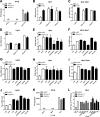
Knockdown of Dicer1 or Ago2 facilitated MHV replication. (A) Mouse Neuro-2a cells were transfected with siRNAs as indicated above and, at 48 h posttransfection, the cells were infected with MHV at an MOI of 0.1 or transfected with 2 μg of poly(I·C). At 16 h postinfection or posttransfection, IFN-β production was determined by ELISA. (B to J) Mouse Neuro-2a cells were cotransfected with 40 nM siRNA and 500-ng protein expression plasmids as indicated and infected with MHV strain A59 at an MOI of 0.1 at 48 h posttransfection. At 16 h postinfection, the culture supernatants were collected and subjected to plaque assays on L2 cells to determine the MHV titers. (B, E, and H) The relative titer folds of MHV were quantified and are shown in the bar diagrams. Total RNAs were extracted from the transfected cells and subjected to RT-PCR to determine the indicated mRNA levels. (C, F, and I) MHV RNA7 level. (D, G, and J) Isg56 mRNA level. (K) L2 cells were transfected with siRNAs as indicated above and, at 48 h posttransfection, the cells were infected with MHV or SeV. At 16 h postinfection, IFN-β production was determined by ELISA. (L) L2 was transfected and infected similarly as described for Neuro-2a cells. The MHV titers were determined by plaque assay. Empty vector (Vec) and siGFP were used as the negative control. The data were normalized to the abundance of endogenous mouse GAPDH mRNA. Error bars indicate the means and standard deviations for triplicate experiments.
FIG 10
FIG 10
Model for the suppression of RNAi in mammalian cells by coronavirus N protein. After the entry and un coating of coronaviral virions, the single-stranded genomic RNA (gRNA) is protected by N proteins and serves as a template for the synthesis of negative-strand gRNA and a set of subgenomic RNA (sgRNA). The (−)gRNA and (−)sgRNA are replicated to generate full-length gRNA and a set of (+)sgRNA. The virus-derived dsRNA could be generated during viral transcription and replication. sgRNA and gRNA sequences may also form an intramolecular hairpin structure. These viral dsRNAs may be recognized by Dicer, consequently triggering antiviral RNAi. Coronavirus N proteins may repress the RNAi at three different stages as indicated.

Similar articles

Cited by

References

    1. Carthew RW, Sontheimer EJ. 2009. Origins and mechanisms of miRNAs and siRNAs. Cell 136:642–655. doi:10.1016/j.cell.2009.01.035. - DOI - PMC - PubMed
    1. Ding SW. 2010. RNA-based antiviral immunity. Nat Rev Immunol 10:632–644. doi:10.1038/nri2824. - DOI - PubMed
    1. Umbach JL, Cullen BR. 2009. The role of RNAi and microRNAs in animal virus replication and antiviral immunity. Genes Dev 23:1151–1164. doi:10.1101/gad.1793309. - DOI - PMC - PubMed
    1. Takeuchi O, Akira S. 2009. Innate immunity to virus infection. Immunol Rev 227:75–86. doi:10.1111/j.1600-065X.2008.00737.x. - DOI - PMC - PubMed
    1. Parameswaran P, Sklan E, Wilkins C, Burgon T, Samuel MA, Lu R, Ansel KM, Heissmeyer V, Einav S, Jackson W, Doukas T, Paranjape S, Polacek C, FBdos Santos Jalili R, Babrzadeh F, Gharizadeh B, Grimm D, Kay M, Koike S, Sarnow P, Ronaghi M, Ding SW, Harris E, Chow M, Diamond MS, Kirkegaard K, Glenn JS, Fire AZ. 2010. Six RNA viruses and forty-one hosts: viral small RNAs and modulation of small RNA repertoires in vertebrate and invertebrate systems. PLoS Pathog 6:e1000764. doi:10.1371/journal.ppat.1000764. - DOI - PMC - PubMed

Publication types

MeSH terms

LinkOut - more resources