. 2015 Sep;89(17):8982-98.
doi: 10.1128/JVI.01220-15. Epub 2015 Jun 17.

Role of Host Cell p32 in Herpes Simplex Virus 1 De-Envelopment during Viral Nuclear Egress

Role of Host Cell p32 in Herpes Simplex Virus 1 De-Envelopment during Viral Nuclear Egress

Zhuoming Liu et al. J Virol. 2015 Sep.

Abstract

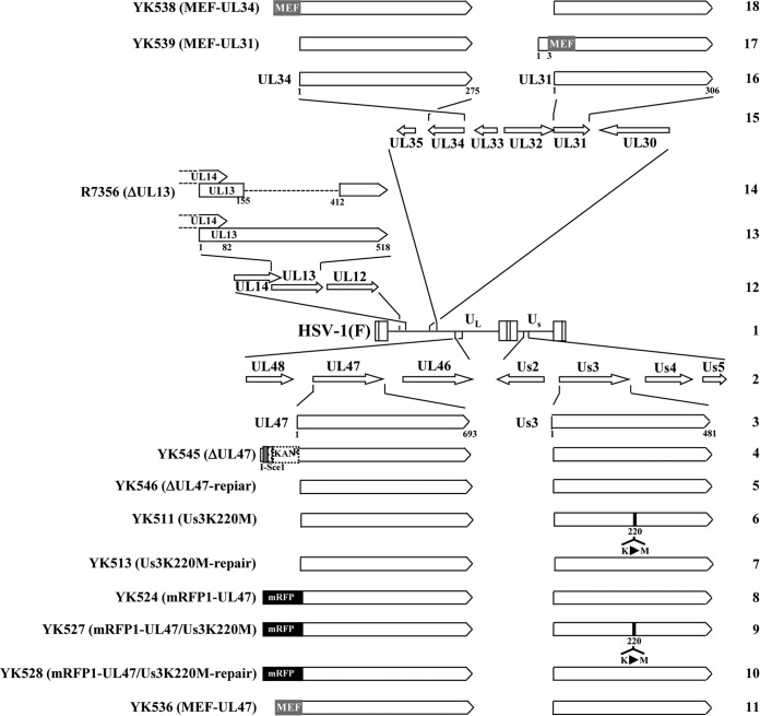
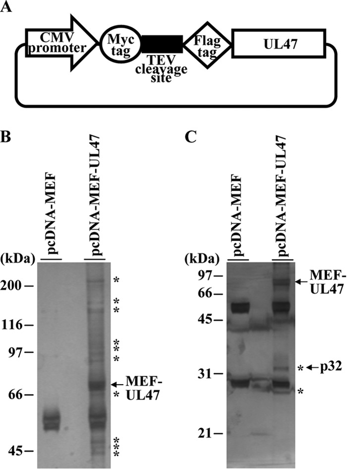
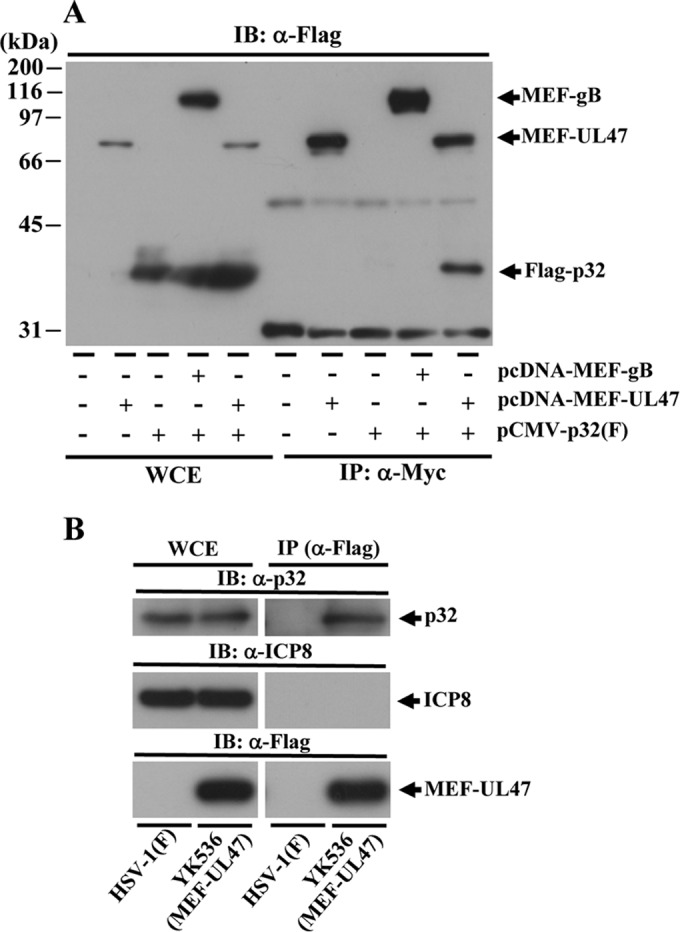
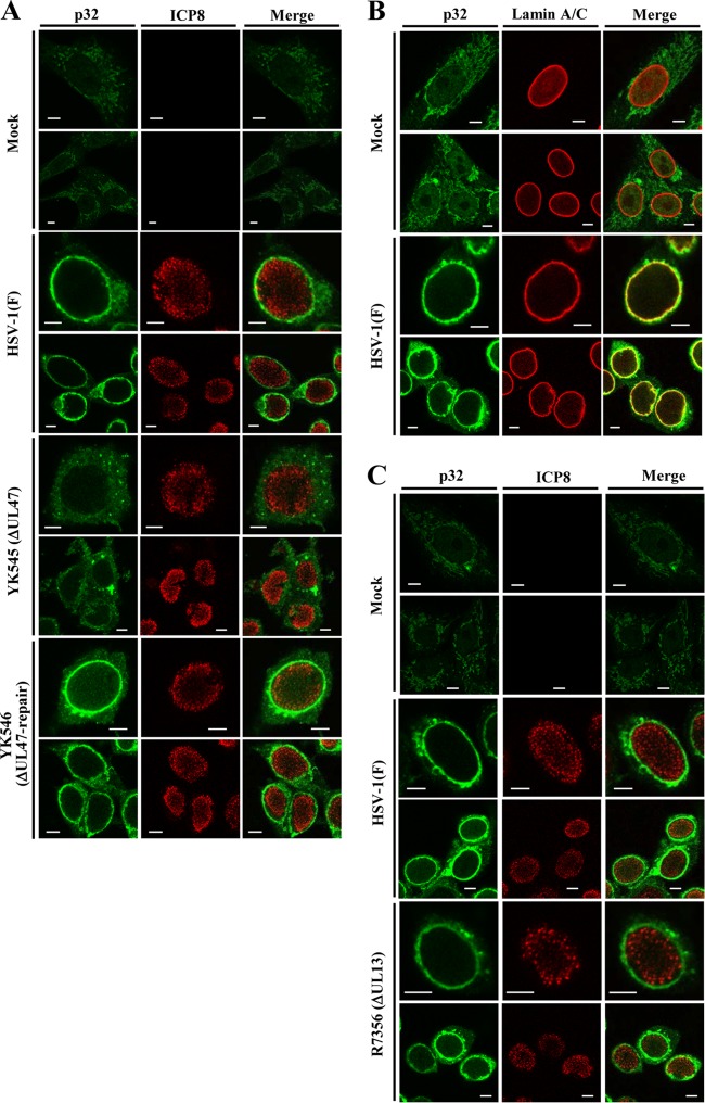
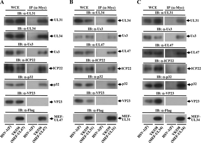
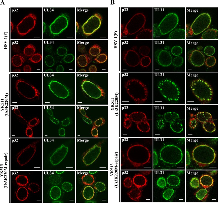
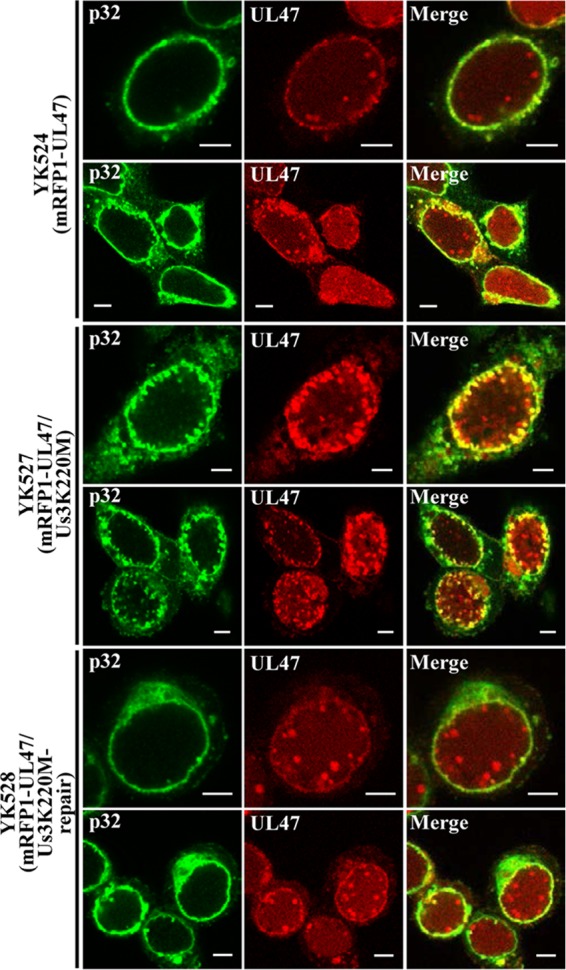
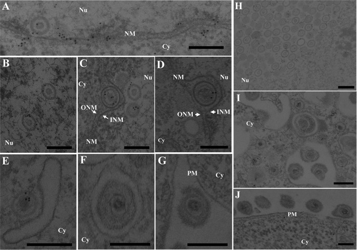
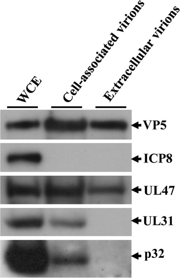
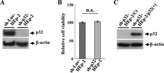
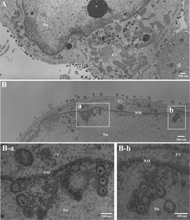
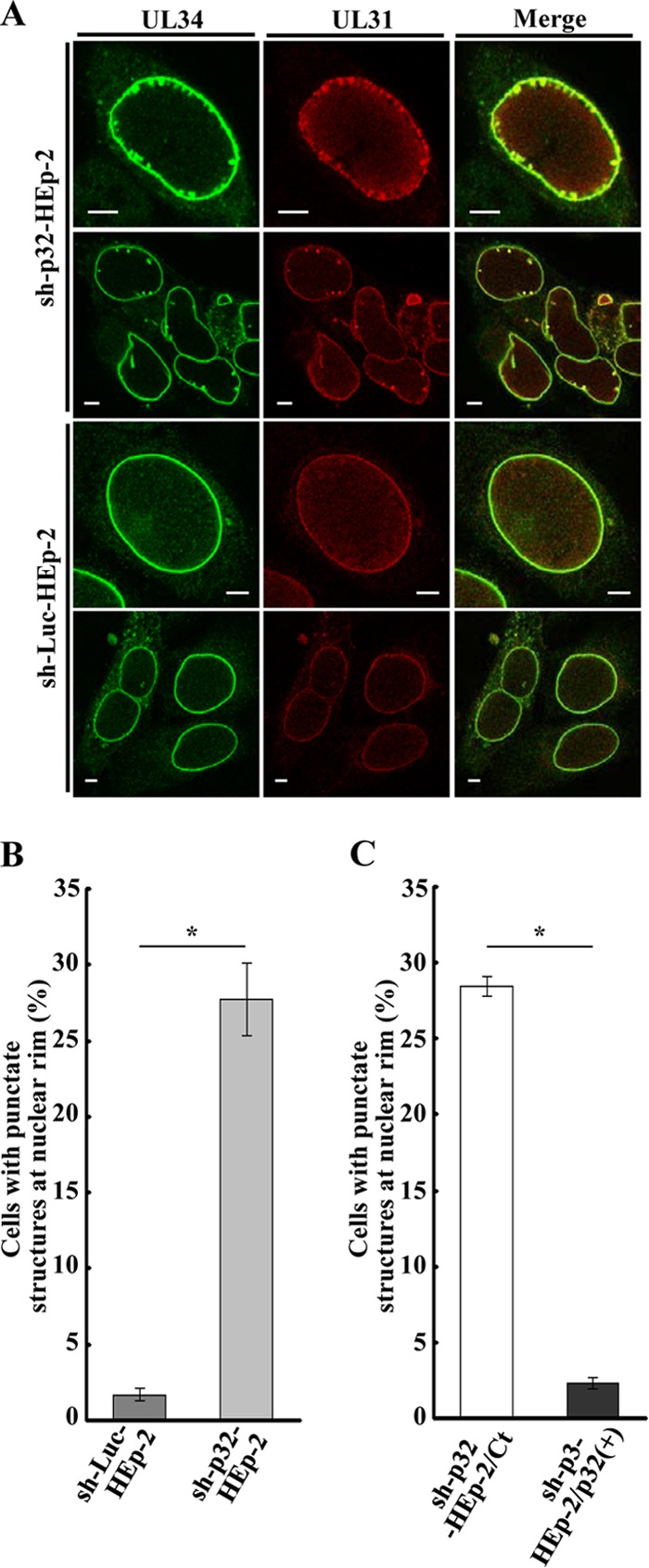
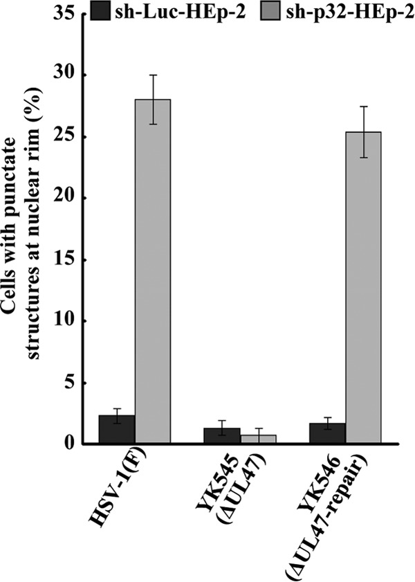
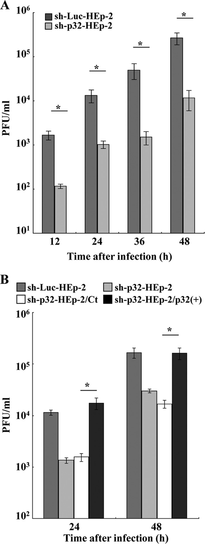
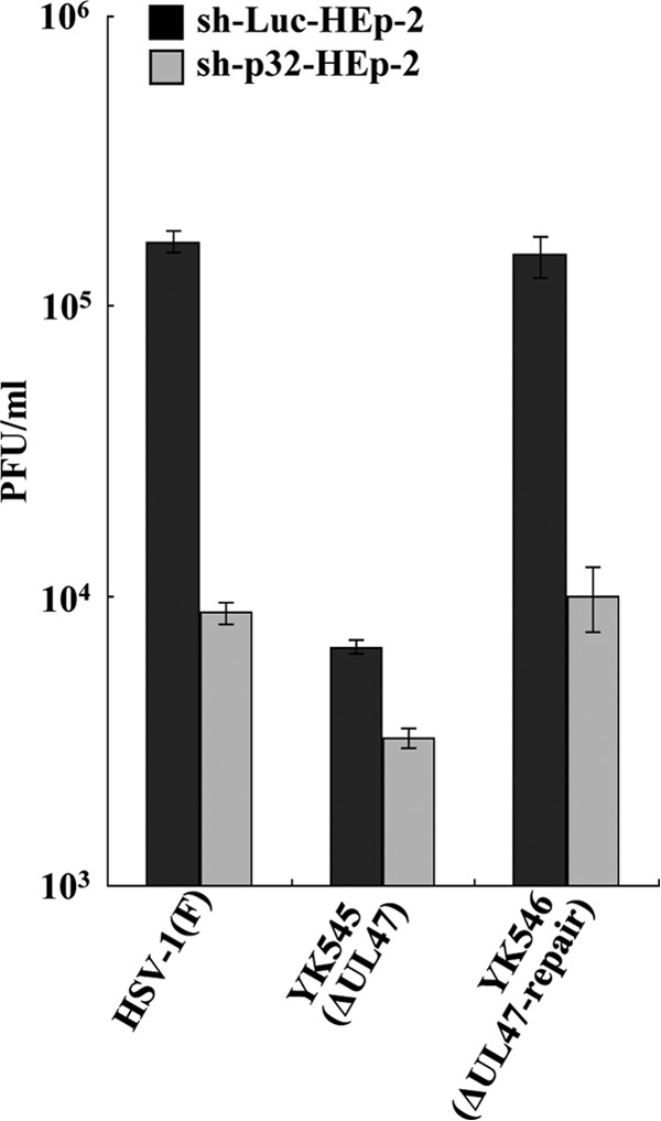
To clarify the function(s) of the herpes simplex virus 1 (HSV-1) major virion structural protein UL47 (also designated VP13/14), we screened cells overexpressing UL47 for UL47-binding cellular proteins. Tandem affinity purification of transiently expressed UL47 coupled with mass spectrometry-based proteomics technology and subsequent analyses showed that UL47 interacted with cell protein p32 in HSV-1-infected cells. Unlike in mock-infected cells, p32 accumulated at the nuclear rim in HSV-1-infected cells, and this p32 recruitment to the nuclear rim required UL47. p32 formed a complex(es) with HSV-1 proteins UL31, UL34, Us3, UL47, and/or ICP22 in HSV-1-infected cells. All these HSV-1 proteins were previously reported to be important for HSV-1 nuclear egress, in which nucleocapsids bud through the inner nuclear membrane (primary envelopment) and the enveloped nucleocapsids then fuse with the outer nuclear membrane (de-envelopment). Like viral proteins UL31, UL34, Us3, and UL47, p32 was detected in primary enveloped virions. p32 knockdown reduced viral replication and induced membranous invaginations adjacent to the nuclear rim containing primary enveloped virions and aberrant localization of UL31 and UL34 in punctate structures at the nuclear rim. These effects of p32 knockdown were reduced in the absence of UL47. Therefore, the effects of p32 knockdown in HSV-1 nuclear egress were similar to those of the previously reported mutation(s) in HSV-1 regulatory proteins for HSV-1 de-envelopment during viral nuclear egress. Collectively, these results suggested that p32 regulated HSV-1 de-envelopment and replication in a UL47-dependent manner. IMPORTANCE In this study, we have obtained data suggesting that (i) the HSV-1 major virion structural protein UL47 interacted with host cell protein p32 and mediated the recruitment of p32 to the nuclear rim in HSV-1-infected cells; (ii) p32 was a component of the HSV-1 nuclear egress complex (NEC), whose core components were UL31 and UL34; and (iii) p32 regulated HSV-1 de-envelopment during viral nuclear egress. It has been reported that p32 was a component of human cytomegalovirus NEC and was required for efficient disintegration of nuclear lamina, which has been thought to facilitate HSV-1 primary envelopment during viral nuclear egress. Thus, p32 appeared to be a core component of herpesvirus NECs, like UL31 and UL34 homologs in other herpesviruses, and to play multiple roles in herpesvirus nuclear egress.


Figures

FIG 1
FIG 1
Schematic diagrams of the genome structures of wild-type HSV-1(F) and the relevant domains of the recombinant viruses used in this study. Line 1, wild-type HSV-1(F) genome; line 2, domains with the UL46 to UL48 and Us2 to Us5 genes; line 3, domains with the UL47 and Us3 genes; lines 4 to 11, recombinant viruses with mutations in the UL47 and/or Us3 genes; line 12, domains with the UL12 to UL14 genes; line 13, domains with the UL13 gene and a part of the UL14 gene; line 14, recombinant virus with a null mutation in the UL13 gene; line 15, domains with the UL30 to UL35 genes; line 16, domains of the UL34 and UL31 genes; line 17, recombinant virus encoding MEF-tagged UL31; line 18, recombinant virus encoding MEF-tagged UL34.
FIG 2
FIG 2
Identification of cellular proteins interacting with HSV-1 UL47. (A) Schematic diagram of expression plasmid pcDNA-MEF-UL47 encoding UL47 fused to an MEF tag. (B and C) 293T cells were transfected with the empty vector pcDNA-MEF or plasmid pcDNA-MEF-UL47, harvested, and immunoprecipitated with anti-Myc antibody and anti-Flag antibody. Immunoprecipitates were separated in 7.5% (B) or 12% (C) denaturing gels and silver stained. Bands marked with asterisks were excised, digested, and analyzed by mass spectrometry. The arrow marks MEF-UL47 and p32. Molecular mass markers are indicated on the left.
FIG 3
FIG 3
Interaction of UL47 with p32. (A) 293T cells were mock transfected or transfected with pcDNA-MEF-UL47 alone, pCMV-p32(F) encoding Flag-tagged p32 alone, or pCMV-p32(F) in combination with either pcDNA-MEF-UL47 or pcDNA-MEF-gB. At 2 days posttransfection, cells were harvested, immunoprecipitated (IP) with anti-Myc antibody, and analyzed by immunoblotting (IB) with anti-Flag antibody. WCE, whole-cell extract. Molecular mass markers are indicated on the left. (B) Vero cells infected with wild-type HSV-1(F) or YK536 (MEF-UL47) at an MOI of 5 for 18 h were harvested, immunoprecipitated with anti-Flag antibody, and analyzed by immunoblotting with anti-p32 antibody, anti-ICP8 antibody, or anti-Flag antibody.
FIG 4
FIG 4
Localization of p32 in HSV-1-infected cells and effect of UL47 and UL13 on p32 localization. (A) Vero cells mock infected or infected with wild-type HSV-1(F), YK545 (ΔUL47), or YK546 (ΔUL47-repair) at an MOI of 3 were fixed at 18 h postinfection, permeabilized, stained with anti-p32 antibody or anti-ICP8 antibody, and examined by confocal microscopy. (B) Vero cells mock infected or infected with wild-type HSV-1(F) at an MOI of 3 were fixed at 18 h postinfection, permeabilized, stained with anti-p32 antibody or anti-lamin A/C antibody, and examined by confocal microscopy. (C) Experiments were done by the same procedure as for panel A), except R7356 (DUL13) was used instead of YK545 (ΔUL47) and YK546 (ΔUL47-repair). Scale bar, 5 μm.
FIG 5
FIG 5
Interactions among UL47, p32, UL31, UL34, ICP22, and Us3 in HSV-1-infected cells. Vero cells infected with wild-type HSV-1(F) (A to C) and YK536 (MEF-UL47) (A), YK539 (MEF-UL31) (B), or YK538 (MEF-UL34) (C) at an MOI of 5 for 18 h were harvested, immunoprecipitated with anti-Myc antibody, and analyzed by immunoblotting with the indicated antibodies.
FIG 6
FIG 6
Effect of Us3 kinase activity on localization of p32, UL31, and UL34 in HSV-1-infected cells. Vero cells were infected with wild-type HSV-1(F), YK511 (Us3K220M), or YK513 (Us3K220M-repair) at an MOI of 3, fixed at 18 h postinfection, permeabilized, stained with anti-p32 antibody in combination with anti-UL34 (A) or anti-UL31 (B) antibody, and examined by confocal microscopy. Scale bar, 5 μm.
FIG 7
FIG 7
Effect of Us3 kinase activity on localization of p32 and mRFP1-UL47 in HSV-1-infected cells. Vero cells were infected with YK524 (mRFP1-UL47), YK527 (mRFP1-UL47/Us3K220M), or YK528 (mRFP1-UL47/Us3K220M-repair) at an MOI of 3, fixed at 18 h postinfection, permeabilized, stained with anti-p32 antibody, and examined by confocal microscopy. Scale bar, 5 μm.
FIG 8
FIG 8
Localization of p32 in HSV-1-infected cells by immunoelectron microscopy. Vero cells were infected with wild-type HSV-1(F) at an MOI of 5, fixed at 18 h postinfection, embedded, sectioned, stained with rabbit anti-p32 polyclonal antibody followed by goat anti-rabbit IgG conjugated to 10-nm gold particles, and examined by transmission electron microscopy. Nu, nucleus; Cy, cytoplasm; NM, nuclear membrane; INM, inner nuclear membrane; ONM, outer nuclear membrane; PM, plasma membrane. p32 was detected along the nuclear membrane (A), on capsids in the nucleus (A to C and H) and cytoplasm (E and I), and on primary enveloped virions in the perinuclear space (C and D), but it was barely detectable on secondary enveloped virions in the cytoplasm (F and I) and in the extracellular space (G and J). Bars, 200 nm.
FIG 9
FIG 9
Detection of p32 in cell-associated and extracellular virions. Cell-associated and extracellular virions were purified and analyzed by immunoblotting with antibodies to the indicated proteins.
FIG 10
FIG 10
Characterization of sh-Luc-HEp-2, sh-p32-HEp-2, sh-p32-HEp-2/Ct, and sh-p32-HEp-2/p32(+) cells. (A) Expression of p32 in sh-Luc-HEp-2 and sh-p32-HEp-2 cells analyzed by immunoblotting with anti-p32 (top) and anti-β-actin (bottom) antibodies. (B) Cell viability of sh-Luc-HEp-2 and sh-p32-HEp-2 cells assayed 24 h after 2 × 10⁴ cells were seeded on 96-well plates. Each value is the mean ± standard error of the results of triplicate experiments and is expressed relative to the mean for sh-Luc-HEp-2 cells, which was normalized to 100%. n.s., not statistically significant. Data are representative of three independent experiments. (C) Expression of p32 in sh-p32-HEp-2/Ct and sh-p32-HEp-2/p32(+) cells analyzed by immunoblotting with anti-p32 (top) and anti-β-actin (bottom) antibodies.
FIG 11
FIG 11
Ultrastructural analysis of the effect of p32 on HSV-1 nuclear egress. sh-Luc-HEp-2 (A) and sh-p32-HEp-2 cells (B) infected with wild-type HSV-1(F) at an MOI of 5 were fixed at 24 h postinfection, embedded, sectioned, stained, and examined by transmission electron microscopy. (B-a and B-b) Higher magnifications of the corresponding boxed areas in panel B showing invagination structures containing primary enveloped virions. Nu, nucleus; Cy, cytoplasm; NM, nuclear membrane.
FIG 12
FIG 12
Effect of p32 on localization of UL34 and UL31 in HSV-1-infected cells. (A) sh-p32-HEp-2 and sh-Luc-HEp-2 cells were infected with wild-type HSV-1(F) at an MOI of 5, fixed at 24 h postinfection, permeabilized, stained with anti-UL34 and anti-UL31 antibodies, and examined by confocal microscopy. (B and C) Quantification of infected cells showing aberrant punctate structures at the nuclear rim. Infected sh-p32-HEp-2 and sh-Luc-HEp-2 cells (B) and infected sh-p32-HEp-2/Ct and sh-p32-HEp-2/p32(+) cells (C) were examined by confocal microscopy as described for panel A, and the percentage of cells with aberrant punctate structures at the nuclear rim was determined for 100-cell samples. Each value is the mean ± standard error of the results of triplicate experiments. Asterisks indicate statistically significant differences (*, P < 0.05). Data are representative of results from three independent experiments.
FIG 13
FIG 13
Effect of p32 and UL47 on localization of UL34 and UL31 in HSV-1-infected cells. sh-Luc-HEp-2 and sh-p32-HEp-2 cells were infected with wild-type HSV-1(F), YK545 (ΔUL47) or YK546 (ΔUL47-repair) at an MOI of 5, fixed at 24 h postinfection, permeabilized, stained with anti-UL34 and anti-UL31 antibodies, and examined by confocal microscopy. The percentage of cells with aberrant punctate structures at the nuclear rim was determined for 100-cell samples. Each value is the mean ± standard error of the results of triplicate experiments. Asterisks indicate statistically significant differences (*, P < 0.05). Data are representative of results from three independent experiments.
FIG 14
FIG 14
Effect of p32 on HSV-1 replication in cell cultures. (A) sh-Luc-HEp-2 and sh-p32-HEp-2 cells were infected with wild-type HSV-1(F) at an MOI of 0.01. At the indicated times postinfection, total virus from cell culture supernatants and infected cells was harvested and assayed on Vero cells. Each value is the mean ± the standard error of the results of triplicate experiments. Asterisks indicate statistically significant differences (*, P < 0.05). Data are representative of results from three independent experiments. (B) sh-Luc-HEp-2, sh-p32-HEp-2, sh-p32-HEp-2/Ct, and sh-p32-HEp-2/p32(+) cells were infected with wild-type HSV-1(F) at an MOI of 0.01. At 24 and 48 h postinfection, total virus from cell culture supernatants and infected cells was harvested and assayed on Vero cells. Each value is the mean ± standard error of the results of triplicate experiments. Asterisks indicate statistically significant differences (*, P < 0.05). Data are representative of results from three independent experiments.
FIG 15
FIG 15
Effect of p32 and UL47 on HSV-1 replication in cell cultures. sh-Luc-HEp-2 and sh-p32-HEp-2 cells were infected with wild-type HSV-1(F), YK545 (ΔUL47), or YK546 (ΔUL47-repair) at an MOI of 0.01. At 24 h postinfection, total virus from cell culture supernatants and infected cells was harvested and assayed on Vero cells. Each value is the mean ± standard error of the results of triplicate experiments. Asterisks indicate statistically significant differences (*, P < 0.05). Data are representative of results from three independent experiments.

Similar articles

Cited by

References

    1. Mettenleiter TC, Muller F, Granzow H, Klupp BG. 2013. The way out: what we know and do not know about herpesvirus nuclear egress. Cell Microbiol 15:170–178. doi:10.1111/cmi.12044. - DOI - PubMed
    1. Johnson DC, Baines JD. 2011. Herpesviruses remodel host membranes for virus egress. Nat Rev Microbiol 9:382–394. doi:10.1038/nrmicro2559. - DOI - PubMed
    1. Reynolds AE, Ryckman BJ, Baines JD, Zhou Y, Liang L, Roller RJ. 2001. U(L)31 and U(L)34 proteins of herpes simplex virus type 1 form a complex that accumulates at the nuclear rim and is required for envelopment of nucleocapsids. J Virol 75:8803–8817. doi:10.1128/JVI.75.18.8803-8817.2001. - DOI - PMC - PubMed
    1. Roller RJ, Zhou Y, Schnetzer R, Ferguson J, DeSalvo D. 2000. Herpes simplex virus type 1 U(L)34 gene product is required for viral envelopment. J Virol 74:117–129. doi:10.1128/JVI.74.1.117-129.2000. - DOI - PMC - PubMed
    1. Roizman B, Knipe DM, Whitley RJ. 2013. Herpes simplex viruses, p 1823–1897. In Knipe DM, Howley PM, Cohen JI, Griffin DE, Lamb RA, Martin MA, Racaniello VR, Roizman B (ed), Fields virology, 6th ed Lippincott Williams & Wilkins, Philadelphia, PA.

Publication types

MeSH terms