. 2015 Sep;89(17):8880-96.
doi: 10.1128/JVI.00354-15. Epub 2015 Jun 17.

Biology of Zika Virus Infection in Human Skin Cells

Affiliations

Biology of Zika Virus Infection in Human Skin Cells

Rodolphe Hamel et al. J Virol. 2015 Sep.

Abstract

Zika virus (ZIKV) is an emerging arbovirus of the Flaviviridae family, which includes dengue, West Nile, yellow fever, and Japanese encephalitis viruses, that causes a mosquito-borne disease transmitted by the Aedes genus, with recent outbreaks in the South Pacific. Here we examine the importance of human skin in the entry of ZIKV and its contribution to the induction of antiviral immune responses. We show that human dermal fibroblasts, epidermal keratinocytes, and immature dendritic cells are permissive to the most recent ZIKV isolate, responsible for the epidemic in French Polynesia. Several entry and/or adhesion factors, including DC-SIGN, AXL, Tyro3, and, to a lesser extent, TIM-1, permitted ZIKV entry, with a major role for the TAM receptor AXL. The ZIKV permissiveness of human skin fibroblasts was confirmed by the use of a neutralizing antibody and specific RNA silencing. ZIKV induced the transcription of Toll-like receptor 3 (TLR3), RIG-I, and MDA5, as well as several interferon-stimulated genes, including OAS2, ISG15, and MX1, characterized by strongly enhanced beta interferon gene expression. ZIKV was found to be sensitive to the antiviral effects of both type I and type II interferons. Finally, infection of skin fibroblasts resulted in the formation of autophagosomes, whose presence was associated with enhanced viral replication, as shown by the use of Torin 1, a chemical inducer of autophagy, and the specific autophagy inhibitor 3-methyladenine. The results presented herein permit us to gain further insight into the biology of ZIKV and to devise strategies aiming to interfere with the pathology caused by this emerging flavivirus.

Importance: Zika virus (ZIKV) is an arbovirus belonging to the Flaviviridae family. Vector-mediated transmission of ZIKV is initiated when a blood-feeding female Aedes mosquito injects the virus into the skin of its mammalian host, followed by infection of permissive cells via specific receptors. Indeed, skin immune cells, including dermal fibroblasts, epidermal keratinocytes, and immature dendritic cells, were all found to be permissive to ZIKV infection. The results also show a major role for the phosphatidylserine receptor AXL as a ZIKV entry receptor and for cellular autophagy in enhancing ZIKV replication in permissive cells. ZIKV replication leads to activation of an antiviral innate immune response and the production of type I interferons in infected cells. Taken together, these results provide the first general insights into the interaction between ZIKV and its mammalian host.


Figures

FIG 1
FIG 1
Primary human fibroblasts are susceptible to ZIKV. (A) Primary fibroblasts infected with ZIKV (MOI = 1) and mock-infected cells were analyzed at different times postinfection for the presence of the viral envelope protein by immunofluorescence with the 4G2 MAb and an FITC-conjugated anti-mouse IgG. (B) Viral replication was determined by plaque assay of culture supernatants of ZIKV-infected cells. (C) Expression of viral RNA was determined by real-time RT-PCR. Data are representative of three independent experiments, each performed in duplicate (error bars represent standard errors of the means [SEM]). The Wilcox-Mann-Whitney test was employed to analyze the differences between sets of data. *, P < 0.05.
FIG 2
FIG 2
ZIKV infects human keratinocytes and induces morphological changes in human skin biopsy specimens. Primary human keratinocytes (A) and human skin biopsy specimens (B) were infected with ZIKV (MOI of 1 and 106 PFU, respectively), and expression of viral RNA was determined at different time points by real-time RT-PCR. Data are representative of three independent experiments, each performed in duplicate (error bars represent SEM). The Wilcox-Mann-Whitney test was employed to analyze the differences between sets of data. *, P < 0.05. (C to E) Microscopic observation of mock (C)- or ZIKV (D and E)-infected human skin biopsy specimens. Small arrows indicate keratinocyte cytoplasmic vacuolation. The large arrow indicates a superficial subcorneal edema, and also cytoplasmic vacuolation. Magnification, ×20. Data are representative of two independent experiments.
FIG 3
FIG 3
Dendritic cells are permissive to ZIKV and DENV. Human immature dendritic cells were infected with ZIKV or DENV (MOI = 1) for 24 hpi, and the intracellular presence of the viral envelope protein was detected using the pan-flavivirus Ab 4G2 by flow cytometry. Mean fluorescence intensities were determined, and percentages of infected cells compared to noninfected cells were calculated. Data are representative of three independent experiments.
FIG 4
FIG 4
Entry receptors involved in ZIKV infection. (A) Expression profiles of HEK293T cells stably expressing DC-SIGN, TIM-1, TIM-4, AXL, or Tyro3 (white histograms) and of parental, nontransfected cells (gray histograms). MFI, mean fluorescence intensity. (B) HEK293T cells expressing the indicated receptors were incubated with ZIKV (MOI = 0.1 and 1), and the percentage of infected cells was determined by measuring the expression of the viral envelope protein by flow cytometry at 24 hpi. Data are representative of three independent experiments.
FIG 5
FIG 5
Involvement of AXL and TIM-1 in ZIKV infection of A549 cells. (A) Cell surface expression levels of AXL, TIM-1, and DC-SIGN on A549 cells, as determined by flow cytometry. Immunofluorescence staining of cells with a specific MAb (white histograms) is superimposed on that with an isotype control MAb (gray histograms). (B) A549 cells were incubated with ZIKV (MOI = 1) for 1 h at 4°C in the presence of neutralizing anti-TIM-1 (5 μg/ml) and/or anti-AXL (10 μg/ml) or with different concentrations of goat IgG, as a control. The percentage of infected cells was measured by flow cytometry and normalized to that in the presence of control IgG. Data shown are representative flow cytometry analysis data (upper panels) and are also presented as means ± SEM for at least three independent experiments (lower panel). (C) A549 cells were transfected with the indicated siRNA, and TIM-1 and AXL expression was assessed by flow cytometry at 24 hpi. (D) Cells were infected with ZIKV (MOI = 1). Infection was normalized to that in nontargeting (siNT) siRNA-transfected cells. To test the significance of the differences, analysis of variance (ANOVA) was performed with GraphPad Prism software. Statistically significant differences between each condition and control cells are denoted by asterisks, which indicate P values of <0.05. Data are representative of three independent experiments.
FIG 6
FIG 6
Expression of AXL permits ZIKV infection of skin fibroblasts. (A) Cell surface expression levels of AXL and TIM-1 on HFF1 cells were monitored by flow cytometry. Immunofluorescence staining of cells with a specific MAb (white histograms) is superimposed on those with an isotype control MAb (gray histograms). (B) HFF1 cells were incubated with ZIKV (MOI = 3) or DENV (MOI = 5) for 1 h at 4°C in the presence of neutralizing anti-AXL or normal goat IgG, as a control. The percentage of infected cells was measured by flow cytometry and normalized to that in the presence of control IgG. Data shown are representative flow cytometry analysis data (upper panels) and are presented as means ± SEM for at least three independent experiments (lower panel). (C) HFF1 cells were transfected with the indicated siRNA for 24 h, and then cells were infected with ZIKV (MOI = 3) or DENV (MOI = 5). Infection was normalized to that in nontargeting (siNT) siRNA-transfected cells. To test the significance of the differences, ANOVA was performed with GraphPad Prism software. Statistically significant differences between each condition and control cells are denoted by asterisks, which indicate P values of <0.05. Data are representative of three independent experiments.
FIG 7
FIG 7
ZIKV induces an innate antiviral response in primary human skin fibroblasts. (A) Primary human fibroblasts were exposed to ZIKV (MOI = 1), and mRNA levels were quantified over time by real-time RT-PCR. Results are expressed as the fold induction of transcripts in ZIKV-infected cells relative to those in mock-infected cells. Data are representative of three independent experiments, each performed in duplicate (error bars represent SEM). The Wilcoxon-Mann-Whitney test was employed to analyze the differences between sets of data. P values of <0.05 were considered significant (*). (B) Cells were exposed to ZIKV (MOI = 1) at the indicated times, and MX1 protein levels were detected by Western blotting using a specific antibody. The immunoblot was stripped and reblotted with an anti-α-tubulin Ab as a control for protein loading. Data are representative of three independent experiments.
FIG 8
FIG 8
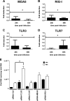
Effects of PRR silencing on ZIKV replication and IFN expression. (A to D) siRNAs specific for MDA5 (siRNA-MDA5), RIG-I (siRNA-RIG-I), TLR3 (siRNA-TLR3), and TLR7 (siRNA-TLR7), as well as a nonspecific siRNA (siRNA-Ctrl), were transfected into HFF1 cells 24 h before infection with ZIKV (MOI = 0.1). Reductions of mRNA levels by siRNAs were confirmed by real-time RT-PCR at 24 and 48 hpi. Results are expressed as the fold induction of expression of transcripts in specific siRNA-transfected cells relative to that in siRNA-Ctrl-transfected cells. The latter value corresponds to 1 on the ordinate of each histogram. Data are representative of two independent experiments, each performed in triplicate, and are normalized according to the 18S mRNA levels in the samples (error bars represent SEM). The Wilcoxon-Mann-Whitney test was used to analyze the differences between sets of data. P values of <0.05 were considered significant (*). (E) Viral copy numbers in siRNA-transfected cells were measured by real-time RT-PCR at 24 and 48 hpi. Statistically significant differences (P values < 0.05) between specific siRNA- and siRNA-Ctrl-transfected cells were determined by ANOVA, using GraphPad Prism software, and are denoted by an asterisk. Data are representative of two independent experiments, each performed in triplicate.
FIG 9
FIG 9
IFNs inhibit ZIKV infection. Primary skin fibroblasts were pretreated with different concentrations of IFN-α, IFN-β, and IFN-γ for 6 h before infection and were then exposed to ZIKV at an MOI of 1. (A to C) Inhibition of viral replication at 24 hpi was measured by real-time RT-PCR. (D to F) Release of viral particles, as quantified by plaque assay of culture supernatants. The statistical significance of the data was determined using ANOVA and GraphPad Prism software; statistically significant differences are denoted by asterisks (P < 0.05). Data are representative of three independent experiments, each performed in duplicate (error bars represent SEM).
FIG 10
FIG 10
Electron microscopic imaging of ZIKV-infected primary fibroblasts. (A) Membrane vesicles with sizes between 70 and 100 nm, observed in intimate association with the endoplasmic reticulum, are indicated by a white arrow. Black arrows indicate the presence of spherical capsids detected in intracellular vacuoles or docked to intracellular membranes. (B) Enlargement of a ZIKV particle. The intracellular electron-dense spherical capsid is 40 nm in diameter. (C) Assembled capsids are transported to the cell surface in intracellular vacuoles. (D) Autophagosomes are frequently detected in infected fibroblasts, and assembled capsids are observed inside this compartment. Data are representative of two independent experiments.
FIG 11
FIG 11
ZIKV induces autophagy in infected skin fibroblasts. (A) Visualization of autophagosome formation by LC3 aggregation in mock- or ZIKV-infected cells and cells treated with Torin 1. Cells were fixed at 24 hpi, and the colocalization of autophagosomes and ZIKV was determined by immunofluorescence, using MAbs specific for LC3 or the viral envelope protein (4G2). Data are representative of three independent experiments. (B and C) Primary human skin fibroblasts were exposed to ZIKV (MOI = 2) in the absence (cells were treated with the vehicle [0.05% dimethyl sulfoxide]) or presence of Torin 1 (B) or 3-MA (C), at the indicated concentrations, and viral replication was quantified by real-time RT-PCR at 24 and 48 hpi. Data are representative of three independent experiments. To test the significance of the differences, ANOVA was performed with GraphPad Prism software. Statistically significant differences between each condition and control cells are denoted by asterisks (P < 0.05).

Similar articles

Cited by

  • Taking AIM at Influenza: The Role of the AIM2 Inflammasome.
    Xu DW, Tate MD. Xu DW, et al. Viruses. 2024 Sep 27;16(10):1535. doi: 10.3390/v16101535. Viruses. 2024. PMID: 39459869 Free PMC article. Review.
  • Zika Virus Infects Human Placental Macrophages.
    Quicke KM, Bowen JR, Johnson EL, McDonald CE, Ma H, O'Neal JT, Rajakumar A, Wrammert J, Rimawi BH, Pulendran B, Schinazi RF, Chakraborty R, Suthar MS. Quicke KM, et al. Cell Host Microbe. 2016 Jul 13;20(1):83-90. doi: 10.1016/j.chom.2016.05.015. Epub 2016 May 27. Cell Host Microbe. 2016. PMID: 27247001 Free PMC article.
  • A Mouse Model of Zika Virus Pathogenesis.
    Lazear HM, Govero J, Smith AM, Platt DJ, Fernandez E, Miner JJ, Diamond MS. Lazear HM, et al. Cell Host Microbe. 2016 May 11;19(5):720-30. doi: 10.1016/j.chom.2016.03.010. Epub 2016 Apr 5. Cell Host Microbe. 2016. PMID: 27066744 Free PMC article.
  • Antagonism of interferon signaling by fibroblast growth factors promotes viral replication.
    Maddaluno L, Urwyler C, Rauschendorfer T, Meyer M, Stefanova D, Spörri R, Wietecha M, Ferrarese L, Stoycheva D, Bender D, Li N, Strittmatter G, Nasirujjaman K, Beer HD, Staeheli P, Hildt E, Oxenius A, Werner S. Maddaluno L, et al. EMBO Mol Med. 2020 Sep 7;12(9):e11793. doi: 10.15252/emmm.201911793. Epub 2020 Jul 27. EMBO Mol Med. 2020. PMID: 32720440 Free PMC article.
  • Insights into mosquito-borne arbovirus receptors.
    Liu J, Quan Y, Tong H, Zhu Y, Shi X, Liu Y, Cheng G. Liu J, et al. Cell Insight. 2024 Aug 24;3(6):100196. doi: 10.1016/j.cellin.2024.100196. eCollection 2024 Dec. Cell Insight. 2024. PMID: 39391003 Free PMC article. Review.

References

    1. Kuno G, Chang GJ, Tsuchiya KR, Karabatsos N, Cropp CB. 1998. Phylogeny of the genus Flavivirus. J Virol 72:73–83. - PMC - PubMed
    1. Moore DL, Causey OR, Carey DE, Reddy S, Cooke AR, Akinkugbe FM, David-West TS, Kemp GE. 1975. Arthropod-borne viral infections of man in Nigeria, 1964–1970. Ann Trop Med Parasitol 69:49–64. - PubMed
    1. Simpson DI. 1964. Zika virus infection in man. Trans R Soc Trop Med Hyg 58:335–338. doi:10.1016/0035-9203(64)90200-7. - DOI - PubMed
    1. Smithburn KC. 1954. Neutralizing antibodies against arthropod-borne viruses in the sera of long-time residents of Malaya and Borneo. Am J Hyg 59:157–163. - PubMed
    1. Fagbami AH. 1979. Zika virus infections in Nigeria: virological and seroepidemiological investigations in Oyo State. J Hyg 83:213–219. doi:10.1017/S0022172400025997. - DOI - PMC - PubMed

Publication types

MeSH terms

Substances