. 2015 Sep;89(17):8897-908.
doi: 10.1128/JVI.00941-15. Epub 2015 Jun 17.

ERdj5 Reductase Cooperates with Protein Disulfide Isomerase To Promote Simian Virus 40 Endoplasmic Reticulum Membrane Translocation

Affiliations

ERdj5 Reductase Cooperates with Protein Disulfide Isomerase To Promote Simian Virus 40 Endoplasmic Reticulum Membrane Translocation

Takamasa Inoue et al. J Virol. 2015 Sep.

Abstract

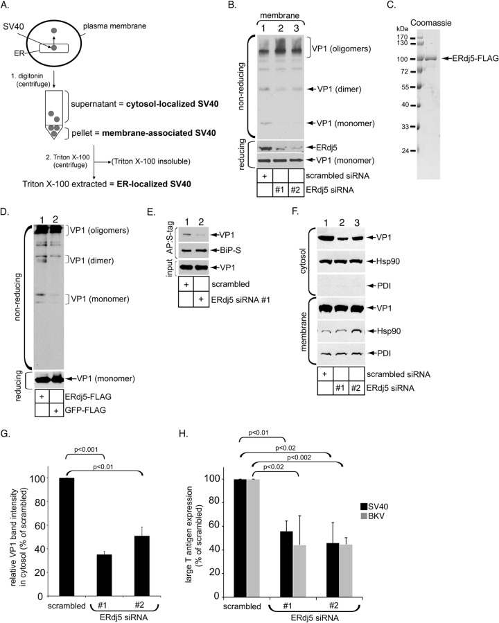
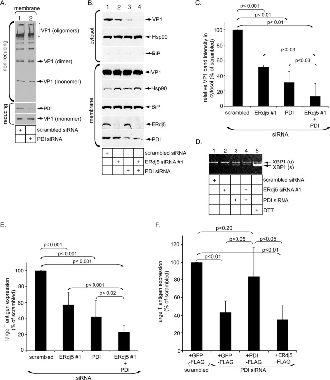
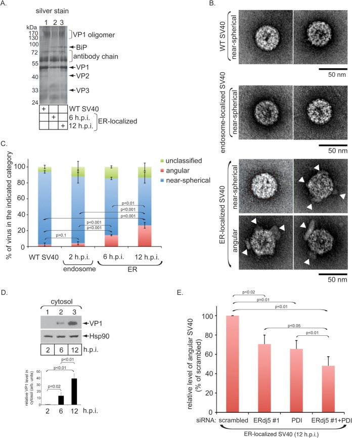
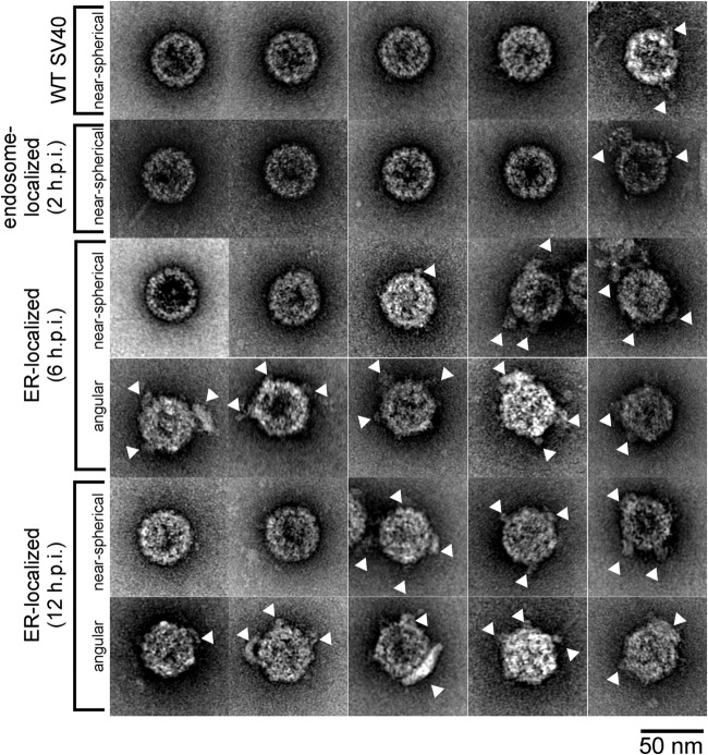
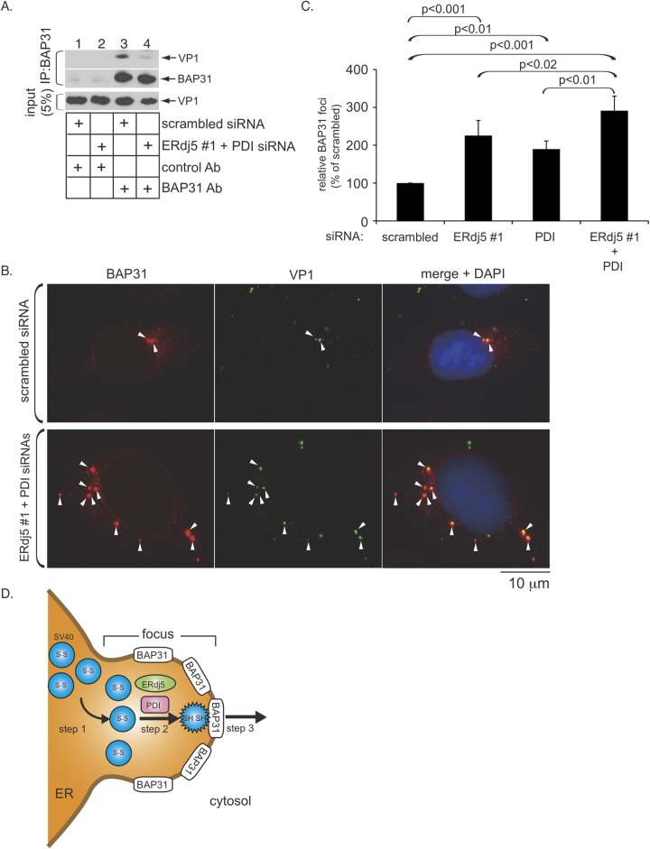
The nonenveloped polyomavirus (PyV) simian virus 40 (SV40) traffics from the cell surface to the endoplasmic reticulum (ER), where it penetrates the ER membrane to reach the cytosol before mobilizing into the nucleus to cause infection. Prior to ER membrane penetration, ER lumenal factors impart structural rearrangements to the virus, generating a translocation-competent virion capable of crossing the ER membrane. Here we identify ERdj5 as an ER enzyme that reduces SV40's disulfide bonds, a reaction important for its ER membrane transport and infection. ERdj5 also mediates human BK PyV infection. This enzyme cooperates with protein disulfide isomerase (PDI), a redox chaperone previously implicated in the unfolding of SV40, to fully stimulate membrane penetration. Negative-stain electron microscopy of ER-localized SV40 suggests that ERdj5 and PDI impart structural rearrangements to the virus. These conformational changes enable SV40 to engage BAP31, an ER membrane protein essential for supporting membrane penetration of the virus. Uncoupling of SV40 from BAP31 traps the virus in ER subdomains called foci, which likely serve as depots from where SV40 gains access to the cytosol. Our study thus pinpoints two ER lumenal factors that coordinately prime SV40 for ER membrane translocation and establishes a functional connection between lumenal and membrane events driving this process.

Importance: PyVs are established etiologic agents of many debilitating human diseases, especially in immunocompromised individuals. To infect cells at the cellular level, this virus family must penetrate the host ER membrane to reach the cytosol, a critical entry step. In this report, we identify two ER lumenal factors that prepare the virus for ER membrane translocation and connect these lumenal events with events on the ER membrane. Pinpointing cellular components necessary for supporting PyV infection should lead to rational therapeutic strategies for preventing and treating PyV-related diseases.


Figures

FIG 1
FIG 1
ERdj5 promotes SV40 disulfide bond reduction, ER membrane transport, and infection. (A) Flow diagram of the ER-to-cytosol transport assay depicting the cell fractionation protocol used in this study. In step 1, SV40-infected cells are treated with digitonin and centrifuged to generate a supernatant fraction (cytosol) containing cytosol-localized SV40 and a pellet fraction (membrane) containing membrane-associated SV40. In step 2, the pellet fraction can be further treated with Triton X-100 to extract ER-localized SV40 (Fig. 3). (B) CV-1 cells transfected with scrambled siRNA, ERdj5 siRNA 1, or ERdj5 siRNA 2 were infected with SV40 and fractionated as outlined above for panel A to generate Triton X-100-extracted material containing ER-localized SV40. Samples were subjected to reducing or nonreducing SDS-PAGE followed by immunoblotting with VP1-specific antibodies. (C) Coomassie staining of C-terminally FLAG-tagged ERdj5 (ERdj5-FLAG) purified from HEK 293T cells. (D) WT SV40 was incubated with ERdj5-FLAG or GFP-FLAG and subjected to nonreducing SDS-PAGE followed by immunoblotting with VP1-specific antibodies. (E) CV-1 cells expressing BiP-S and transfected with either scrambled siRNA or ERdj5 siRNA 1 were infected with SV40 for 6 h, harvested, and fractionated as described above for panel A. The resulting Triton X-100-extracted material containing ER-localized SV40 was incubated with S protein-conjugated agarose beads, and the isolated proteins were subjected to SDS-PAGE followed by immunoblotting with anti-VP1 and anti-S tag antibodies. AP, affinity purification. (F) CV-1 cells transfected with scrambled siRNA, ERdj5 siRNA 1, or ERdj5 siRNA 2 were infected with SV40 for 12 h and subjected to fractionation as outlined above for panel A. The cytosol and membrane fractions were analyzed by reducing SDS-PAGE and immunoblotted with the indicated antibodies. (G) The VP1 band intensity in the cytosol fraction shown in panel F was quantified with ImageJ (NIH). Data represent the means ± standard deviations of data from at least 3 independent experiments. A two-tailed t test was used. (H) Cells transfected with scrambled siRNA, ERdj5 siRNA 1, or ERdj5 siRNA 2 were infected with SV40 for 24 h or BKV for 48 h, fixed, and subjected to immunofluorescence staining using a SV40 or BKV large T antigen antibody. Large T antigen-positive nuclei were scored. Data represent the means ± standard deviations of data from at least 3 independent experiments. A two-tailed t test was used.
FIG 2
FIG 2
ERdj5 cooperates with PDI to stimulate SV40 ER membrane transport and infection. (A) Same as Fig. 1B except that cells were transfected with PDI siRNA. (B) Same as Fig. 1F except that cells were transfected with scrambled siRNA, ERdj5 siRNA 1, PDI siRNA, or ERdj5 siRNA 1 plus PDI siRNA. The resulting cytosol and membrane fractions were analyzed by immunoblotting with the indicated antibodies. (C) The VP1 band intensity in the cytosol fraction in panel A was quantified as described in the legend of Fig. 1G. Data represent the means ± standard deviations of data from at least 3 independent experiments. A two-tailed t test was used. (D) Reverse transcription-PCR analysis of the unspliced (u) and spliced (s) forms of XBP1 mRNA from cells transfected with the indicated siRNAs or treated with DTT. (E) Same as Fig. 1H except that cells were transfected with scrambled siRNA, ERdj5 siRNA 1, PDI siRNA, or ERdj5 siRNA 1 plus PDI siRNA. A two-tailed t test was used. (F) CV-1 cells initially transfected with scrambled or PDI siRNA were subsequently transfected with GFP-FLAG, PDI-FLAG, or ERdj5-FLAG. Cells were then infected with SV40 and subjected to immunofluorescence analyses using anti-FLAG and anti-T antigen antibodies. FLAG and T antigen double-positive cells were counted and analyzed as described above for panel E. Data represent the means ± standard deviations of data from at least 3 independent experiments. A two-tailed t test was used.
FIG 3
FIG 3
ERdj5 and PDI impart structural instability to ER-localized SV40, as revealed by EM analysis. (A) ER-localized SV40 (at 6 and 12 h p.i.) was immunoprecipitated with VP1-specific antibodies from the Triton X-100-extracted material, and the immunoprecipitate was captured by magnetic beads and subjected to SDS-PAGE followed by silver staining. WT SV40 (incubated with a buffer containing Triton X-100) was also subjected to the same immunoprecipitation method. (B) WT SV40, endosome-localized SV40 (at 2 h p.i.) (see Materials and Methods for the method of isolation of endosome-localized virus), and ER-localized SV40 were negatively stained and visualized by EM. Shown are representative images of viruses from each category. White arrowheads point to virus-associated materials. (C) Quantification of WT, endosome-localized (2 h p.i.), and ER-localized (6 and 12 h p.i.) SV40 as near-spherical, angular, or unclassified particle projections. Data represent the means ± standard deviations of data from at least 3 independent experiments. Each experiment was performed with ∼100 viral particles. A two-tailed t test was used. (D) VP1 levels in the cytosol fraction derived from cells infected with SV40 for the indicated times (immunoblots) were quantified (graph). Data represent the means ± standard deviations of data from at least 3 independent experiments. A two-tailed t test was used. (E) ER-localized SV40 (12 h p.i.) isolated from cells transfected with the indicated siRNAs was subjected to EM analysis as described above for panel B. Angular particles were analyzed and scored as described above for panel C. Data are normalized to the level of angular particles found in the scrambled control. A two-tailed t test was used.
FIG 4
FIG 4
Additional representative EM images of WT, endosome-localized (2 h p.i.), and ER-localized (6 and 12 h p.i.) SV40 particles. White arrowheads point to virus-associated materials.
FIG 5
FIG 5
Physical and functional relationship between ERdj5/PDI and the ER membrane protein BAP31. (A) Cells transfected with the indicated siRNAs were infected with SV40 for 16 h, treated with DSP for 30 min, and harvested. The resulting whole-cell lysate was immunoprecipitated (IP) with a control anti-c-Myc tag (9E10) or anti-BAP31 antibody (Ab). The immunoprecipitates were subjected to SDS-PAGE and analyzed by immunoblotting with the indicated antibodies. (B) Cells transfected with the indicated siRNAs were infected with SV40 for 16 h, fixed, and subjected to immunofluorescence staining with anti-BAP31 and -VP1 antibodies. DAPI, 4′,6-diamidino-2-phenylindole. (C) Numbers of cells with at least one BAP31-positive focus in SV40-infected cells transfected with the indicated siRNAs were determined. Data are normalized to values for scrambled siRNA and represent the means ± standard deviations of data from at least 3 independent experiments. A two-tailed t test was used. (D) Model depicting the ERdj5- and PDI-dependent conformational change of SV40 and its relationship to the BAP31 ER membrane protein (see the text).

Similar articles

Cited by

References

    1. Chandran K, Farsetta DL, Nibert ML. 2002. Strategy for nonenveloped virus entry: a hydrophobic conformer of the reovirus membrane penetration protein micro 1 mediates membrane disruption. J Virol 76:9920–9933. doi:10.1128/JVI.76.19.9920-9933.2002. - DOI - PMC - PubMed
    1. Ivanovic T, Agosto MA, Zhang L, Chandran K, Harrison SC, Nibert ML. 2008. Peptides released from reovirus outer capsid form membrane pores that recruit virus particles. EMBO J 27:1289–1298. doi:10.1038/emboj.2008.60. - DOI - PMC - PubMed
    1. Moyer CL, Wiethoff CM, Maier O, Smith JG, Nemerow GR. 2011. Functional genetic and biophysical analyses of membrane disruption by human adenovirus. J Virol 85:2631–2641. doi:10.1128/JVI.02321-10. - DOI - PMC - PubMed
    1. Trask SD, Kim IS, Harrison SC, Dormitzer PR. 2010. A rotavirus spike protein conformational intermediate binds lipid bilayers. J Virol 84:1764–1770. doi:10.1128/JVI.01682-09. - DOI - PMC - PubMed
    1. Wiethoff CM, Wodrich H, Gerace L, Nemerow GR. 2005. Adenovirus protein VI mediates membrane disruption following capsid disassembly. J Virol 79:1992–2000. doi:10.1128/JVI.79.4.1992-2000.2005. - DOI - PMC - PubMed

Publication types

MeSH terms

LinkOut - more resources