. 2015 Jul 31;290(31):19307-18.
doi: 10.1074/jbc.M115.663286. Epub 2015 Jun 17.

Interleukin-35 Inhibits Endothelial Cell Activation by Suppressing MAPK-AP-1 Pathway

Affiliations

Interleukin-35 Inhibits Endothelial Cell Activation by Suppressing MAPK-AP-1 Pathway

Xiaojin Sha et al. J Biol Chem. .

Abstract

Vascular response is an essential pathological mechanism underlying various inflammatory diseases. This study determines whether IL-35, a novel responsive anti-inflammatory cytokine, inhibits vascular response in acute inflammation. Using a mouse model of LPS-induced acute inflammation and plasma samples from sepsis patients, we found that IL-35 was induced in the plasma of mice after LPS injection as well as in the plasma of sepsis patients. In addition, IL-35 decreased LPS-induced proinflammatory cytokines and chemokines in the plasma of mice. Furthermore, IL-35 inhibited leukocyte adhesion to the endothelium in the vessels of lung and cremaster muscle and decreased the numbers of inflammatory cells in bronchoalveolar lavage fluid. Mechanistically, IL-35 inhibited the LPS-induced up-regulation of endothelial cell (EC) adhesion molecule VCAM-1 through IL-35 receptors gp130 and IL-12Rβ2 via inhibition of the MAPK-activator protein-1 (AP-1) signaling pathway. We also found that IL-27, which shares the EBI3 subunit with IL-35, promoted LPS-induced VCAM-1 in human aortic ECs and that EBI3-deficient mice had similar vascular response to LPS when compared with that of WT mice. These results demonstrated for the first time that inflammation-induced IL-35 inhibits LPS-induced EC activation by suppressing MAPK-AP1-mediated VCAM-1 expression and attenuates LPS-induced secretion of proinflammatory cytokines/chemokines. Our results provide insight into the control of vascular inflammation by IL-35 and suggest that IL-35 is an attractive novel therapeutic reagent for sepsis and cardiovascular diseases.

Keywords: cytokine; endothelial cell; endothelial cell activation; inflammation; interleukin-35; lipopolysaccharide (LPS); sepsis; vascular cell adhesion molecule-1 (VCAM-1); vascular inflammation.


Figures

FIGURE 1.
FIGURE 1.
Plasma levels of IL-35 are induced in mice with LPS-induced endotoxemia. A, schematic representation of LPS-induced endotoxemia model (LPS) with intraperitoneal (ip) injection (20 μg/g of body weight)). B–D, detection of IL-35, IL-12p40, and IL-27p28 by ELISAs in the plasma of wild type mice after LPS challenge after 0, 1.5, 4, and 24 h, n = 9–10 in each group. mIL, mouse IL. E, comparison of plasma levels of IL-35, IL-12p40, and IL-27p28 in each time point to further demonstrate the specificity of IL-35 detection. n = 9–10 in each group. The results are expressed as the means ± S.E. NS, not significant. *, p < 0.05; **, p < 0.01; ***, p < 0.001.
FIGURE 2.
FIGURE 2.
Plasma levels of IL-35 are induced in patients with sepsis. A, human IL-35 levels in the plasma samples from patients with sepsis (n = 6) and healthy controls (n = 14). C, general information about patients with sepsis. B, general information of healthy controls. The results are expressed as the means ± S.E. *, p < 0.05.
FIGURE 3.
FIGURE 3.
IL-35 suppresses cytokine and chemokine expression in the plasma of wild type mice challenged with LPS. A, in the left panel, mouse cytokine/chemokine arrays were performed to detect the expression changes of 40 inflammatory cytokines/chemokines in the plasma from mice treated with LPS, LPS plus rIL-35, and WT control mice. Plasma samples in each group were pooled from three mice. In the right panel, the arrangement of the mouse cytokine/chemokine array was shown. Cytokines and chemokines that were induced by LPS are in bold. rIL-35-inhibited cytokines and chemokines are labeled with asterisks. B and C, quantification of cytokine and chemokine expressions. The variations of the manufacturer's designated positive control spots between each array were used to determine the confidence interval of nonspecific variations between samples. *, p < 0.05.
FIGURE 4.
FIGURE 4.
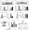
IL-35 inhibits LPS-induced leukocyte adhesion in vivo and in vitro. A, leukocyte adhesion to cremaster post-capillary venules of control, LPS-treated, and LPS plus rIL-35 co-treated mice after 4-h treatment was visualized using intravital microscopy (n = 6 in each group, scale bar = 20 μm). B, quantitative analysis was performed by enumerating rhodamine 6G-labeled leukocytes that adhered to mouse cremaster muscle post-capillary venule endothelium (n = 6 in each group). C, mouse lung histological analysis in control, LPS-treated, and LPS plus rIL-35 co-treated mice was performed after hematoxylin and eosin (HE) staining (scale bar in upper panel = 200 μm; scale bar in lower panel = 20 μm), and the inflammatory cell adhesion to endothelium was further indicated by the arrows (n = 6 in each group). D, the adhesion of MPO-positive neutrophil granulocytes and macrophages to the vascular wall was detected in the lung paraffin-embedded sections of the control mice (left panel), LPS-treated mice (middle panel), and LPS and rIL-35 co-treated mice (right panel) after 4-h treatment (n = 6 mice in each group). Scale bars: 200 μm (upper panel), 20 μm (lower panel) (n = 6 in each group). E, exodus of inflammatory cells into the mouse lungs was counted in the BALF of mice (n = 6 in each group). F, schematic representation of monocyte adhesion assay. G and H, the adhesion of non-stimulated, fluorescence-labeled human peripheral blood mononuclear cells/THP1 cells to HAECs stimulated by LPS and LPS plus rIL-35 was quantified. The result shown is representative of 3 independent experiments. untreat, untreated. The results are expressed as the means ± S.E. *, p < 0.05; ***, p < 0.001.
FIGURE 5.
FIGURE 5.
IL-35 inhibits LPS-induced up-regulation of VCAM-1 in vivo and in vitro. A, upper panel, the expressions of endothelial cell adhesion molecules VCAM-1 and ICAM-1 in mouse aortas were determined by Western blot analyses after control, LPS, and LPS plus rIL-35 treatments. VCAM-1 and ICAM-1 quantifications were presented in the lower panel. (n = 4 in each group). B, upper panel, VCAM-1 and ICAM-1 expressions in mouse lungs were determined by Western blot analyses. The quantitative data of VCAM-1 and ICAM-1 were presented in the lower panel (n = 3 in each group). C and D, VCAM-1 and ICAM-1 expressions in non-treated, LPS-treated, and LPS plus IL-35 co-treated HAECs were quantified by using flow cytometry. The quantitative data of the -fold changes of VCAM-1+ cell percentage and ICAM-1+ cell percentage in each treatment group over the control group are presented to the right of representative flow cytometry data. The result shown is representative of 3 independent experiments. E, the expressions of IL-35 receptor subunits gp130 and IL-12Rβ2 in HAECs were determined by Western blot analysis. F, inhibition of LPS up-regulation of VCAM-1 by IL-35 in HAECs was reversed by anti-gp130 antibody and anti-IL-12Rβ2 antibody but not by IgG controls. The results shown are representative of 3 independent experiments. G, schematic representation of IL-35 receptor formats including two types of homodimers, IL-12Rβ2-IL-12Rβ2 and gp130-gp130, and a heterodimer of IL-12Rβ2-gp130 (upper panel). IL-35 receptor formats can be blocked by anti-IL-12Rβ2 antibody and anti-gp130 antibody (lower panel). The results are expressed as the means ± S.E. NS, not significant. *, p < 0.05; **, p < 0.01; ***, p < 0.001.
FIGURE 6.
FIGURE 6.
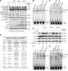
IL-35 inhibits LPS-induced activation of MAPK-AP1 pathway, which mediates LPS-induced VCAM-1 up-regulation in human aortic endothelial cells. A, the expressions of proteins involved in the MAPK pathway were analyzed with Western blots in HAECs stimulated by LPS and LPS plus IL-35 treatments for 5, 15, and 30 min. The relative mean pixel density ratios of phosphorylated (p) kinase over total kinase proteins were shown. B, HAECs were treated with LPS for 1.5 h with or without IL-35 pretreatment for 18 h, and EMSA was used to detect the binding of transcription factor AP-1 in the nuclear extracts. C, EMSA was used to determine the specificity of AP-1 binding to the AP-1 consensus oligonucleotides using consensus competitor oligonucleotides, mutant competitor oligonucleotides, and supershift antibodies for AP-1 subunits c-Fos and c-Jun. D, AP-1-binding sites were found in the promoters of VCAM-1 gene, proinflammatory cytokine genes, and chemokine genes that were suppressed by IL-35. N.D., not determined; N.F., not found; PMID, PubMed IDs of the studies. E, protein expressions of phosphorylated (p)-IκBα, total IκBα, and β-actin were analyzed using Western blots. The relative mean pixel density ratios of phosphorylated over total proteins were shown. F, EMSAs were used to detect the binding of transcription factor NF-κB in the nuclear extracts in HAECs. G, EMSA was used to determine the specificity of NF-κB binding to the NF-κB consensus oligonucleotide using competitor oligonucleotides, mutant oligonucleotides, and supershift antibodies for NF-κB subunits p50 and p65. The result shown is representative of 3 independent experiments.
FIGURE 7.
FIGURE 7.
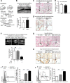
IL-35 inhibits LPS-induced monocyte adhesion to endothelial cells in EBI3-deficient mice in vivo. A, schematic representation on two possibilities concerning whether EBI3−/− mice have increased LPS-induced EC activation. B, leukocyte adhesion to cremaster post-capillary venules in living WT mice and EBI3−/− mice was visualized using intravital microscopy in the upper panel (n = 3 male mice for each group, scale bar = 20 μm). Quantification was shown in the lower panel. Data are representative of 3 independent experiments. C, mouse lung histological analyses were performed by hematoxylin and eosin (HE) staining in WT control mice and EBI3−/− mice (scale bar in upper panel = 200 μm; scale bar in lower panel = 20 μm) in non-stimulated conditions. D, MPO-positive adhesion of neutrophil/granulocyte and macrophages to the vascular wall in lung was examined (scale bar in upper panel = 200 μm; scale bar in lower panel = 20 μm) in non-stimulated conditions. E, leukocyte numbers in the peripheral blood of WT mice and EBI3−/− mice were quantified (WT mice, n = 11; EBI3−/− mice, n = 12). F, leukocyte adhesion to cremaster post-capillary venules in live WT mice and EBI3−/− mice challenged with LPS was examined using intravital microscopy in the upper panel. The pictures were captured from the venules of LPS-challenged WT mice, LPS-challenged EBI3−/− mice, and LPS plus rIL-35 co-treated EBI3−/− mice. Scale bar = 20 μm. In the lower column graph, quantification was performed by enumerating rhodamine 6G-labeled leukocytes that adhered to mouse cremaster muscle post-capillary venule endothelium per minute (n = 6 in each group). G, mouse lung histological analysis was performed by H&E staining in LPS-treated WT control mice (left panel), LPS-treated EBI3−/− mice (middle panel), and LPS plus IL-35 co-treated EBI3−/− mice (right panel). Upper pictures, scale bar = 200 μm; lower pictures, scale bar = 20 μm. The adhesion of inflammatory cells to endothelium is indicated by the arrows (n = 6 in each group). H, MPO-positive neutrophil granulocyte and macrophage adhesions to the vascular wall in lung were examined. Scale bar = 200 μm (top panel), scale bar = 20 μm (bottom panel) (n = 6). I and J, VCAM-1 and ICAM-1 expressions in non-treated, LPS-treated, and LPS plus IL-27 co-treated HAECs were quantified using flow cytometry. The quantitative data of the -fold changes of VCAM-1+ cell percentage and ICAM-1+ cell percentage in each treatment group over the control group are presented to the right of representative flow cytometry data. The results shown are representative of 3 independent experiments. The results are expressed as the means ± S.E. NS, not significant. *, p < 0.05; ***, p < 0.001.
FIGURE 8.
FIGURE 8.
A new working model. LPS can induce inflammatory response in endothelial cells by engaging Toll-like receptor 4 (TLR4) and activating the MAPK-AP-1 and NF-κB pathways. IL-35 is induced by LPS presumably in regulatory T cells, B cells, and dendritic cells at the same time, which suppresses LPS-induced endothelial cell activation by inhibiting expressions of VCAM-1 in endothelial cells, proinflammatory cytokines, and chemokines in plasma and monocyte adhesion to endothelium, thereby controlling inflammation scale in sepsis. In IL-35 subunit EBI3 gene-deficient (EBI3−/−) mice, endogenous IL-35 could not be induced by LPS, but exogenous IL-35 could still suppress LPS-induced inflammatory response in endothelial cells because of intact and functional IL-35 receptors and its signaling pathway(s) in EBI3−/− mice.

Similar articles

Cited by

References

    1. Angus D. C., Linde-Zwirble W. T., Lidicker J., Clermont G., Carcillo J., Pinsky M. R. (2001) Epidemiology of severe sepsis in the United States: analysis of incidence, outcome, and associated costs of care. Crit. Care Med. 29, 1303–1310 - PubMed
    1. Wood K. A., Angus D. C. (2004) Pharmacoeconomic implications of new therapies in sepsis. PharmacoEconomics 22, 895–906 - PubMed
    1. Stoll L. L., Denning G. M., Weintraub N. L. (2004) Potential role of endotoxin as a proinflammatory mediator of atherosclerosis. Arterioscler. Thromb. Vasc. Biol. 24, 2227–2236 - PubMed
    1. Xiong Z., Yan Y., Song J., Fang P., Yin Y., Yang Y., Cowan A., Wang H., Yang X. F. (2009) Expression of TCTP antisense in CD25(high) regulatory T cells aggravates cuff-injured vascular inflammation. Atherosclerosis 203, 401–408 - PMC - PubMed
    1. Ait-Oufella H., Salomon B. L., Potteaux S., Robertson A. K., Gourdy P., Zoll J., Merval R., Esposito B., Cohen J. L., Fisson S., Flavell R. A., Hansson G. K., Klatzmann D., Tedgui A., Mallat Z. (2006) Natural regulatory T cells control the development of atherosclerosis in mice. Nat. Med. 12, 178–180 - PubMed

Publication types

MeSH terms