. 2015 Jul 2;523(7558):83-7.
doi: 10.1038/nature14545. Epub 2015 Jun 17.

Spatiotemporal control of a novel synaptic organizer molecule

Affiliations

Spatiotemporal control of a novel synaptic organizer molecule

Kelly Howell et al. Nature. .

Abstract

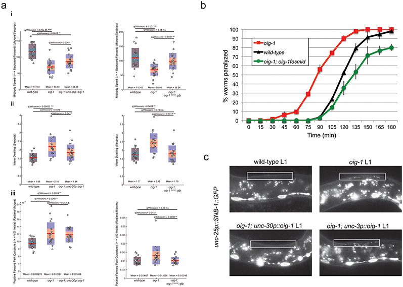
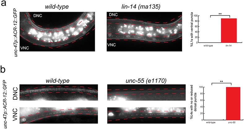
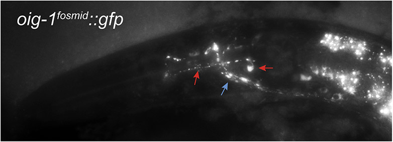
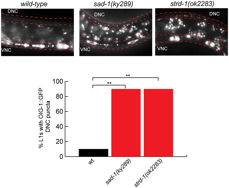
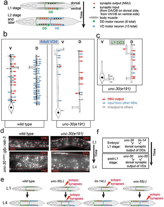
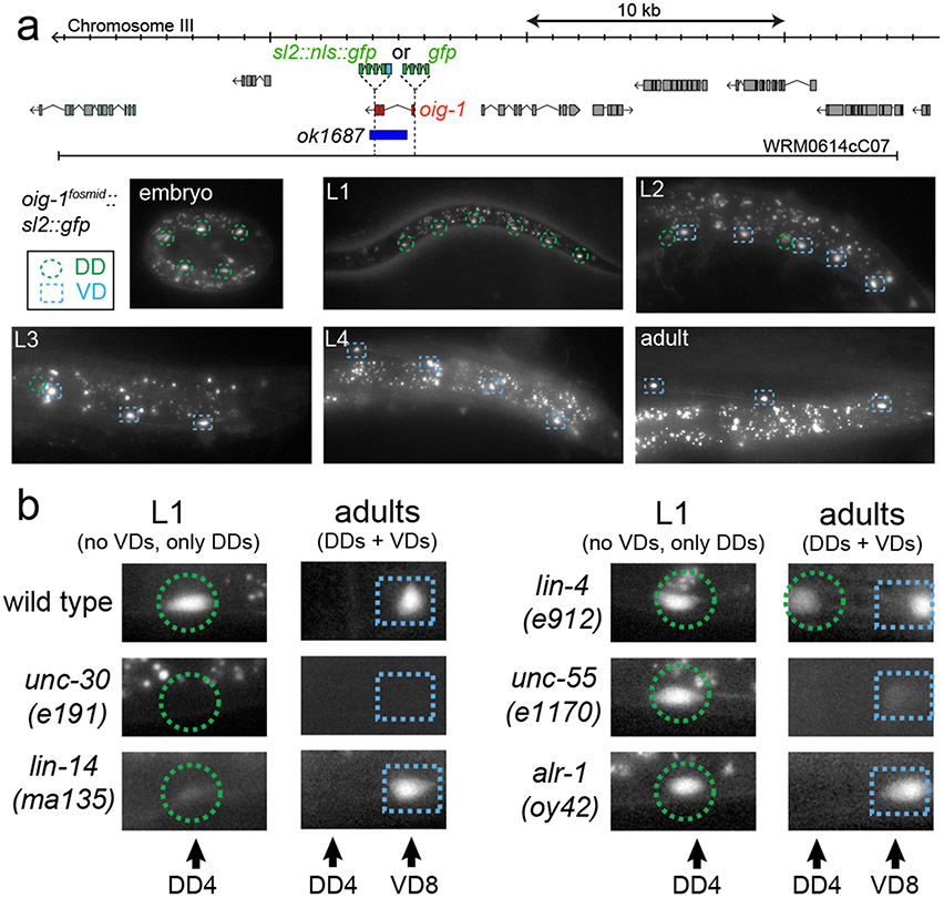
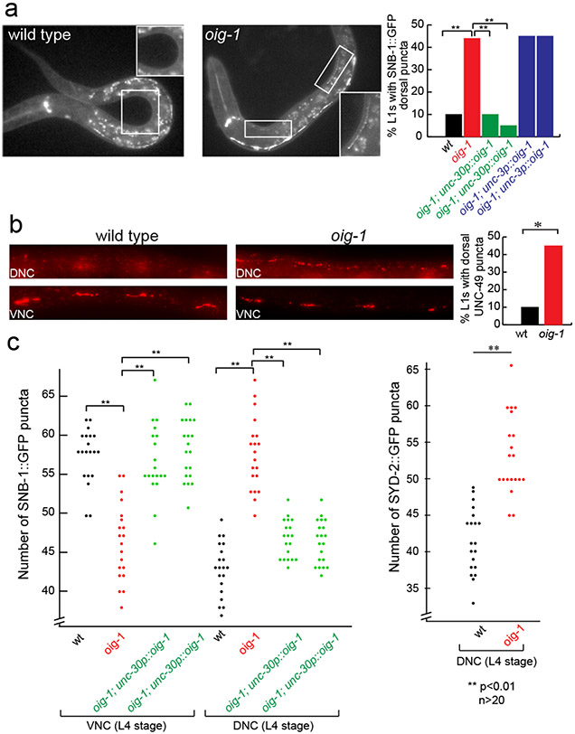
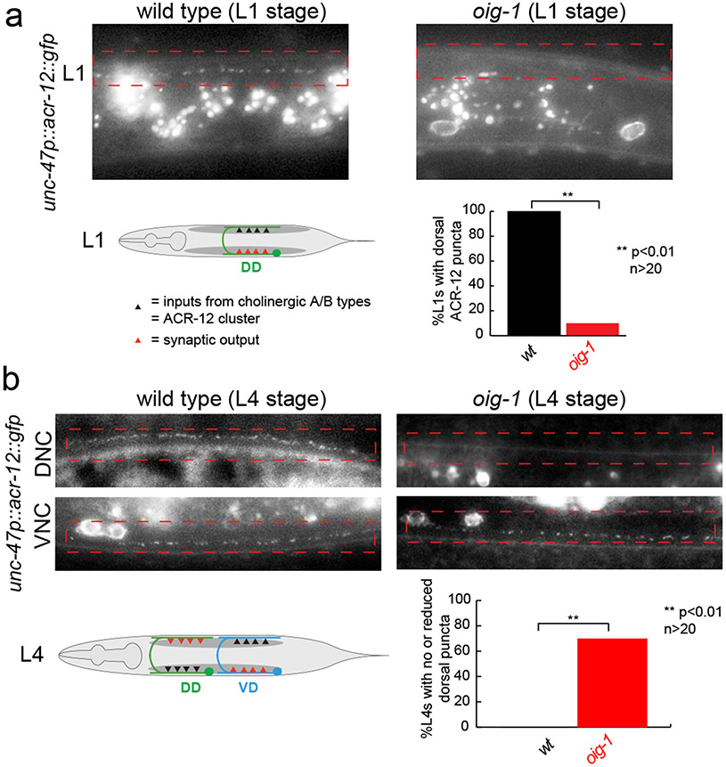
Synapse formation is a process tightly controlled in space and time. How gene regulatory mechanisms specify spatial and temporal aspects of synapse formation is not well understood. In the nematode Caenorhabditis elegans, two subtypes of the D-type inhibitory motor neuron (MN) classes, the dorsal D (DD) and ventral D (VD) neurons, extend axons along both the dorsal and ventral nerve cords. The embryonically generated DD motor neurons initially innervate ventral muscles in the first (L1) larval stage and receive their synaptic input from cholinergic motor neurons in the dorsal cord. They rewire by the end of the L1 moult to innervate dorsal muscles and to be innervated by newly formed ventral cholinergic motor neurons. VD motor neurons develop after the L1 moult; they take over the innervation of ventral muscles and receive their synaptic input from dorsal cholinergic motor neurons. We show here that the spatiotemporal control of synaptic wiring of the D-type neurons is controlled by an intersectional transcriptional strategy in which the UNC-30 Pitx-type homeodomain transcription factor acts together, in embryonic and early larval stages, with the temporally controlled LIN-14 transcription factor to prevent premature synapse rewiring of the DD motor neurons and, together with the UNC-55 nuclear hormone receptor, to prevent aberrant VD synaptic wiring in later larval and adult stages. A key effector of this intersectional transcription factor combination is a novel synaptic organizer molecule, the single immunoglobulin domain protein OIG-1. OIG-1 is perisynaptically localized along the synaptic outputs of the D-type motor neurons in a temporally controlled manner and is required for appropriate selection of both pre- and post-synaptic partners.


Figures

Extended Data Figure 1:
Extended Data Figure 1:. Electron microscopical analysis of unc-30(e191) mutants
a: Reconstructions of a VD4 and a DD2 MN from an unc-30(e191) animal compared to the same neurons in a wild-type animal. Cell bodies (large black dots) are all situated in the ventral cord. Processes emanate anteriorly (up on plots) from the cell body and run along the ventral cord. Lateral branches leave the ventral cord (v) and run round to the dorsal cord (d) as a circumferential commissure (broken horizontal process in the plots). Commissures from unc-30 mutant type-D neurons are situated in the same regions as those of their wild-type counterparts. However the cell bodies of DD neurons are often displaced anteriorly in the mutants, with the consequence that DD neurons have shorter processes in the ventral cord (c). Processes in the dorsal cord run anteriorly in mutant animals, whereas they branch with the main branch running posteriorly in wild-type animals. Neuromuscular junctions (NMJs) in unc-30 mutants are made predominantly in the dorsal cord by both the DDs and VDs, whereas in the wild type, only DD neurons innervate dorsal muscles. The synaptic inputs to the DD and VD neurons in mutant animals (inward pointing arrows for chemical synapses and "T" for gap junctions) are generally abnormal. The reconstructed DD2 neuron received synapses from several unidentified processes on the ventral side (depicted with a "?"). These processes do not belong to VA or VB neurons, the normal pre-synaptic partners of DD, as all the local VA and VB neurons were identified. From the location and synaptic behavior of these processes, it is probable that they belong to interneurons which span the length of the cord and do not usually innervate D-type neurons. An Asterisk ("*") indicates that a synapse has multiple post-synaptic elements. A total of six VDs and three DDs were reconstructed. Each reconstruction covered around 2000 EM sections corresponding to a length of 100μ along the body of the animal. The unc-30(e191) mutation does not affect MN cell body position or the synaptic behaviour of the DA/DB neurons, except in regard to their synapses to D-type neurones; this made it possible to unambiguously identify D type neurones from their positions and by eliminating other identified classes of MNs. Electron microscopy and reconstructions of micrographs of serial sections were performed as described in . b: Electron micrographs. The processes of DD and VD neurons normally run subjacent to the bounding basal lamina of the ventral cord immediately dorsal to the axons of the VA and VB neurons (a). NMJs are made through the basal lamina onto muscle arms (m). In unc-30(e191) animals, the axons of the DD and VD neurons wander round the cord and do not run in defined locations; the configuration shown in (b) is typical but not stereotyped. Very few NMJs are made in the ventral cord by the DD or VD neurons in unc-30 mutants; those that are made look rather small (c). Atypical synapses (d) are often made onto DD or VD processes from neurons such as the touch receptor neuron AVM (6). It is probable that these synapses are not normally found as the processes of AVM and DD or VD do not normally run alongside each other in wild-type animals. Both the DD and VD MNs make NMJs to dorsal muscles in the dorsal cord of unc-30 mutants, whereas in wild-type animals, only DD neurons do so and VD neurons innervate ventral muscles in the ventral cord. (e) & (f) show NMJs made by VD (e) and DD (f) neurons in the dorsal cord of an unc-30(e191) mutant. Scale bars 1μ.
Extended Data Figure 2:
Extended Data Figure 2:. RAB-3 is ectopically localized in lin-14, unc-55, and oig-1 mutants.
a: Presynaptic marker RAB-3 ectopically localizes to the dorsal nerve cord (DNC; marked in red) in lin-14 mutant animals. RAB-3::GFP puncta (from otEx5663, unc-30p::GFP::RAB-3) localize mostly to the ventral nerve cord (VNC; marked in red) in wild-type L1 animals (left). Ectopic RAB-3::GFP puncta localize mostly to the dorsal nerve cord in 95% of lin-14 L1 animals (right, scored in progeny from lin-14 null animals carrying a lin-14 rescue array . Ventral and dorsal nerve cords are indicated by red dotted lines. L1 animals were obtained by hypochlorite-treating gravid adult animals and letting embryos hatch and arrest in M9 for 16-18 hours. n>20 for each strain scored. b: RAB-3 ectopically localizes to the dorsal nerve cord in unc-55 L4 mutant animals. RAB-3::GFP puncta localizes to both the ventral (VNC) and dorsal (DNC) nerve cord in 100% of wild-type L4 animals (left). RAB-3::GFP puncta localize mostly to the dorsal nerve cord in 100% of unc-55 L4 animals (right). Ventral and dorsal nerve cords are indicated by red dotted lines. Signals between the nerve cords are autofluorescence from the gut. n>20 for each strain scored. c: RAB-3 ectopically localizes to the dosal nerve cord in oig-1 mutants. RAB-3 normally localizes to the ventral nerve cord (VNC, marked in red) in wild-type L1 animals (top left). Ectopic RAB-3::GFP puncta localize to the dorsal nerve cord in 55% of oig-1 L1 animals (top right, compared to 20% of wild-type animals). L1 animals were obtained by hypochlorite-treating gravid adult animals and letting embryos hatch and arrest in M9 for 16-18 hours. n>20 for each strain scored. In wild-type L4 animals, more RAB-3::GFP puncta are localized in the VNC than in the DNC of the animal (bottom, black dots). Conversely, in oig-1 mutants, more RAB-3::GFP puncta are localized in the DNC than in the VNC (bottom, red dots). **p<0.01, n=20 for each strain.
Extended Data Figure 3:
Extended Data Figure 3:. Independence of transcription factor activities
a: Expression of unc-30 is not affected by loss of lin-14 or unc-55. A 2.4kb unc-30 promoter gfp fusion reporter is expressed in the DD MNs (green circles) in wild-type L1 animals; this expression is not affected in lin-14(−) mutant animals (scored in progeny from lin-14 null mothers carrying a lin-14 rescue array ). An unc-30 fosmid-based reporter, kindly provided by the Transgeneome project, is expressed in the DD (green circles) and VD (blue squares) MNs (DD4 to VD10 shown) in wild-type L4 animals; this expression is not affected in unc-55(e1170) L4 animals. n>20 for each genotype. b: Expression of a lin-14 fosmid-based reporter construct is unaffected by loss of unc-30. lin-14 is expressed in the DA, DB, and DD MNs in the VNC at the L1 stage (Average number of VNC cells=15); this expression is not affected in unc-30(e191) mutant L1s (Average number of VNC cells=15);. n>20 for each genotype. Loss of unc-30 also does not affect unc-55 expression, as shown by .
Extended Data Figure 4:
Extended Data Figure 4:. Deletion of a putative UNC-30 binding site results in loss of oig-1 expression in the D-type neurons.
Regions of the oig-1 promoter were fused to gfp to analyze expression. (+) indicates robust expression of the reporter construct in the specified cell type, whereas (−) indicates loss of expression in the specified cell type. Twenty worms at both the L1 and L4 stage were scored for each line. Expression of a 1kb promoter reporter (prom 1) recapitulates expression of the oig-1fosmid::gfp reporter in the D-type MNs (see Fig.2). This region contained 3 elements that exactly match the UNC-30 consensus binding site (TAATC, purple box, ) and multiple others that are a partial match to the UNC-30 binding site (magenta and blue boxes). Further deletion of this prom 1 defined a minimal 125bp element that is sufficient to drive oig-1 expression in the D-type MNs (prom 6). This element contains two sites that partially match the UNC-30 binding sequence. Deletion of the AAATC site in the context of the 1kb promoter (prom 9) has no effect on oig-1 expression in the D-type MNs. Deletion of the TAAAC site in the context of the 1kb promoter reporter (prom 10) results in complete loss of oig-1 expression specifically in the D-type MNs.
Extended Data Figure 5.
Extended Data Figure 5.. oig-1 mutants defects and their rescue.
a: oig-1 mutants display locomotory defects. Locomotion of L4 animals was analyzed with tracking assays (Yemini et al 2013). The graphs on the left side of each panel correspond to assays comparing wild-type, oig-1 mutant, and oig-1; unc-30p::oig-1 animals. The graphs on the right side of each panel correspond to assays comparing wild-type, oig-1 mutant, and oig-1; oig-1fosmid::gfp animals. Twenty animals (each dot on a plot) were tracked for each genotype for both comparisons. Mean and Q values are indicated. Note that in a previously published analysis of a large panel of available mutants, the same set of locomotory defects that we describe here for oig-1 mutants were found to be affected in unc-55 mutants , albeit in a stronger manner than oig-1 mutants. Also note that the very strong locomotory defects unc-30 defects are qualitatively very different from oig-1 mutants, but this is to be expected since unc-30 mutant do not only show the synaptic defects that we describe here, but also lack the neurotransmitter GABA , thereby disabling any neuromuscular signaling. i: The midbody speed of oig-1 mutant animals is significantly lower than that of wild-type animals. This defect is partially rescued (statistically different from oig-1 mutants but also from wild-type animals) by expressing unc-30p::oig-1 in oig-1 animals (left graph). The lower midbody speed of oig-1 mutants is completely rescued (statistically different from oig-1 mutants but not from wild-type animals) by expressing the oig-1fosmid::gfp in oig-1 mutants. ii: oig-1 mutants exhibit more dwelling than wild-type L4 animals. This defect is partially rescued (statistically different from oig-1 mutants but also from wild-type animals) by expressing unc-30p::oig-1 in oig-1 animals (left graph). The increased dwelling of oig-1 mutants is completely rescued (statistically different from oig-1 mutants but from not wild-type animals) by expressing the oig-1fosmid::gfp in oig-1 mutants. iii: oig-1 mutants exhibit an increased path curvature compared to wild-type animals. This defect is not rescued by expressing unc-30p::oig-1 in oig-1 animals (left graph). The increased path curvature of oig-1 mutants is completely rescued (statistically different from oig-1 mutants but not from wild-type animals) by expressing the oig-1fosmid::gfp in oig-1 mutants. b: Aldicarb-sensitivity defects in oig-1 mutants. oig-1 mutant young adult animals (red squares), which display aberrant GABAergic synapses in both the ventral and dorsal cord, show hypersensitivity to aldicarb-induced paralysis compared to wild-type (black triangles). Expression of oig-1fosmid::gfp from a multicopy transgenic array (green circles) does not only rescue the oig-1 mutant phenotype, but even results in a slight hyposensitivity to aldicarb. Worms were tested every 15 minutes for paralysis by touching the head and tail three times each. n=20 for each strain, repeated 3 times. c: Expression of oig-1 in the D-type neurons rescues ectopic DD synapses in the dorsal nerve cord. At the L1 stage when only the DD MNs are present, SNB-1::GFP (from juIs1-unc-25p::SNB-1::GFP) localizes to the ventral nerve cord (VNC) in wild-type animals (top left). Ectopic SNB-1::GFP puncta localize to the dorsal nerve cord (DNC) of oig-1 mutant L1s (top right). This phenotype is rescued by expressing oig-1 in the D-type MNs (unc-30p::oig-1, otEx4955, bottom left), but not by expressing oig-1 in the neighboring cholinergic MNs (unc-3p::oig-1, otEx4942, bottom right). White boxes indicate the dorsal nerve cord.
Extended Data Figure 6:
Extended Data Figure 6:. ACR-12 is mislocalized in lin-14 and unc-55 mutants.
Cholinergic innervation to the D-type MNs is visualized with an unc-47p::ACR-12::GFP reporter transgene, maintained in an acr-12(ok367) mutant background. . a: ACR-12 puncta localization is affected by loss of lin-14. In wild-type L1 animals, ACR-12 puncta are observed only in the DD neurons in the dorsal (DNC) nerve cord (left). In lin-14 mutant L1 animals (scored in progeny from lin-14 null animals carrying a lin-14 rescue array ), ACR-12 puncta are detected in the ventral nerve cord (VNC) of the DD MNs. Quantification of this data is represented in the graph. Some dorsal puncta in the DD MNs were still observed in 83% of the lin-14 mutant L1s that had puncta in the ventral nerve cord. L1 animals were obtained by hypochlorite-treating gravid adult animals and letting embryos hatch and arrest in M9 for 16-18 hours. n > 20 for each strain scored, **p < 0.01. b: ACR-12 puncta localization is affected by loss of unc-55. In wild-type L4 animals, ACR-12 puncta are observed in both the ventral (VNC) and dorsal (DNC) nerve cords (left). In unc-55 mutant L4 animals, ACR-12 puncta are observed mostly in the ventral nerve cord of unc-55 mutants. Ventral and dorsal nerve cords are marked by red dotted lines. Quantification of this data is represented in the graph. n>20 for each strain, **p < 0.01.
Extended Data Figure 7:
Extended Data Figure 7:. The IgC2 domain is necessary for OIG-1 function.
At the L1 stage when only the DD MNs are present, ectopic SNB-1::GFP puncta (from juIs1-unc-25p::SNB-1::GFP) localize to the dorsal nerve cord (DNC) of oig-1 mutant L1s (red bar) but not in wild-type animal (black bar) (see Fig. 3A). Based on an alignment with the hidden Markov model (HMM) Ig domain (top), a highly conserved residue (W75) and a nonconserved residue (E64) in the OIG-1 Ig domain were mutated in the context of an unc-30p::oig-1 transgene that is able to rescue the L1 ectopic synapse defects (see Fig. 3A). The unc-30p::oig-1E64A transgenes (otEx6212, otEx6213) were still able to rescue the synaptic defects of oig-1 L1 animals (green bars), whereas the unc-30p::oig-1W75A transgenes (otEx6214,otEx6215) had no rescue ability (blue bars). L1 animals were obtained by hypochlorite-treating gravid adult animals and letting embryos hatch and arrest in M9 for 16-18 hours. n>20 for each strain scored, **p < 0.01, *p < 0.05.
Extended Data Figure 8:
Extended Data Figure 8:. OIG-1 localization in other neuron types.
OIG-1::GFP (from oig-1fosmid::gfp) localizes in a punctate manner along axons in the nerve ring (blue arrow) and along a pair of neurons in the pharynx, tentatively identified as the M2 MNs (red arrows point to cell body and process). These neurons form synapses onto pharyngeal muscles along their processes, and these processes also show punctate localization of OIG-1.
Extended Data Figure 9:
Extended Data Figure 9:. OIG-1 is mislocalized in sad-1 and strd-1 mutants.
OIG-1::GFP (from oig-1fosmid::gfp) localizes to the ventral nerve cord (VNC) of wild-type L1 animals (left). In sad-1 mutants (middle), OIG-1::GFP is ectopically localized to the dorsal side (DNC) of L1 animals. In strd-1 mutants, OIG-1::GFP is ectopically localized to the dorsal side of L1 animals. Quantification of the data is shown in graph. n > 20 for each strain scored. **p<.01.
Figure 1:
Figure 1:. Loss of unc-30 disrupts the synaptic connectivity of the DD and VD MNs
a: Schematic of DD rewiring . b: Reconstruction of a VD4 MN from an unc-30(e191) adult animal compared to the same neuron in a wild-type animal. Extended Data Fig. 1 shows a more detailed presentation of the EM data. c: Reconstructed DD3 neuron from an unc-30(e191) L1 larva showing aberrant NMJs in the dorsal cord (D). Previous reconstructions of a wild-type L1 using the same techniques and personnel showed that a reconstructed DD3 made no NMJs on dorsal muscles and 9 NMJs on ventral muscles . d: Presynaptic marker RAB-3 ectopically localizes mostly to ventral cord in wild-type L1 animals (16/20 animals), but ectopically in the dorsal nerve cord (DNC; outlined in red) in unc-30 mutant animals (19/20). At the L4 stage, in which presynaptic specialization are observed in both ventral nerve cord (VNC; outlined in red) and DNC (19/20 animals), unc-30 mutants show few specializations in the VNC (20/20 animals). E,F: Summary of synapse formation defects in unc-30, lin-14, and unc-55 mutants (E) and genetic interpretation (F).
Figure 2:
Figure 2:. Expression of oig-1 correlates with the inhibition of dorsal synapse formation and is controlled by unc-30, lin-14 and unc-55.
a: oig-1 locus, oig-1 fosmid-based reporters (SL2::NLS::GFP fusion to assess gene regulation; GFP fusion after signal sequence to assess protein localization), deletion allele and oig-1fosmid::sl2::nls::gfp expression pattern (similar results with 2 independent lines). b: oig-1 expression is regulated by the transcription factors unc-30 (0/20 animal express the reporter at any stage), lin-14 (3/20 L1 stage animals express the reporter but at reduced levels; scored in progeny from lin-14 null mothers carrying a lin-14 rescue array , n=20), and unc-55 (0/20 animal express the reporter in VD MNs; 20/20 animals express reporter in DD neurons), but not by alr-1 (20/20 animal express the reporter), in which VD identity but not synaptic wiring is affected . In lin-4 animals, oig-1 expression in DDs persists into the adult stage (20/20 adult animal express the reporter).
Figure 3:
Figure 3:. Aberrant D-type MN synapse formation in oig-1 mutants
a: Ectopic DD synapses in the dorsal nerve cord of oig-1 mutant L1s. b: Ectopic UNC-49 localization in oig-1 mutant L1s, as assessed by UNC-49 antibody staining. UNC-17 staining was used as a control to identify the ventral and dorsal nerve cords of the animals (not shown). c: Ectopic VD synapses in the dorsal nerve cord of L4 staged oig-1 mutants. For each marker in each strain, puncta in the posterior of L4 animals from DD4 to VD11 were scored.
Figure 4:
Figure 4:. OIG-1 also controls cholinergic innervation into the DD and VD neurons.
Cholinergic innervation to the D-type MNs is visualized with an unc-47p::ACR-12::GFP reporter transgene, maintained in an acr-12(ok367) mutant background , in L1 stage animals (panel a) and L4 stage animals (panel b).
Figure 5:
Figure 5:. OIG-1 localization and summary.
a: OIG-1 localization as assessed by with an oig-1 translational reporter fusion protein shown in Fig.2a. cc = coelomocytes, which take up secreted proteins, including synaptically localized proteins . b: OIG-1::GFP localizes perisynaptically as indicated by co-staining with UNC-10 (left panels) and UNC-49 (right panels) antibodies. c: OIG-1 is targeted to presynaptic specializations when ectopically expressed in cholinergic neurons. d: Summary.

Comment in

Similar articles

Cited by

References

    1. White JG, Albertson DG & Anness MA Connectivity changes in a class of motoneurone during the development of a nematode. Nature 271, 764–6 (1978). - PubMed
    1. Eastman C, Horvitz HR & Jin Y Coordinated transcriptional regulation of the unc-25 glutamic acid decarboxylase and the unc-47 GABA vesicular transporter by the Caenorhabditis elegans UNC-30 homeodomain protein. J Neurosci 19, 6225–34 (1999). - PMC - PubMed
    1. Jin Y, Hoskins R & Horvitz HR Control of type-D GABAergic neuron differentiation by C. elegans UNC-30 homeodomain protein. Nature 372, 780–3 (1994). - PubMed
    1. Hallam SJ & Jin Y lin-14 regulates the timing of synaptic remodelling in Caenorhabditis elegans. Nature 395, 78–82 (1998). - PubMed
    1. Ruvkun G & Giusto J The Caenorhabditis elegans heterochronic gene lin-14 encodes a nuclear protein that forms a temporal developmental switch. Nature 338, 313–9 (1989). - PubMed

REFERENCES FOR ONLINE MATERIAL

    1. Yeh E, Kawano T, Weimer RM, Bessereau JL & Zhen M Identification of genes involved in synaptogenesis using a fluorescent active zone marker in Caenorhabditis elegans. J Neurosci 25, 3833–41 (2005). - PMC - PubMed
    1. Tursun B, Cochella L, Carrera I & Hobert O A toolkit and robust pipeline for the generation of fosmid-based reporter genes in C. elegans. PLoS ONE 4, e4625 (2009). - PMC - PubMed
    1. Yemini E, Jucikas T, Grundy LJ, Brown AE & Schafer WR A database of Caenorhabditis elegans behavioral phenotypes. Nat Methods 10, 877–9 (2013). - PMC - PubMed
    1. Gendrel M, Rapti G, Richmond JE & Bessereau JL A secreted complement-control-related protein ensures acetylcholine receptor clustering. Nature 461, 992–6 (2009). - PubMed
    1. Duerr JS, Han HP, Fields SD & Rand JB Identification of major classes of cholinergic neurons in the nematode Caenorhabditis elegans. J Comp Neurol 506, 398–408 (2008). - PubMed

Publication types

MeSH terms

LinkOut - more resources