. 2015 Jun 29;212(7):991-9.
doi: 10.1084/jem.20142290. Epub 2015 Jun 15.

A dural lymphatic vascular system that drains brain interstitial fluid and macromolecules

Affiliations

A dural lymphatic vascular system that drains brain interstitial fluid and macromolecules

Aleksanteri Aspelund et al. J Exp Med. .

Abstract

The central nervous system (CNS) is considered an organ devoid of lymphatic vasculature. Yet, part of the cerebrospinal fluid (CSF) drains into the cervical lymph nodes (LNs). The mechanism of CSF entry into the LNs has been unclear. Here we report the surprising finding of a lymphatic vessel network in the dura mater of the mouse brain. We show that dural lymphatic vessels absorb CSF from the adjacent subarachnoid space and brain interstitial fluid (ISF) via the glymphatic system. Dural lymphatic vessels transport fluid into deep cervical LNs (dcLNs) via foramina at the base of the skull. In a transgenic mouse model expressing a VEGF-C/D trap and displaying complete aplasia of the dural lymphatic vessels, macromolecule clearance from the brain was attenuated and transport from the subarachnoid space into dcLNs was abrogated. Surprisingly, brain ISF pressure and water content were unaffected. Overall, these findings indicate that the mechanism of CSF flow into the dcLNs is directly via an adjacent dural lymphatic network, which may be important for the clearance of macromolecules from the brain. Importantly, these results call for a reexamination of the role of the lymphatic system in CNS physiology and disease.


Figures

Figure 1.
Figure 1.
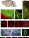
Terminally differentiated lymphatic vessels in the dura mater of the brain. Visualization of CNS lymphatic vasculature using Prox1-GFP reporter mice with DiI counterstaining for blood vasculature, Vegfr3+/LacZ reporter mice and immunofluorescence for PECAM1, and the lymphatic markers PROX1, LYVE1, PDPN, CCL21, and VEGFR3, as indicated. White arrowheads denote lymphatic vessels, yellow arrowheads denote the skull exit sites, and asterisks denote valves. (A) A schematic image of the various areas analyzed. The letters in bold refer to the corresponding images below. MMA, middle meningeal artery; PPA, pterygopalatine artery; RGV, retroglenoid vein; RRV, rostral rhinal vein; SS, sigmoid sinus; SSS, superior sagittal sinus; TV, transverse vein. (B) Lymphatic vessels running down along the SS and exiting the skull. (C) Lymphatic vessels running down along the proximal MMA branches. (D) Lymphatic vessels around the RGV with some vessels exiting the skull. (E–H) Whole-mount LYVE1 immunofluorescence of the skull top and base. (E) Lymphatic vessels along the SSS and the distal parts of the anterior MMA branch extending toward the bregma. (F) Lymphatic vessels along the SSS, bifurcating into the TVs at the confluence of sinuses. (G) Lymphatic vessels exiting the skull along the optic (II) and the trigeminal (V) nerves and through the cribriform plate (CP). CN, cranial nerve. (H) Lymphatic vessels associated with the glossopharyngeal (IX), vagus (X), and accessory (XI) nerves. XII, hypoglossal nerve. (I and J) Stereomicrographs of tissues in a Vegfr3+/LacZ reporter mouse showing the skull exit of dural lymphatic vessels along the PPA (I) and through the CP into a nasal concha. OB, olfactory bulb area. (K) Immunofluorescence of thick skull section for PECAM1, PROX1, and CCL21. bv, blood vessel; sas, subarachnoid space. (L–P) Whole-mount immunofluorescence staining of superior sagittal lymphatic vessels with antibodies against PECAM1 (L), LYVE1 (M), PDPN (N), CCL21 (O), VEGFR3 (P), and PROX1 (M–P). LYVE1 and PECAM1 colocalization is indicated with the dashed lines. n = 2–3 per staining. Data are from two to three independent experiments. Bars: (B–H and L–P) 100 µm; (K) 50 µm.
Figure 2.
Figure 2.
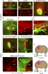
Dura mater lymphatic vessels drain brain ISF into dcLNs. (A–J) Analysis of lymphatic outflow routes of cerebral ISF by fluorescent stereomicroscopy in Prox1-GFP (green) mice 1 h after PEG-IRDye (red) injection into the brain parenchyma without (A–F) and with (G–J) ligation of the efferent lymphatic vessel of the dcLN. See K for schematic illustration of the experimental setup and summary of the results with and without ligation. (A and B) dcLNs and scLNs (both indicated with arrowheads) showing preferential filling of the ipsilateral dcLN but no filling in the scLNs. (C) Drainage into the ipsilateral dcLN via the efferent carotid lymphatic vessels (arrowheads). CCA, common carotid artery. (D) Internal carotid artery (ICA) and adjacent lymphatic vessels (white arrowheads) immediately below the osseous skull, showing drainage from the skull (yellow arrowhead). (E and F) Lymphatic vessels around the pterygopalatine artery (PPA), showing tracer uptake by the dura mater lymphatic vessels (arrowheads) only in the basal parts of the skull, nearby their exit site. MMA, middle meningeal artery. (G) Placement of a suture around the efferent lymphatic vessel (asterisk) of the dcLN. Arrowheads, afferent lymphatic vessels. (H) Afferent lymphatic vessel of the dcLN after ligation (asterisk), showing bulging of the afferent vessels (arrowheads). (I and J) Lymphatic vessels around the posterior branch of the MMA, showing increased filling of lymphatic vessels after ligation, extending above the retroglenoid vein (RGV) level. n = 2–3/group. Data are representative of two independent experiments. Bars: (A–E and G–J) 500 µm; (F) 100 µm.
Figure 3.
Figure 3.
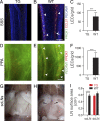
Absence of dural lymphatic vasculature in K14-VEGFR3-Ig TG mice. (A–F) Analysis of dura mater lymphatic vasculature in K14-VEGFR3-Ig TG and WT littermate control mice. (A–C) Immunofluorescence of the superior sagittal lymphatic vessels (arrowheads) for PECAM1, PROX1, and CCL21 (A and B) and quantification of PROX1+/CCL21+ lymphatic ECs (LECs)/grid (C). (D–F) Immunofluorescence of the pterygopalatine and middle meningeal lymphatic vessels (arrowheads) for PECAM1 and PROX1 (D and E) and quantification of PROX1+ LECs/grid (F). (G–I) Stereomicroscopic photographs showing the absence of the scLNs (arrows) in the TG mice (G and H) and quantification of the (mean left/right) scLN and dcLN surface areas (I). Micrographs of the dcLNs are shown in Fig. 4 C. (A–F) n = 3 (TG) and 4 (WT). (G and H) n = 4/group. Data are representative of two independent experiments. Bars: (A, B, D, and E) 100 µm; (G and H) 2 mm. Error bars indicate SD. Statistical analysis: two-tailed Student’s t test (C and F) and two-way ANOVA followed by Šídák’s post-hoc test (I). ***, P < 0.001; ****, P < 0.0001.
Figure 4.
Figure 4.
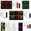
Lack of dural lymphatic vessels compromises CNS macromolecule clearance. Analysis of A488-OVA distribution 2 h after intraparenchymal injection in K14-VEGFR3-Ig TG mice and WT littermate controls. (A and B) Representative false color maps and quantification of the epifluorescence efficiency in the brain using IVIS imaging. (C and D) Representative images and quantification of the fluorescence in the dcLNs (indicated by arrows). (E and F) Representative fluorescent images of the A488-OVA tracer (indicated by arrowheads) accumulation in the LYVE1-stained lymphatic vessels around the PPA and MMA, with quantification of the A488-OVA–positive signal. Note the partial leakage of the tracer from the vessels caused by the perfusion fixation. (G) Fluorescent images of brain sections stained with DAPI and antibodies against endomucin (EMCN), showing the A488-OVA tracer distribution in the glymphatic system. (H) Plot profile analysis of the fluorescence along the indicated lines in G, showing A488-OVA signal in the subendothelial and perivascular spaces (arrows) in both TG and WT mice. (I) Immunofluorescent images of dcLNs stained with DAPI and antibodies against LYVE1. (J) Quantification of the LYVE1+ area in the dcLNs in TG mice and WT littermate controls. (A, B, and G–J) n = 4 (TG) and 3 (WT). (C–F) n = 3 (TG) and 4 (WT). Data are representative of two independent experiments. Bars: (C) 2 mm; (E) 100 µm; (G) 8 µm; (I) 1,000 µm. Error bars indicate SD. Statistical analysis: two-tailed Student’s t test. **, P < 0.01; ***, P < 0.001.
Figure 5.
Figure 5.
Lack of dural lymphatic vasculature inhibits CSF uptake into the dcLNs. (A) Schematic illustration of the experimental setup. (B) Representative fluorescent images of the dcLN in TG and WT mice 30 min after PEG-IRDye injection into the cisterna magna. AF, green channel autofluorescence. Bar, 1,000 µm. (C) Quantification of the dcLN fluorescence. n = 6 (TG) and 5 (WT). Data are representative of two independent experiments. Error bars indicate SD. Statistical analysis: two-tailed Student’s t test. *, P < 0.05.

Comment in

  • Implications of the discovery of brain lymphatic pathways.
    Iliff JJ, Goldman SA, Nedergaard M. Iliff JJ, et al. Lancet Neurol. 2015 Oct;14(10):977-9. doi: 10.1016/S1474-4422(15)00221-5. Lancet Neurol. 2015. PMID: 26376966 Free PMC article. No abstract available.
  • Lymphatic vessels of the dura mater: a new discovery?
    Bucchieri F, Farina F, Zummo G, Cappello F. Bucchieri F, et al. J Anat. 2015 Nov;227(5):702-3. doi: 10.1111/joa.12381. Epub 2015 Sep 18. J Anat. 2015. PMID: 26383824 Free PMC article. No abstract available.
  • Lymphatics in the Brain?!
    Choy C, Jandial R. Choy C, et al. Neurosurgery. 2016 Feb;78(2):N14. doi: 10.1227/01.neu.0000479890.79747.0d. Neurosurgery. 2016. PMID: 26779792 No abstract available.

Similar articles

Cited by

References

    1. Alitalo A.K., Proulx S.T., Karaman S., Aebischer D., Martino S., Jost M., Schneider N., Bry M., and Detmar M.. 2013. VEGF-C and VEGF-D blockade inhibits inflammatory skin carcinogenesis. Cancer Res. 73:4212–4221. 10.1158/0008-5472.CAN-12-4539 - DOI - PubMed
    1. Andres K.H., von Düring M., Muszynski K., and Schmidt R.F.. 1987. Nerve fibres and their terminals of the dura mater encephali of the rat. Anat. Embryol. (Berl.). 175:289–301. 10.1007/BF00309843 - DOI - PubMed
    1. Aspelund A., Tammela T., Antila S., Nurmi H., Leppänen V.-M., Zarkada G., Stanczuk L., Francois M., Mäkinen T., Saharinen P., et al. . 2014. The Schlemm’s canal is a VEGF-C/VEGFR-3-responsive lymphatic-like vessel. J. Clin. Invest. 124:3975–3986. 10.1172/JCI75395 - DOI - PMC - PubMed
    1. Choi I., Chung H.K., Ramu S., Lee H.N., Kim K.E., Lee S., Yoo J., Choi D., Lee Y.S., Aguilar B., and Hong Y.-K.. 2011. Visualization of lymphatic vessels by Prox1-promoter directed GFP reporter in a bacterial artificial chromosome-based transgenic mouse. Blood. 117:362–365. 10.1182/blood-2010-07-298562 - DOI - PMC - PubMed
    1. Deane R., Sagare A., and Zlokovic B.V.. 2008. The role of the cell surface LRP and soluble LRP in blood-brain barrier Aβ clearance in Alzheimer’s disease. Curr. Pharm. Des. 14:1601–1605. 10.2174/138161208784705487 - DOI - PMC - PubMed

Publication types