. 2015 Jun 12;10(6):e0128951.
doi: 10.1371/journal.pone.0128951. eCollection 2015.

RNA-Seq Analysis of Human Trigeminal and Dorsal Root Ganglia with a Focus on Chemoreceptors

Affiliations

RNA-Seq Analysis of Human Trigeminal and Dorsal Root Ganglia with a Focus on Chemoreceptors

Caroline Flegel et al. PLoS One. .

Abstract

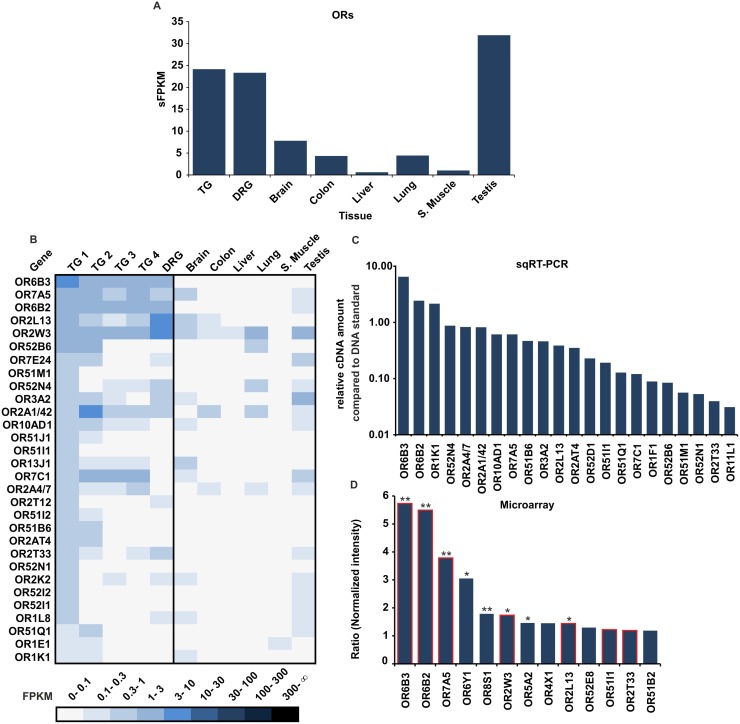
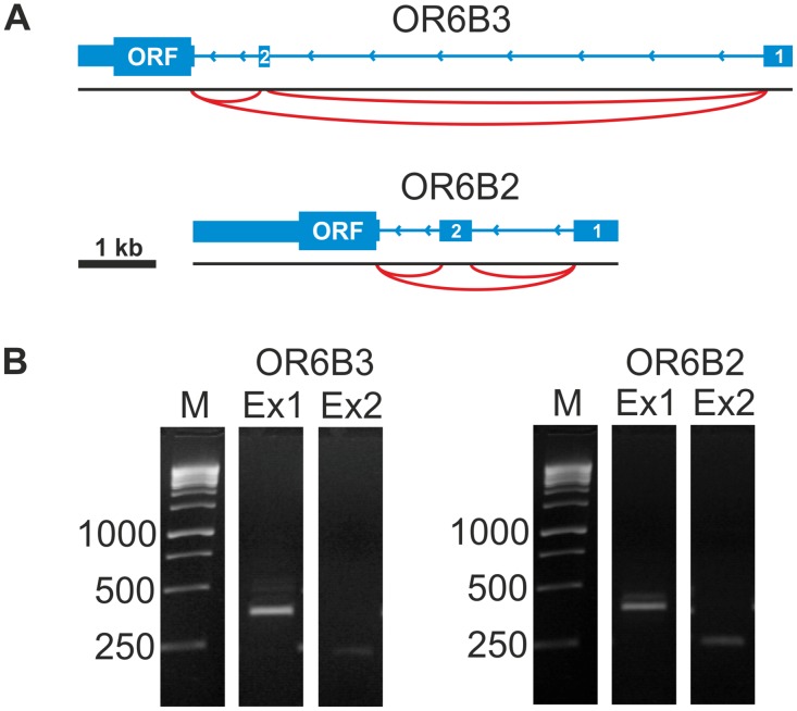
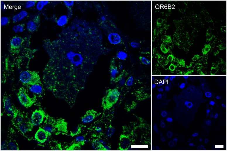
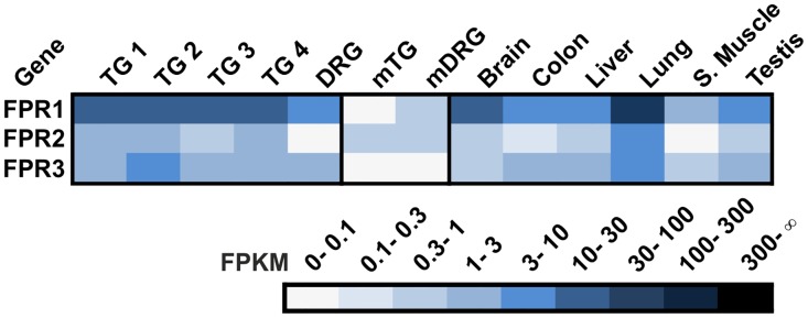
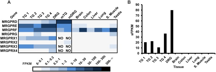
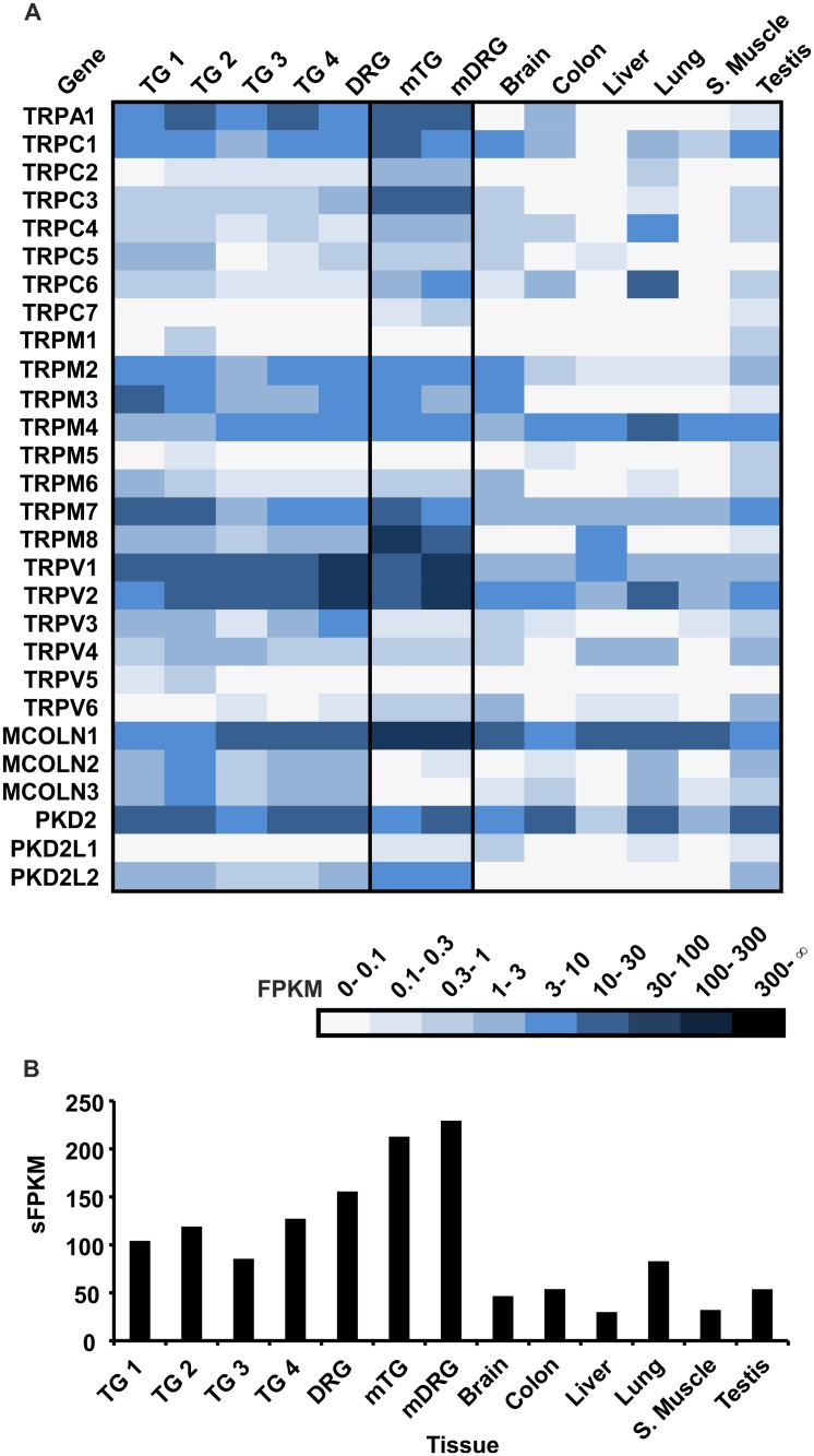
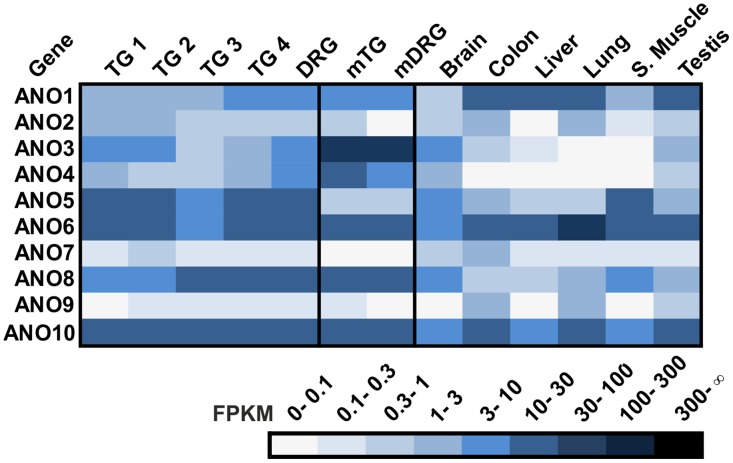
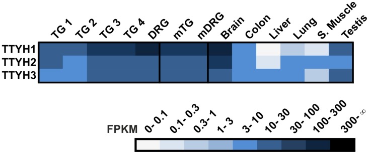
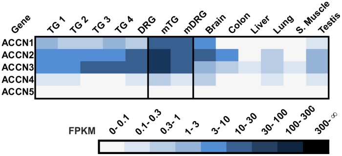
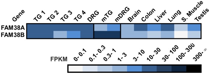
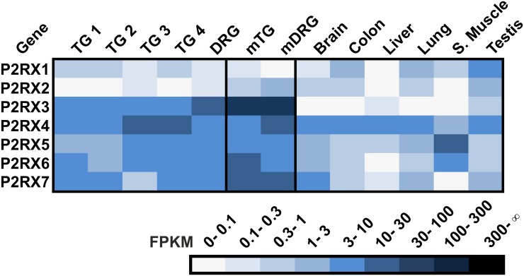
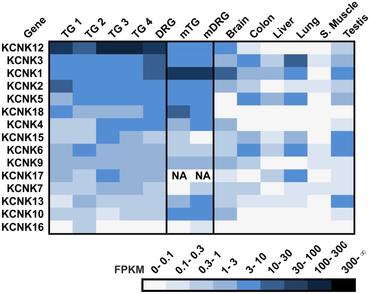
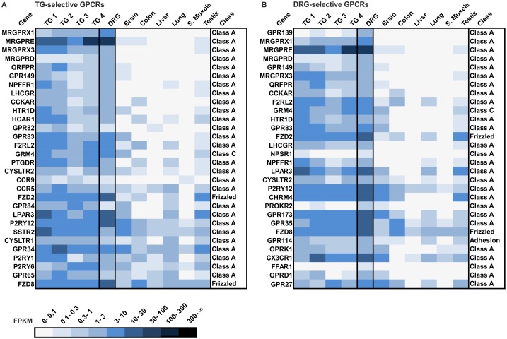
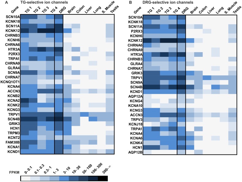
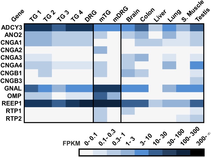
The chemosensory capacity of the somatosensory system relies on the appropriate expression of chemoreceptors, which detect chemical stimuli and transduce sensory information into cellular signals. Knowledge of the complete repertoire of the chemoreceptors expressed in human sensory ganglia is lacking. This study employed the next-generation sequencing technique (RNA-Seq) to conduct the first expression analysis of human trigeminal ganglia (TG) and dorsal root ganglia (DRG). We analyzed the data with a focus on G-protein coupled receptors (GPCRs) and ion channels, which are (potentially) involved in chemosensation by somatosensory neurons in the human TG and DRG. For years, transient receptor potential (TRP) channels have been considered the main group of receptors for chemosensation in the trigeminal system. Interestingly, we could show that sensory ganglia also express a panel of different olfactory receptors (ORs) with putative chemosensory function. To characterize OR expression in more detail, we performed microarray, semi-quantitative RT-PCR experiments, and immunohistochemical staining. Additionally, we analyzed the expression data to identify further known or putative classes of chemoreceptors in the human TG and DRG. Our results give an overview of the major classes of chemoreceptors expressed in the human TG and DRG and provide the basis for a broader understanding of the reception of chemical cues.


Conflict of interest statement

Competing Interests: The authors have declared that no competing interests exist.

Figures

Fig 1
Fig 1. OR expression in the human TG and DRG.
A The bar diagram shows the summarized expression (sFPKM) of detected OR transcripts. The sFPKM values of OR gene transcripts in the TG and DRG are in the same range as the summarized expression values found in testis tissue. B The heatmap shows FPKM values for the 30 most abundant OR transcripts found in the TG 1 sample compared with other TG and DRG samples and reference tissues (brain, colon, liver, lung, s.muscle, and testis). Darker colors indicate higher FPKM values, and white indicates the absence of detectable transcripts. The ORs were sorted according to the FPKM values found in the TG 1 sample. As described in [41], the FPKM values of OR2A4 and OR2A7 as well as that of OR2A1 and OR2A42, which share high sequence similarity (99–100%), were analyzed together. C Validation of RNA-Seq results using semi qRT-PCR. The diagram shows the summarized semi qRT-PCR compared with 3 ng of genomic DNA. We investigated 22 different ORs that showed either high, moderate, or low expression in TG 1. D Microarray analysis of the ORs in TG 1. Intensity values for the TG 1 sample were normalized to the reference tissues (liver, heart, s. muscle, skin). Given are all ORs with ratios >1.5. ORs were also detected by RNA-Seq as indicated by red frames. t-test: *p<0.05; **p<0.00013 (after Bonferroni correction).
Fig 2
Fig 2. Analysis of highly expressed OR genes in the human TG and DRG.
A Schematic representation of the newly identified 5’UTRs of OR6B3 and OR6B2. The gene is indicated by blue bars (exon) and thin lines (intron). The coding exon is indicated by ORF (open reading frame) and splice junctions as red arcs. We detected two unannotated 5’UTRs for OR6B3 and OR6B2. B 5’UTR-validation of OR-transcripts using RT-PCR with intron-spanning primers in the DRG sample. The newly identified 5’UTRs of the OR transcripts were confirmed by RT-PCR with a forward primer located in the identified exon and a reverse primer located in the ORF of the respective OR. OR6B3: Ex1 (forward primer in exon 1 of 5’UTR and reverse in OR6B3 ORF); Ex2 (forward primer in exon 2 of 5’UTR and reverse primer in OR6B3 ORF). OR6B2: Ex1 (forward primer in exon 1 of 5’UTR and reverse primer in OR6B2 ORF); Ex2 (forward primer in exon 2 of 5’UTR and reverse primer in OR6B2 ORF). The amplified PCR products were confirmed by Sanger sequencing.
Fig 3
Fig 3. The OR6B2 protein is localized to satellite cells in human DRG.
Immunohistochemical staining of human DRG sections with the OR6B2 antibody indicated OR6B2 protein expression in the neuron-surrounding satellite cells. DAPI staining (blue) was used to determine the number and localization of cell nuclei. The secondary antibody alone did not produce any staining (S4 Fig). Scale bars: 10 μm.
Fig 4
Fig 4. Expression of FPRs in the human TG and DRG.
Expression of FPR transcripts could be detected in human (TG and DRG) and murine (mTG and mDRG) sensory ganglia and other reference tissues (brain, colon, liver, lung, s. muscle, and testis).
Fig 5
Fig 5. Expression of MRGPRs in the human TG and DRG.
A MRGPR transcripts were detected in the TG and DRG, whereas they were mostly absent in the reference tissues investigated (brain, colon, liver, lung, s. muscle, and testis). Only MRGPRF transcripts could be detected in all reference tissues. NO = no ortholog B Investigation of the sFPKM values of all MRGPRs with the exception of MRGPRF in all tissues. The expression of MRGPRs is restricted to the sensory ganglia investigated.
Fig 6
Fig 6. Expression of TRPs in the human TG and DRG.
A TRP transcripts were detected in the human TG and DRG. B Investigation of the sFPKM values of the TRP transcripts across all tissues. The expression of TRP is higher in human sensory ganglia than in the human reference tissues (brain, colon, liver, lung, s. muscle, and testis) but lower than that in the mTG and mDRG.
Fig 7
Fig 7. Expression of ANOs in the human TG and DRG.
Transcription of ANO channel members was detected in the TG and DRG.
Fig 8
Fig 8. Expression of TTYHs in the human TG and DRG.
The transcription of all TTYH channel members was detected in the TG and DRG. The expression of the TTYH genes is higher in the human sensory ganglia than the human reference tissues with the exception of the brain and testis.
Fig 9
Fig 9. Expression of ACCN in the human TG and DRG.
ACCNs 1–3 are highly expressed in sensory ganglia.
Fig 10
Fig 10. Expression of piezo receptors in the human TG and DRG.
FAM38A and B are highly expressed in both sensory ganglia.
Fig 11
Fig 11. Expression of P2XRs in the human TG and DRG.
The ionotropic purinergic receptors are highly expressed in sensory ganglia. Five of the seven P2XRs are expressed higher in the TG and DRG than in any other tissue.
Fig 12
Fig 12. Expression of KCNK channels in the human TG and DRG.
All KCNK genes are expressed in the TG and DRG. The KCNK genes are sorted by the mean of their expression values across all human sensory ganglia.
Fig 13
Fig 13. Expression analysis of the 30 most selectively expressed GPCRs in the human TG and DRG.
A The heatmap shows the 30 most selectively expressed GPCR transcripts in the TG in comparison with the DRG and reference tissues (brain, colon, liver, lung, s. muscle, and testis). Genes are ranked according to their selective expression in the TG (Quotient of the mean FPKM value (TG1-4) and the mean FPKM value for all reference tissues; only the transcripts detected in all four TG samples with FPKM >0.1 are taken into account). MRGPRs are the most selective GPCRs that have been detected. B Shown are the 30 most selectively expressed GPCRs in the DRG. Genes are ranked according to their selective expression in DRG. The ranking was calculated by the quotient of the FPKM value of DRG (FPKM >0.1) and the mean FPKM value for all reference tissues. GPR139 is the most specific GPCR followed by three MRGPRs, which have been selectively detected in the human DRG.
Fig 14
Fig 14. Expression analysis of the 30 most selectively expressed ion channels in the human TG and DRG.
A The heatmap shows the 30 most selectively expressed ion channels in the TG compared with the DRG and reference tissues (brain, colon, liver, lung, s.muscle, and testis). Genes are ranked according to their selective expression in the TG (Quotient of the mean FPKM value (TG1-4) and the mean FPKM value of all reference tissues, only those genes expressed in all four TG with FPKM >0.1 were taken into account), and of which SCN10A is the most selectively expressed ion channel in the human TG. B Shown are the 30 most selectively expressed ion channels in the DRG. The genes are ranked according to their selective expression in the DRG. The ranking was calculated by the quotient of the FPKM value in the DRG (FPKM >0.1) and the mean FPKM value for all reference tissues. SCN10A is the most selective ion channel detected in the DRG.
Fig 15
Fig 15. Expression of olfactory signal transduction components in the human TG and DRG.
Expression pattern of signal transduction components including adenylyl cyclase III (ADCY3), the calcium-activated chloride channel (ANO2), the classical olfactory and rod CNG channel subunits (CNGA1, CNGA2, CNG3, CNGA4, CNGB1 and CNGB3), and Gαolf (GNAL). We also investigated the expression of accessory proteins including receptor-transporting proteins (RTP1 and RTP2) and receptor-enhancing proteins 1 (REEP1) as well as the expression of olfactory marker protein (OMP), a specific marker for olfactory sensory neurons.

Similar articles

Cited by

References

    1. Laska M, Distel H, Hudson R. Trigeminal perception of odorant quality in congenitally anosmic subjects. Chem. Senses 1997; 22(4):447–56. - PubMed
    1. Viana F. Chemosensory properties of the trigeminal system. ACS Chem Neurosci 2011; 2(1):38–50. 10.1021/cn100102c - DOI - PMC - PubMed
    1. Schöbel N, Radtke D, Kyereme J, Wollmann N, Cichy A, Obst K, et al. Astringency Is a Trigeminal Sensation That Involves the Activation of G Protein-Coupled Signaling by Phenolic Compounds. Chem. Senses 2014. - PubMed
    1. Silver WL, Farley LG, Finger TE. The effects of neonatal capsaicin administration on trigeminal nerve chemoreceptors in the rat nasal cavity. Brain Res. 1991; 561(2):212–6. - PubMed
    1. Caterina MJ, Schumacher MA, Tominaga M, Rosen TA, Levine JD, Julius D. The capsaicin receptor: a heat-activated ion channel in the pain pathway. Nature 1997; 389(6653):816–24. - PubMed

Publication types

Grants and funding

Caroline Flegel was funded by Heinrich und Alma Vogelsang Stiftung. Hanns Hatt was funded by DFG-Sonderforschungsbereich 642 “GTP- and ATP dependent membrane processes.” The funders had no role in the study design, data collection and analysis, decision to publish, or preparation of the manuscript.