DW-F5: A novel formulation against malignant melanoma from Wrightia tinctoria
- PMID: 26061820
- PMCID: PMC4650611
- DOI: 10.1038/srep11107
DW-F5: A novel formulation against malignant melanoma from Wrightia tinctoria
Erratum in
-
Corrigendum: DW-F5: A novel formulation against malignant melanoma from Wrightia tinctoria.Sci Rep. 2015 Aug 3;5:12662. doi: 10.1038/srep12662. Sci Rep. 2015. PMID: 26237232 Free PMC article. No abstract available.
Abstract
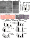
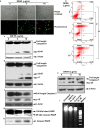
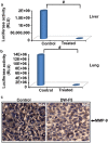
Wrightia tinctoria is a constituent of several ayurvedic preparations against skin disorders including psoriasis and herpes, though not yet has been explored for anticancer potential. Herein, for the first time, we report the significant anticancer properties of a semi-purified fraction, DW-F5, from the dichloromethane extract of W. tinctoria leaves against malignant melanoma. DW-F5 exhibited anti-melanoma activities, preventing metastasis and angiogenesis in NOD-SCID mice, while being non-toxic in vivo. The major pathways in melanoma signaling mediated through BRAF, WNT/β-catenin and Akt-NF-κB converging in MITF-M, the master regulator of melanomagenesis, were inhibited by DW-F5, leading to complete abolition of MITF-M. Purification of DW-F5 led to the isolation of two cytotoxic components, one being tryptanthrin and the other being an unidentified aliphatic fraction. The overall study predicts Wrightia tinctoria as a candidate plant to be further explored for anticancer properties and DW-F5 as a forthcoming drug formulation to be evaluated as a chemotherapeutic agent against malignant melanoma.
Figures


Similar articles
-
Corrigendum: DW-F5: A novel formulation against malignant melanoma from Wrightia tinctoria.Sci Rep. 2015 Aug 3;5:12662. doi: 10.1038/srep12662. Sci Rep. 2015. PMID: 26237232 Free PMC article. No abstract available.
-
Topical application of serine proteases from Wrightia tinctoria R. Br. (Apocyanaceae) latex augments healing of experimentally induced excision wound in mice.J Ethnopharmacol. 2013 Aug 26;149(1):377-83. doi: 10.1016/j.jep.2013.06.056. Epub 2013 Jul 6. J Ethnopharmacol. 2013. PMID: 23838477
-
Deciphering the Mechanism of Action of Wrightia tinctoria for Psoriasis Based on Systems Pharmacology Approach.J Altern Complement Med. 2017 Nov;23(11):866-878. doi: 10.1089/acm.2016.0248. Epub 2017 Jun 12. J Altern Complement Med. 2017. PMID: 28604055
-
In vitro antifungal activity of indirubin isolated from a South Indian ethnomedicinal plant Wrightia tinctoria R. Br.J Ethnopharmacol. 2010 Oct 28;132(1):349-54. doi: 10.1016/j.jep.2010.07.050. Epub 2010 Aug 5. J Ethnopharmacol. 2010. PMID: 20691774
-
A review on phytochemical, pharmacological, and pharmacognostical profile of Wrightia tinctoria: Adulterant of kurchi.Pharmacogn Rev. 2014 Jan;8(15):36-44. doi: 10.4103/0973-7847.125528. Pharmacogn Rev. 2014. PMID: 24600194 Free PMC article. Review.
Cited by
-
Alternative Options for Skin Cancer Therapy via Regulation of AKT and Related Signaling Pathways.Int J Mol Sci. 2020 Sep 18;21(18):6869. doi: 10.3390/ijms21186869. Int J Mol Sci. 2020. PMID: 32962182 Free PMC article. Review.
-
Heteronemin, a marine natural product, sensitizes acute myeloid leukemia cells towards cytarabine chemotherapy by regulating farnesylation of Ras.Oncotarget. 2018 Apr 6;9(26):18115-18127. doi: 10.18632/oncotarget.24771. eCollection 2018 Apr 6. Oncotarget. 2018. PMID: 29719594 Free PMC article.
-
Evaluation of in vitro anti-cancer potential and apoptotic profile of ethanolic plant extract of Wrightia tinctoria against oral cancer cell line.J Oral Maxillofac Pathol. 2024 Apr-Jun;28(2):211-215. doi: 10.4103/jomfp.jomfp_32_24. Epub 2024 Jul 11. J Oral Maxillofac Pathol. 2024. PMID: 39157850 Free PMC article.
-
Cucurbitacin B, Purified and Characterized From the Rhizome of Corallocarpus epigaeus Exhibits Anti-Melanoma Potential.Front Oncol. 2022 Jun 8;12:903832. doi: 10.3389/fonc.2022.903832. eCollection 2022. Front Oncol. 2022. PMID: 35756619 Free PMC article.
-
Targeting receptor tyrosine kinase signaling: Avenues in the management of cutaneous squamous cell carcinoma.iScience. 2023 May 5;26(6):106816. doi: 10.1016/j.isci.2023.106816. eCollection 2023 Jun 16. iScience. 2023. PMID: 37235052 Free PMC article. Review.
References
-
- Chandrashekar R., Adake P., Rao S. & Santanusaha S. WRIGHTIA TINCTORIA: AN OVERVIEW. J. Drug Deliv. 3, 196–198 (2013).
-
- Mitra S., Seshadri S., Venkataranganna M. & Gopumadhavan S. Reversal Of Parakeratosis, A Feature Of Psoriasis By Wrightia tinctoria (In Emulsion)-Histological Evaluation Based On Mouse Tail Test. Ind. J. Dermatol. 43, 102–104 (1998).
-
- Krishnamoorthy J., Ranganathan S., Shankar S. G. & Ranjith M. Dano: A herbal solution for dandruff. Afr. J. Biotechnol. 5, 960–962 (2006).
-
- Lakshman D. K., Rao K., Madhavi B., Kumar D. S. & Banji D. Anti oxidation activity of Wrightia tinctoria Roxb bark and Schrebera swietenoides Roxb bark extract. J. Pharm. Res. 4, 396 (2011).
-
- Sathyanarayanan S. et al. Preliminary phytochemical screening and study of antiviral activity and cytotoxicity of Wrightia tinctoria. Int. J. Chem. Sci. 7, 1–5 (2009).
Publication types
MeSH terms
Substances
LinkOut - more resources
Full Text Sources
Other Literature Sources
Research Materials
Miscellaneous

