. 2015 Jul 15;128(14):2497-508.
doi: 10.1242/jcs.168724. Epub 2015 Jun 8.

TORC2 mediates the heat stress response in Drosophila by promoting the formation of stress granules

Affiliations

TORC2 mediates the heat stress response in Drosophila by promoting the formation of stress granules

Irena Jevtov et al. J Cell Sci. .

Abstract

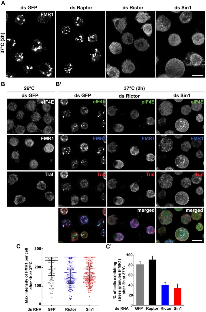
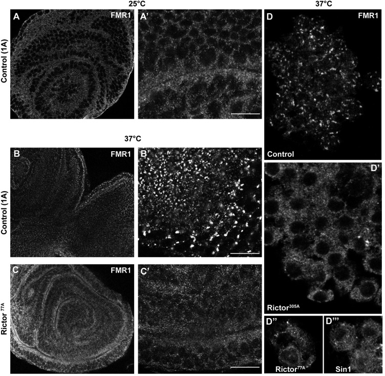
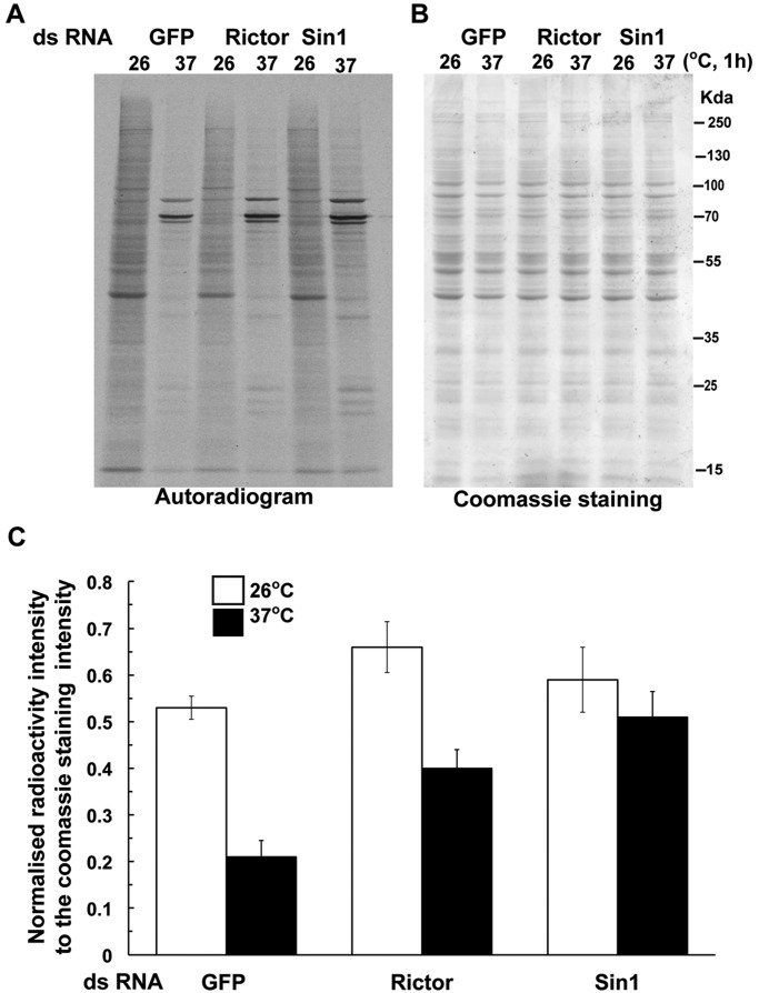
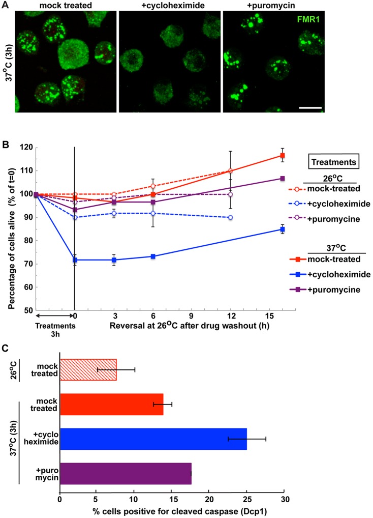
The kinase TOR is found in two complexes, TORC1, which is involved in growth control, and TORC2, whose roles are less well defined. Here, we asked whether TORC2 has a role in sustaining cellular stress. We show that TORC2 inhibition in Drosophila melanogaster leads to a reduced tolerance to heat stress, whereas sensitivity to other stresses is not affected. Accordingly, we show that upon heat stress, both in the animal and Drosophila cultured S2 cells, TORC2 is activated and is required for maintaining the level of its known target, Akt1 (also known as PKB). We show that the phosphorylation of the stress-activated protein kinases is not modulated by TORC2 nor is the heat-induced upregulation of heat-shock proteins. Instead, we show, both in vivo and in cultured cells, that TORC2 is required for the assembly of heat-induced cytoprotective ribonucleoprotein particles, the pro-survival stress granules. These granules are formed in response to protein translation inhibition imposed by heat stress that appears to be less efficient in the absence of TORC2 function. We propose that TORC2 mediates heat resistance in Drosophila by promoting the cell autonomous formation of stress granules.

Keywords: Akt; Drosophila S2 cells; Heat stress; Heat-shock protein; PKB; Rictor; SAPK; Sin1; Stress granules; TORC2; Translation.


Conflict of interest statement

Competing interests

The authors declare no competing or financial interests.

Figures

Fig. 1.
Fig. 1.
Rictor and Sin1 mutant alleles. (A) Schematic representation of the Rictor locus and the mutant alleles Rictor77A and Rictor305A. (B) PCR product of the Rictor open reading frame (ORF) amplified from cDNA of Rictor mutant and control flies. In the deletion mutants, the length of the Rictor ORF is 757 bp shorter than in the control, resulting in a premature stop codon after 58 amino acids. (C) Western blot visualization of Akt phosphorylation (p-Akt) on S505 in lines 1A (control), the Rictor77A mutant, the Sin1 mutant, the double Rictor77A; Sin1 mutant and in the Rictor77A carrying a rescuing a Rictor transgene (da>Rictor). Note that p-Akt is reduced in Rictor and Sin1 single mutants and in Rictor; Sin1 double mutants. Reduced phosphorylation is rescued with ubiquitous expression of Rictor.
Fig. 2.
Fig. 2.
TORC2 mutant flies and larvae are sensitive to heat stress. (A) Mobility curve of control (1A) and Rictor mutants (77A and 305A) adult flies exposed to 37°C, expressed as number of mobile flies versus time elapsed. Note that both Rictor mutants behave similarly and that they are different from the control line (n=80 for each genetic background). (B) Control (1A, n=79), Rictor (77A, n=83; 305A, n=74) and Sin1 (n=103) mutant larvae were exposed to 37°C for 2 h and the number of mobile versus immobile (paralyzed) larvae was counted. The graph shows the mean±s.d. of three independent experiments. (C) Quantification of the sensitivity to heat stress of control flies (1A, n=117, red curve), Rictor77A (n=131, blue curve) and Rictor305A (n=99, green curve) and of the rescue by the ubiquitous expression of Rictor (by means of tub-Gal4 [tub>Rictor (n=31, yellow curve); Rictor77A; tub>Rictor (n=24, violet curve); Rictor305A; tub>Rictor (n=9, turquoise curve)]. P<0.01, tub>Rictor versus control; P<0.001, Rictor77A; tub>Rictor versus Rictor77A; P<0.001, Rictor305A; tub>Rictor versus Rictor305A. This is expressed as the probability of being paralyzed. Experiment is performed as in A but at 38.5°C. P-value for any mutant versus control is P<0.001. (D) Quantification of the sensitivity to heat stress of control flies (1A, n=186, red curve), y w (n=40, violet curve), Sin1 mutant (n=14, green curve), and Rictor305A; Sin1 double mutants (n=31, turquoise curve) as above. P-values for differences between Sin1 and Rictor; Sin1 mutants compared to the control are P<0.001.
Fig. 3.
Fig. 3.
Akt phosphorylation on S505 and stability upon heat stress are TORC2 dependent. (A) Western blot of Akt phosphorylated on S505 (p-Akt)and total Akt in lysates of control and Rictor77A homozygous mutant larvae upon heat exposure for up to 2 h. Note that in Rictor mutants, the heat-induced Akt phosphorylation is not observed and that Akt is lost upon heat stress. (B) Western blot of p-Akt in lysates of S2 cells upon heat stress at 37°C for increasing time (up to 2 h). (C) Western blot of p-Akt in GFP-, Rictor- and Sin1-depleted S2 cells exposed at 37°C for 2 h. Note that p-Akt does not increase upon loss of TORC2 function. The lower band in this blot is non specific. (D) Western blot of total Akt in GFP-, Rictor- and Sin1-depleted S2 cells exposed to 26°C and 37°C for 2 h. Note that the Akt level is similar for all conditions at 26°C as well as for mock-depleted conditions at 37°C but dramatically drops in heat-exposed Rictor- and Sin1-depleted cells.
Fig. 4.
Fig. 4.
HSP gene expression and translation is not compromised in Rictor mutants. (A) Western blot of phosphorylated p38 (p-p38), phosphorylated JNK (p-Jnk) and phosphorylated Erk1/2 (p-Erk) of control and TORC2 mutant larvae upon heat stress. Note that their phosphorylation does not change. (B) Quantitative RT-PCR of HSP gene expression upon heat treatment in control and Rictor77A mutant larvae. The data represent the average of two biological replicates from larvae raised and treated identically. The expression upon heat is represented relative to HSP expression levels under normal conditions. (C) Western blot of HSP83 and HSP70 in extracts of control (1A) and Rictor305A larvae exposed to 37°C for indicated time. (D) Western blot of HSP83 and HSP70 in lysates of mock-, Rictor- and Sin1-depleted S2 cells exposed to 37°C for the indicated time. Note that less lysate was loaded for HSP70 after exposure to 37°C to better appreciate possible differences.
Fig. 5.
Fig. 5.
Stress granule formation is delayed in TORC2 depleted S2 cells. (A) Immunofluorescence visualization of endogenous FMR1 in S2 cells depleted for GFP (control, dsGFP), Raptor (TORC1), Rictor and Sin1 (TORC2) and heat stressed at 37°C for 2 h. Note that in heat-stressed mock- and Raptor-depleted cells, FMR1 is found in stress granules, whereas in Rictor- and Sin1-depleted cells, FMR1 remains largely cytoplasmic. (B,B′) Immunofluorescence visualization of endogenous FMR1, eIF4E and Tral (P-bodies) in GFP-depleted S2 cells grown at 26°C (B) and in GFP-, Rictor- and Sin1-depleted S2 cells heat stressed at 37°C for 2 h (B′). Note that eIF4E colocalizes with FMR1 in stress granules upon heat stress and that Tral localization is not affected by heat stress and by depletion of the TORC2 components. Scale bars: 10 μm. (C­,C′) Quantification of stress granule formation in GFP-, Raptor-, Rictor- and Sin1-depleted S2 cells. The maximum FMR1 intensity per cell upon exposure at 37°C for 1 h (C) is represented. The error bar is the mean±interquartile range. The bar representation in C′ displays the mean±s.d. percentage of cells exhibiting stress granules in GFP-, Raptor-, Rictor- and Sin1-depleted cells upon exposure at 37°C for 2 h from three independent experiments. P<0.0005, Rictor-depleted versus GFP; P<0.005, Sin1-depleted versus GFP.
Fig. 6.
Fig. 6.
Stress granule formation is impaired in Drosophila tissues. (A–C′) Immunofluorescence visualization of FMR1 in imaginal discs of control larvae at 25°C (A,A′), larvae exposed to 37°C for 2 h (B,B′) and Rictor77A mutant larvae exposed at 37°C for 2 h (C,C′), in low (left) and high (right) magnification. (D) Immunofluorescence visualization of FMR1 in clusters of hemocytes from control (D), Rictor305A (D′), Rictor77A (D″) and Sin1 (D‴) mutant third-instar larvae heat stressed for 2 h. Note that stress granule formation is reduced in mutants for TORC2 components. Scale bars: 10 μm.
Fig. 7.
Fig. 7.
Heat-induced protein translation inhibition is not impaired upon TORC2 loss of function. (A) Autoradiogram of total proteins radiolabelled with a pulse of [S]methionine for 15 min from lysates of mock (GFP)-, Rictor- and Sin1-depleted S2 cells after 55 min at 26°C and 37°C analysed using 10% SDS-PAGE. Note that the 26°C and 37°C patterns are similar for all cells. (B) Coomassie gel of fractionated proteins for the samples analysed in A. (C) Quantification of the autoradiography normalized to the Coomassie staining at 26°C and 37°C for mock-, Rictor- and Sin1-depleted S2 cells (as performed in A). Results are mean±s.d. (n=2).
Fig. 8.
Fig. 8.
Stress granule formation is a pro-survival mechanism. (A) Immunofluorescence visualization of FMR1 in S2 cells incubated at 37°C for 3 h in the presence of cycloheximide and puromycine. Note that cycloheximide treatment completely blocks stress granule formation whereas puromycine does not. Scale bar: 10 µm. (B) Graph of cell survival (as percentage of cell that remain alive, mean±s.d., n=3) after 3 h incubation at 26°C or 37°C in the presence of drugs (treatment is as in A) followed by incubation at 26°C in the absence of drug. P-values between all the treatments (except for cycloheximide at 37°C) are above 0.13 making the difference insignificant. The P-value between cycloheximide at 37°C and the other treatments is below 0.0000001. (C) Percentage of cells positive for cleaved caspase 3 during treatment as in A. Results are mean±s.d. (n=3).

Similar articles

Cited by

References

    1. Anderson P. and Kedersha N. (2008). Stress granules: the Tao of RNA triage. Trends Biochem. Sci. 33, 141-150. 10.1016/j.tibs.2007.12.003 - DOI - PubMed
    1. Andjelkovic M., Jones P. F., Grossniklaus U., Cron P., Schier A. F., Dick M., Bilbe G. and Hemmings B. A. (1995). Developmental regulation of expression and activity of multiple forms of the Drosophila RAC protein kinase. J. Biol. Chem. 270, 4066-4075. 10.1074/jbc.270.8.4066 - DOI - PubMed
    1. Arimoto K., Fukuda H., Imajoh-Ohmi S., Saito H. and Takekawa M. (2008). Formation of stress granules inhibits apoptosis by suppressing stress-responsive MAPK pathways. Nat. Cell Biol. 10, 1324-1332. 10.1038/ncb1791 - DOI - PubMed
    1. Bang O.-S., Ha B.-G., Park E. K. and Kang S.-S. (2000). Activation of Akt is induced by heat shock and involved in suppression of heat-shock-induced apoptosis of NIH3T3 cells. Biochem. Biophys. Res. Commun. 278, 306-311. 10.1006/bbrc.2000.3805 - DOI - PubMed
    1. Beauchamp E. M. and Platanias L. C. (2013). The evolution of the TOR pathway and its role in cancer. Oncogene 32, 3923-3932. 10.1038/onc.2012.567 - DOI - PubMed

Publication types

MeSH terms

Substances