. 2015 Jun 3;10(6):e0127057.
doi: 10.1371/journal.pone.0127057. eCollection 2015.

Use of autoantigen-loaded phosphatidylserine-liposomes to arrest autoimmunity in type 1 diabetes

Affiliations

Use of autoantigen-loaded phosphatidylserine-liposomes to arrest autoimmunity in type 1 diabetes

Irma Pujol-Autonell et al. PLoS One. .

Abstract

Introduction: The development of new therapies to induce self-tolerance has been an important medical health challenge in type 1 diabetes. An ideal immunotherapy should inhibit the autoimmune attack, avoid systemic side effects and allow β-cell regeneration. Based on the immunomodulatory effects of apoptosis, we hypothesized that apoptotic mimicry can help to restore tolerance lost in autoimmune diabetes.

Objective: To generate a synthetic antigen-specific immunotherapy based on apoptosis features to specifically reestablish tolerance to β-cells in type 1 diabetes.

Methods: A central event on the surface of apoptotic cells is the exposure of phosphatidylserine, which provides the main signal for efferocytosis. Therefore, phosphatidylserine-liposomes loaded with insulin peptides were generated to simulate apoptotic cells recognition by antigen presenting cells. The effect of antigen-specific phosphatidylserine-liposomes in the reestablishment of peripheral tolerance was assessed in NOD mice, the spontaneous model of autoimmune diabetes. MHC class II-peptide tetramers were used to analyze the T cell specific response after treatment with phosphatidylserine-liposomes loaded with peptides.

Results: We have shown that phosphatidylserine-liposomes loaded with insulin peptides induce tolerogenic dendritic cells and impair autoreactive T cell proliferation. When administered to NOD mice, liposome signal was detected in the pancreas and draining lymph nodes. This immunotherapy arrests the autoimmune aggression, reduces the severity of insulitis and prevents type 1 diabetes by apoptotic mimicry. MHC class II tetramer analysis showed that peptide-loaded phosphatidylserine-liposomes expand antigen-specific CD4+ T cells in vivo. The administration of phosphatidylserine-free liposomes emphasizes the importance of phosphatidylserine in the modulation of antigen-specific CD4+ T cell expansion.

Conclusions: We conclude that this innovative immunotherapy based on the use of liposomes constitutes a promising strategy for autoimmune diseases.


Conflict of interest statement

Competing Interests: The authors have declared that no competing interests exist.

Figures

Fig 1
Fig 1. Liposome features.
A) Cryogenic transmission electron microscopy images of PSAB-liposomes. Bar = 0.2 μm. B) Time course analysis of the capture of 100 μM OG488 labeled PS-liposomes (OG488 PS-liposome) by DCs at 37°C (white squares) and at 4°C (black squares). Results are expressed as mean±SD of three independent experiments (***p<0.001, **p<0.01, Two-way ANOVA). C) Flow cytometry contour plots of the uptake of PS-liposomes (OG488+) by DCs (CD11c+). From left to right, control DCs, DCs cocultured with OG488 PS-liposome at 4°C and at 37°C. One representative experiment of three is shown. Percentage of liposome capture (thick line) is referred to CD11c+ cell subset (thin line).
Fig 2
Fig 2. Effects of the capture of PS-liposomes in DCs phenotype.
A) DCs viability assessed by annexin V and 7aad staining. White symbols represent iDCs, before (triangles) and after the capture of PS-liposomes (squares) or PSAB-liposomes (circles), 24 hours after culture. Black symbols represent viability of mature DCs (mDCs) before (triangles) and after the capture of PS-liposomes (squares) or PSAB-liposomes (circles) after proinflammatory stimulus (LPS). Lines show the mean of at least eight independent experiments. B) Median of fluorescence intensity (MFI) for CD86, CD40, MHC Class I and MHC Class II membrane expression on DCs before and after liposome capture (white symbols) and after exposure to LPS (black symbols). Lines show the mean of at least four independent experiments. Comparisons within each group and between paired maturation conditions showed significant differences (*p<0.05, Wilcoxon test). C) Quantification of the PGE2 production by immature DCs (iDCs) in culture medium (med, white triangles), loaded with PS-liposomes (PS-lipo, white squares) or PSAB-liposomes (PSAB-lipo, white circles), after 24 hours of culture. Data are represented as pg/106 cells. Lines show the mean of a minimum of seven independent experiments. Comparisons between groups showed significant differences (**p<0.01 and *p<0.05, Wilcoxon test).
Fig 3
Fig 3. Impaired DCs’ ability to induce autologous T cell proliferation after PS-liposomes capture, and cytokine secretion.
A) Autologous proliferation of T cells (c.p.m. for 3H thymidine assay) after stimulation induced by iDCs, before (white triangles) and after the capture of PS-liposomes (white squares) or PSAB-liposomes (white circles), with insulin (20 μg/ml) at a ratio of 1:10 for 6 days. Black symbols represent proliferation induced by mature DCs (mDCs) before (triangles) and after the capture of PS-liposomes (squares) or PSAB-liposomes (circles), previously activated with proinflammatory stimuli LPS (100 ng/ml). Lines show the mean of seven independent experiments. Comparisons within each group and between paired maturation conditions showed significant differences (*p<0.05, Wilcoxon test). B) Cytokine production in T cell proliferation experiments. Levels of IFN-γ and IL-17A were measured in supernatants from T cell proliferation experiments induced by iDCs, DCs loaded with empty liposomes (PS-lipo) and DCs loaded with liposomes with insulin peptides (PSAB-lipo) in basal conditions (white bars) or after 24 hours with LPS (black bars). Results are expressed as mean±SEM from four independent experiments. Comparisons within each group and between paired maturation conditions were not able to detect significant differences (p<0.05, Wilcoxon test). “<d” means values below the standard.
Fig 4
Fig 4. Tracking of PS-liposomes.
Histogram of ex vivo relative fluorescent signal (RFU, Relative Fluorescence Units/g tissue) in organs from NOD mice 24 hours after the administration of labeled PS-liposomes (Alexa Fluor 750). PAT, perigonadal adipose tissue; K, kidney; S, spleen; P, pancreas; PLN, pancreatic lymph nodes; MLN, mesenteric lymph nodes; L, liver; MDLN, mediastinal lymph nodes; T, thymus. Results are mean±SEM of three independent experiments.
Fig 5
Fig 5. Immunotherapy using PS-liposomes filled with insulin peptides decreases T1D incidence.
Cumulative incidence (percentage) of T1D in NOD mice treated with PSAB-liposomes (PSAB-lipo, circles, n = 12), PS-liposomes (PS-lipo, squares, n = 18), and sham group (triangles, n = 26). Significant differences were found when compared group treated with PSAB-liposomes versus sham group (*p≤0.05, Kaplan-Meier log-rank analysis).
Fig 6
Fig 6. Reduction in insulitis following administration of PSAB-liposomes.
A) Insulitis score from non-diabetic mice at the end of the follow-up (30 weeks), sham (n = 4), PS-liposomes (PS-lipo, n = 3) and PSAB-liposomes containing autoantigens (PSAB-lipo, n = 6). Results are mean±SD (*p≤0.05, Mann Whitney test). B) Percentage of islets in each of the infiltration categories: White = 0, no insulitis; Dotted = 1, peri-insular; Striped = 2, mild insulitis (<25% of the infiltrated islet); Squared = 3, 25–75% of the islet infiltrated; Black = 4, >75% islet infiltration.
Fig 7
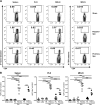
Fig 7. Antigen-specific CD4+ T cell expansion 4 days after i.p. administration of PS2.5mi-liposomes to NOD mice.
A) Representative flow cytometry contour plots of the percentage of 2.5mi+ CD4+ T cells in the spleen, pancreatic lymph nodes (PLN) and mediastinal lymph nodes (MDLN) (gated on CD19-, CD8-, F4/80-, CD11c-, PI- and CD4+ cells) in the sham group and after the administration of PS2.5mi-liposomes or PC2.5mi-liposomes. Left panel: Ag7/2.5mi tetramer staining. Right panel: control staining with Ag7/GPI282–292 tetramer. B) Detection of 2.5mi+ CD4+ T cells in the spleen, PLN and MDLN from 4–8 mice pooled from 4 independent experiments after the administration of saline solution (white triangles, n = 6), 2.5mi peptide (white rhombus, n = 5), PS-liposomes (white squares, n = 4), PS2.5mi-liposomes (white circles, n = 8), PC-liposomes (black squares, n = 5) or PC2.5mi-liposomes (black circles, n = 4). Comparisons between groups showed significant differences (***p<0.001, One-way ANOVA and Bonferroni multiple comparison test).

Similar articles

Cited by

References

    1. Steinman RM, Turley S, Mellman I, Inaba K. The induction of tolerance by dendritic cells that have captured apoptotic cells. The Journal of experimental medicine. 2000;191(3):411–6. - PMC - PubMed
    1. Hochreiter-Hufford A, Ravichandran KS. Clearing the dead: apoptotic cell sensing, recognition, engulfment, and digestion. Cold Spring Harbor perspectives in biology. 2013;5(1):a008748 10.1101/cshperspect.a008748 . - DOI - PMC - PubMed
    1. Fadok VA, Bratton DL, Henson PM. Phagocyte receptors for apoptotic cells: recognition, uptake, and consequences. The Journal of clinical investigation. 2001;108(7):957–62. 10.1172/JCI14122 - DOI - PMC - PubMed
    1. Nagata S, Hanayama R, Kawane K. Autoimmunity and the clearance of dead cells. Cell. 2010;140(5):619–30. 10.1016/j.cell.2010.02.014 . - DOI - PubMed
    1. Eizirik DL, Colli ML, Ortis F. The role of inflammation in insulitis and beta-cell loss in type 1 diabetes. Nature reviews Endocrinology. 2009;5(4):219–26. 10.1038/nrendo.2009.21 . - DOI - PubMed

Publication types

Grants and funding

This work was supported by a grant from Spanish Government (FIS PI12/00195). IPA was supported by AGAUR, Generalitat de Catalunya. MVP and RA are supported by the Health Dept. of the Catalan Government, Generalitat de Catalunya. Special thanks to Ms. M.A. Cardus and her family for their generous donation. The funders had no role in study design, data collection and analysis, decision to publish, or preparation of the manuscript.