. 2015 Jul 17;290(29):18124-18133.
doi: 10.1074/jbc.M115.645549. Epub 2015 Jun 2.

Lipopolysaccharide Primes the NALP3 Inflammasome by Inhibiting Its Ubiquitination and Degradation Mediated by the SCFFBXL2 E3 Ligase

Affiliations

Lipopolysaccharide Primes the NALP3 Inflammasome by Inhibiting Its Ubiquitination and Degradation Mediated by the SCFFBXL2 E3 Ligase

SeungHye Han et al. J Biol Chem. .

Abstract

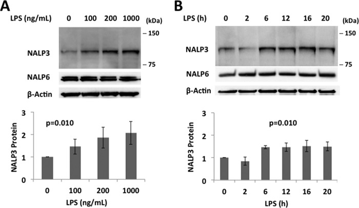
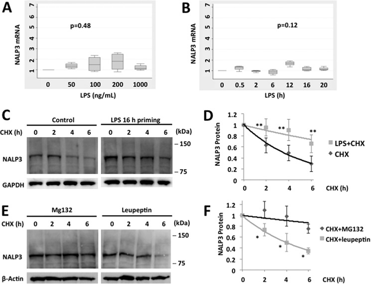
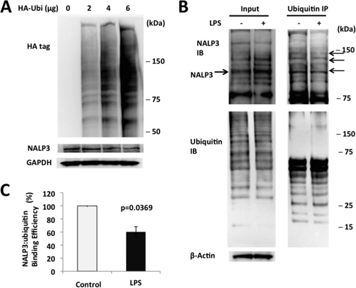
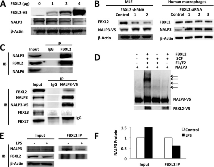
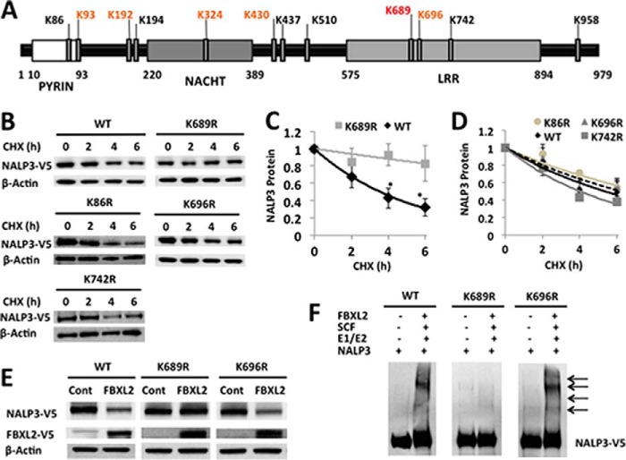
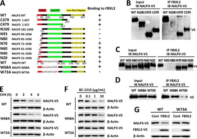
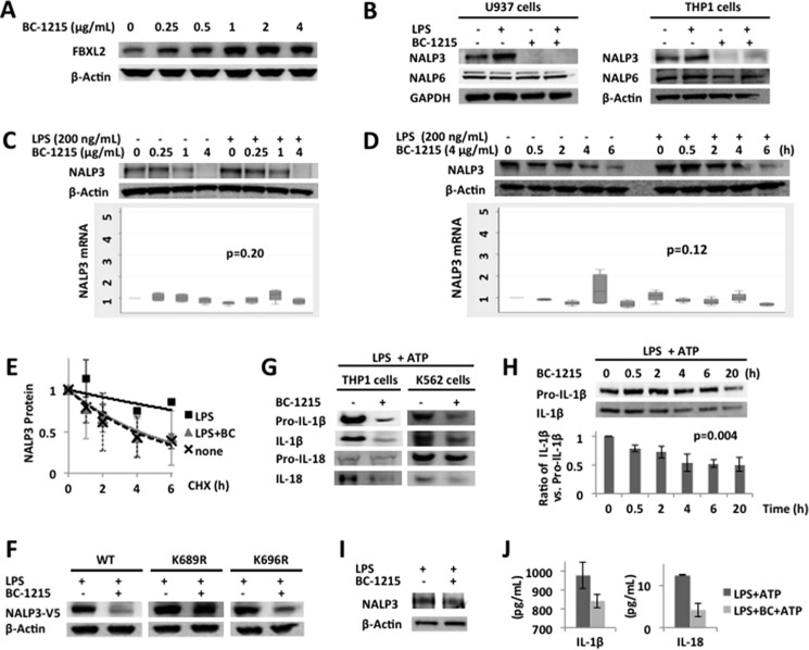
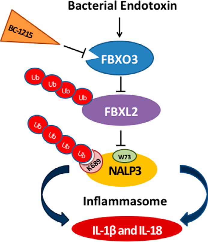
The inflammasome is a multiprotein complex that augments the proinflammatory response by increasing the generation and cellular release of key cytokines. Specifically, the NALP3 inflammasome requires two-step signaling, priming and activation, to be functional to release the proinflammatory cytokines IL-1β and IL-18. The priming process, through unknown mechanisms, increases the protein levels of NALP3 and pro-IL-1β in cells. Here we show that LPS increases the NALP3 protein lifespan without significantly altering steady-state mRNA in human cells. LPS exposure reduces the ubiquitin-mediated proteasomal processing of NALP3 by inducing levels of an E3 ligase component, FBXO3, which targets FBXL2. The latter is an endogenous mediator of NALP3 degradation. FBXL2 recognizes Trp-73 within NALP3 for interaction and targets Lys-689 within NALP3 for ubiquitin ligation and degradation. A unique small molecule inhibitor of FBXO3 restores FBXL2 levels, resulting in decreased NALP3 protein levels in cells and, thereby, reducing the release of IL-1β and IL-18 in human inflammatory cells after NALP3 activation. Our findings uncover NALP3 as a molecular target for FBXL2 and suggest that therapeutic targeting of the inflammasome may serve as a platform for preclinical intervention.

Keywords: IL-1; NALP3; cytokine induction; inflammasome; inflammation; innate immunity.


Figures

FIGURE 1.
FIGURE 1.
LPS priming increases NALP3 protein in human monocyte U937 cells. A and B, immunoblots for NALP3, NALP6 (negative control), and β-actin in lysates from human monocytes, U937 cells (total 3 × 10⁶ cells), treated with LPS for 16 h as indicated (A) or treated with LPS (200 ng/ml) for the indicated periods of time (B). The relative densitometries of NALP3 protein for each immunoblot are shown in the bottom panel. Data are mean ± S.E. of three to five independent experiments. The p values were determined by a nonparametric test for trend.
FIGURE 2.
FIGURE 2.
LPS priming increases NALP3 protein levels by prolonging its half-life. Box plots of messenger RNA expression –fold increase of NALP3 in LPS primed human monocytes, U937 cells. A and B, cells were primed with LPS for 16 h as indicated (A) or with LPS (200 ng/ml) for the indicated periods of time (B). Data are from four independent experiments. The p values were determined by a Kruskal-Wallis test. C, U937 cells were incubated with CHX (40 μg/ml) for various times with or without LPS priming (200 ng/ml for 16 h). D, densitometric plot of NALP3 protein decay versus time of CHX exposure with a best fit line. The half-life was ∼4 h in an unchallenged condition and >6 h after LPS priming. E, U937 cells were incubated with CHX (40 μg/ml) and MG132 (20 μg/ml) or leupeptin (20 μg/ml), and immunoblotting for NALP3 was performed. F, decay curves over time in the presence of MG132 or leupeptin were best fitted. Data are mean ± S.E. of five to eight independent experiments. The p values were determined by a Mann-Whitney test comparing two groups at each time point. *, p < 0.05; **, p < 0.01.
FIGURE 3.
FIGURE 3.
NALP3 is degraded through the ubiquitin proteasome system. A, MLE cells were transfected with either the control plasmid or HA-ubiquitin (HA-Ubi) plasmid for 24 h. Cells were collected and assayed for HA, NALP3, and β-actin (loading control) by immunoblotting. B, A549 cells were incubated with or without LPS (200 ng/ml for 16 h), and cells were lysed and used to detect NALP3 or ubiquitin (left panels, input). Top right panel, ubiquitin was immunoprecipitated (IP), followed by NALP3 immunoblotting (IB) showing reduced intensity of several bands (arrows). C, the relative densitometric intensities of NALP3 bands are shown reflecting reduced association between NALP3 and ubiquitin by 40% after LPS exposure. The bar graph represents mean ± S.D. The p value was determined by Mann-Whitney test. Data are representative of at least two to three independent experiments.
FIGURE 4.
FIGURE 4.
FBXL2 triggers NALP3 degradation. A, MLE cells were transfected with increasing amounts of FBXL2 plasmid for 24–48 h. Cells were collected and assayed for V5 (FBXL2), NALP3, and β-actin by immunoblotting. B, MLE cells were transfected with several FBXL2 lentiviral shRNAs (4 μg). Human primary macrophages were also transfected with several siRNAs to FBXL2 (500 nm) for gene silencing. Shown are protein levels of FBXL2 after transfection of shRNAs or siRNAs and corresponding levels of NALP3 protein. Compared with a control RNA, despite a modest reduction in FBXL2 levels, increased NALP3 levels in the shRNA or siRNA groups were observed. C, U937 cells were lysed, and FBXL2 proteins were coimmunoprecipitated (IP) followed by NALP3, FBXL2, or NALP6 immunoblotting (IB, top panel). In addition, Beas2B cells were transfected with NALP3-V5 prior to IP for V5 and then, NALP3 and various F box proteins were immunoblotted (bottom blot). IgG was used as a control in all IP experiments, and inputs are the IBs of 10% of the cell lysates before performing IP. Note that an upper IgG chain band was detected in the FBXL7 immunoblot. D, in vitro ubiquitination assays showing that NALP3 is a substrate for FBXL2-mediated ubiquitination. E, A549 cells were incubated with or without LPS (200 ng/ml) for 16 h, and cells were lysed. FBXL2 protein was coimmunoprecipitated; first IP for FBXL2 followed by NALP3, FBXL2, or β-actin immunoblotting. F, the relative association between FBXL2 and NALP3 after LPS was quantified using densitometric analysis of the bands in E. The binding capacity between NALP3 and FBXL2 is reduced by ∼45–50% after LPS exposure. Data are representative of at least two to three independent experiments.
FIGURE 5.
FIGURE 5.
Lys-689 is a NALP3 ubiquitination acceptor site. A, full-length and point mutant constructs encoding human NALP3 protein were cloned into a pcDNA3.1D/V5-His vector. B, MLE cells were transfected with either the pcDNA-NALP3 full-length plasmid (4 μg) or point mutant plasmid for 48 h. Cells were exposed to CHX (40 μg/ml) at different time points for a half-life study. Cells were collected and assayed for NALP3 (V5) and β-actin (loading control) by immunoblotting. Representative images are shown. C and D, densitometric plots of NALP3 protein decay versus time of CHX exposure with best fit lines. The half-life was prolonged with a K689R mutant by >6 h (C), whereas WT full-length protein or other mutants exhibited a t½ of ∼4 h (D). Data represent the mean ± S.E. of three to four independent experiments. The p values were determined by a Mann-Whitney test comparing two groups at each time point. *, p < 0.05. E, MLE cells were cotransfected with FBXL2/empty (cont) plasmid (2 μg) and either the WT NALP3 plasmid or point mutant plasmids (2 μg) for 48 h. Cells were collected and assayed for NALP3 (V5) and β-actin (loading control) by immunoblotting. F, in vitro ubiquitination assays show that a K689R NALP3 mutant is less polyubiquitinated compared with WT NALP3 or a K696R mutant. The data in E and F are representative of two to three independent experiments.
FIGURE 6.
FIGURE 6.
Trp-73 within NALP3 is required for FBXL2 interaction. A, several deletional or point mutants of NALP3 were designed and cloned into a pcDNA3.1D/V5-HIS vector. B–D, FBXL2 protein was immunoprecipitated (IP) from cell lysates using an FBXL2 antibody and coupled to protein A/G-agarose resin. FBXL2 resins were then incubated with in vitro-synthesized products expressing HIS-V5-NALP3 mutants. After washing, proteins were eluted and immunoblotted (IB) for NALP3-V5. E, HeLa cells were transfected with NALP3 WT or mutant plasmids, and the half-life was determined via CHX treatment at 40 μg/ml. F, NALP3 WT or mutants (4 μg) were transfected into HeLa cells and treated with BC-1215 for 16 h. Cells were then collected and processed for V5 immunoblotting. G, HeLa cells were cotransfected with NALP3 WT or W73A mutant (2 μg) with or without the FBXL2 plasmid (2 μg). Cells were then collected and immunoblotted for V5 and β-actin. Cont, control.
FIGURE 7.
FIGURE 7.
An FBXO3 inhibitor reduces NALP3 abundance, thereby decreasing cytokine release. A, U937 cells were treated with BC-1215 at different concentrations for 16 h. Cells were collected for FBXL2 and β-actin immunoblotting. B, U937 and THP1 cells (3 × 10⁶ cells each) were incubated with LPS (200 ng/ml) or BC-1215 (8 μg/ml) for 16 h. Cells were collected and lysed for NALP3, NALP6 (negative control), and GAPDH/β-actin immunoblotting. C and D, U937 cells (3 × 10⁶ cells) were primed with LPS (200 ng/ml) in combination with different concentrations of BC-1215 for 16 h as indicated (C) or primed with LPS (200 ng/ml for 16 h) and then exposed to BC-1215 (4 μg/ml) in fresh culture medium for the indicated periods of time (D). Cells were collected to measure NALP3 and β-actin protein (top panels) and mRNA levels (bottom panels). Box plots of the -fold increase of steady-state NALP3 mRNA are shown. Data represent four independent experiments. The p values were determined by a Kruskal-Wallis test. E, U937 cells were incubated in LPS (200 ng/ml) with or without BC-1215 (4 μg/ml for 16 h). Cells were then exposed to CHX (40 μg/ml) at different time points for a half-life study. Immunoblotting for NALP3 and GAPDH (loading control) was performed. Densitometric plots of adjusted NALP3 protein decay over time under different conditions were best fitted. The half-life of LPS-primed NALP3 protein was reduced with BC-1215 treatment comparable with native conditions. Data are mean ± S.E. of two independent experiments. F, MLE cells were transfected either with WT or point mutant NALP3 plasmids for 48 h. LPS (40 μg/ml) was then added to the medium with or without BC-1215 (20 μg/ml) for 18 h. Cells were collected and lysed for NALP3 (V5-tagged) and β-actin (loading control) immunoblotting. Levels of K689R, the point mutant for a putative ubiquitin acceptor site within the NALP3 protein, did not decrease with BC-1215 compared with WT or mutant NALP3. Data are representative of two independent experiments. G, THP1 and K562 cells (3 × 10⁶ cells each) were incubated with LPS (200 ng/ml) with or without BC-1215 (8 μg/ml) for 20 h and then pulsed with ATP (5 mm for 20 min). Culture medium was collected for immunoblotting of pro-IL-1β, IL-1β, pro-IL-18, and IL-18. H, THP1 cells (3 × 10⁶ cells each) were incubated with LPS (200 ng/ml) for 20 h and different time periods of BC-1215 (8 μg/ml), as indicated, in the same volume of culture medium. Cells were then exposed to ATP (5 mm for 20 min). Culture medium was collected for immunoblotting of pro-IL-1β and IL-1β. The ratio of IL-1β versus pro-IL-1β by densitometry is shown (bottom panel). Data represent mean ± S.D. of two independent experiments. The p value was determined by nonparametric test for trend. I, primary human alveolar macrophages were obtained from a healthy volunteer. Human alveolar macrophages (3 × 10⁵ cells) were incubated with BC-1215 (4 μg/ml) for 2 h and then exposed to LPS (100 ng/ml) for 16 h. Cells were lysed and collected for immunoblotting for NALP3 and β actin. J, primary human alveolar macrophages (3 × 10⁵ cells) from the same subject as in I were incubated with BC-1215 (4 μg/ml) for 2 h and then exposed to LPS (100 ng/ml) for 16 h. ATP (5 mm) was added for 15 min before collecting cell culture supernatants for ELISA. Data represent the mean ± S.D. of duplicate measurements.
FIGURE 8.
FIGURE 8.
Molecular regulation of NALP3 inflammasome protein stability mediated by F-box proteins. Endotoxin robustly increases NALP3 protein levels in human inflammatory cells and subsequently induces cytokine synthesis and release. The F-box protein FBXL2 serves as a sentinel inhibitor of NALP3 by mediating its polyubiquitination (Ub) through docking at Trp-73 and ubiquitin ligation at Lys-689, resulting in proteasomal degradation in cells. Another E3 ligase component, FBXO3, targets FBXL2 for its ubiquitination and degradation (8). A small-molecule FBXO3 inhibitor, BC-1215, reduces the levels of NALP3 protein after LPS priming by restoring FBXL2 levels. BC-1215 subsequently decreases proinflammatory cytokine release after the NALP3 inflammasome is activated by a second stimulus.

Similar articles

Cited by

References

    1. Franchi L., Muñoz-Planillo R., Núñez G. (2012) Sensing and reacting to microbes through the inflammasomes. Nat. Immunol. 13, 325–332 - PMC - PubMed
    1. Makabe H., Kojika M., Takahashi G., Matsumoto N., Shibata S., Suzuki Y., Inoue Y., Endo S. (2012) Interleukin-18 levels reflect the long-term prognosis of acute lung injury and acute respiratory distress syndrome. J. Anesth. 26, 658–663 - PubMed
    1. Dolinay T., Kim Y. S., Howrylak J., Hunninghake G. M., An C. H., Fredenburgh L., Massaro A. F., Rogers A., Gazourian L., Nakahira K., Haspel J. A., Landazury R., Eppanapally S., Christie J. D., Meyer N. J., Ware L. B., Christiani D. C., Ryter S. W., Baron R. M., Choi A. M. (2012) Inflammasome-regulated cytokines are critical mediators of acute lung injury. Am. J. Respir. Crit. Care Med. 185, 1225–1234 - PMC - PubMed
    1. Bauernfeind F. G., Horvath G., Stutz A., Alnemri E. S., MacDonald K., Speert D., Fernandes-Alnemri T., Wu J., Monks B. G., Fitzgerald K. A., Hornung V., Latz E. (2009) Cutting edge: NF-κB activating pattern recognition and cytokine receptors license NLRP3 inflammasome activation by regulating NLRP3 expression. J. Immunol. 183, 787–791 - PMC - PubMed
    1. Hershko A., Ciechanover A. (1998) The ubiquitin system. Annu. Rev. Biochem. 67, 425–479 - PubMed

Publication types

MeSH terms

LinkOut - more resources