. 2015 Aug;35(16):2761-70.
doi: 10.1128/MCB.00215-15. Epub 2015 Jun 1.

IRE1α-Dependent Decay of CReP/Ppp1r15b mRNA Increases Eukaryotic Initiation Factor 2α Phosphorylation and Suppresses Protein Synthesis

Affiliations

IRE1α-Dependent Decay of CReP/Ppp1r15b mRNA Increases Eukaryotic Initiation Factor 2α Phosphorylation and Suppresses Protein Synthesis

Jae-Seon So et al. Mol Cell Biol. 2015 Aug.

Abstract

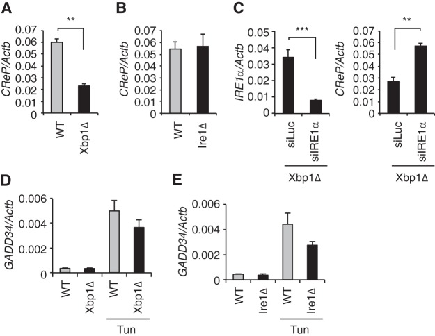
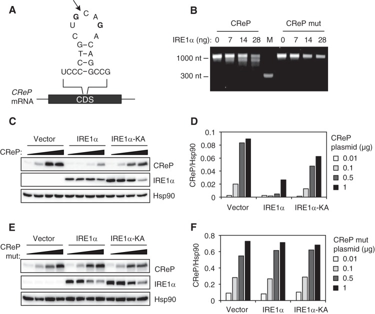
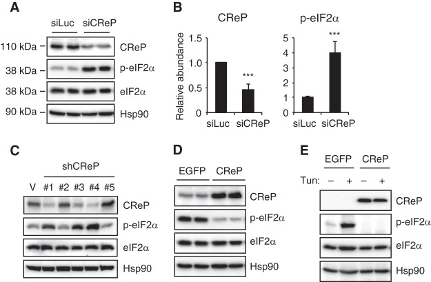
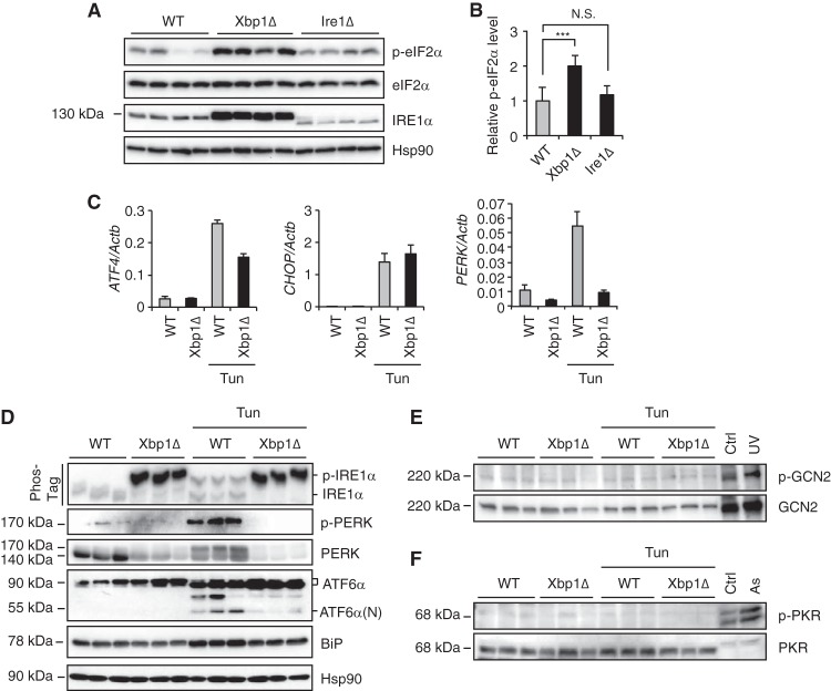
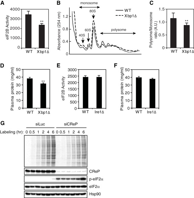
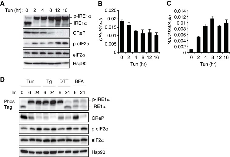
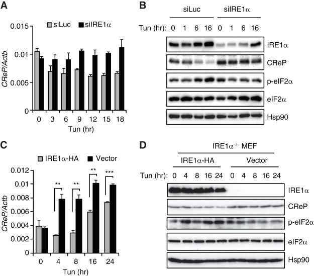
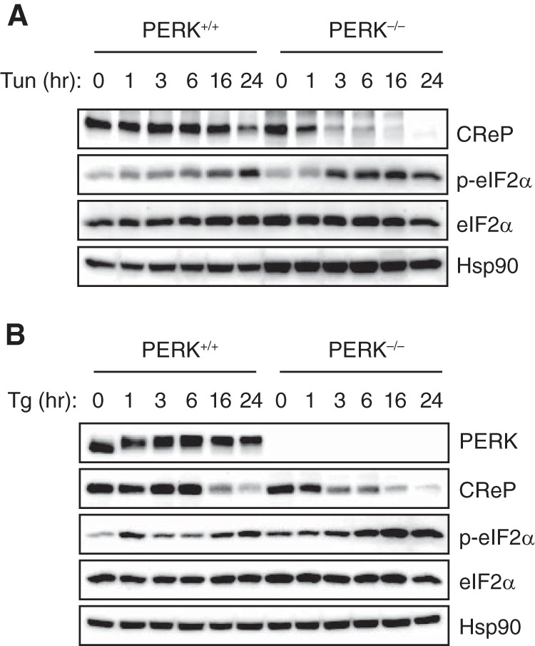
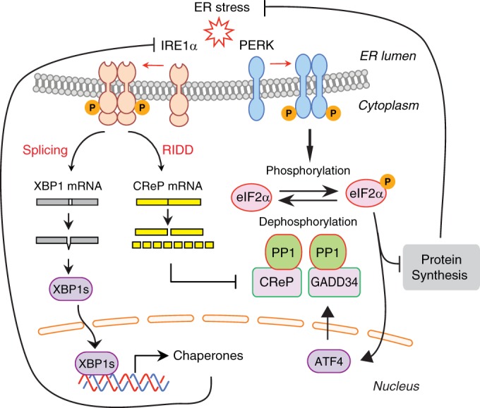
The unfolded protein response (UPR) regulates endoplasmic reticulum (ER) homeostasis and protects cells from ER stress. IRE1α is a central regulator of the UPR that activates the transcription factor XBP1s through an unconventional splicing mechanism using its endoribonuclease activity. IRE1α also cleaves certain mRNAs containing XBP1-like secondary structures to promote the degradation of these mRNAs, a process known as regulated IRE1α-dependent decay (RIDD). We show here that the mRNA of CReP/Ppp1r15b, a regulatory subunit of eukaryotic translation initiation factor 2α (eIF2α) phosphatase, is a RIDD substrate. eIF2α plays a central role in the integrated stress response by mediating the translational attenuation to decrease the stress level in the cell. CReP expression was markedly suppressed in XBP1-deficient mice livers due to hyperactivated IRE1α. Decreased CReP expression caused the induction of eIF2α phosphorylation and the attenuation of protein synthesis in XBP1-deficient livers. ER stress also suppressed CReP expression in an IRE1α-dependent manner, which increased eIF2α phosphorylation and consequently attenuated protein synthesis. Taken together, the results of our study reveal a novel function of IRE1α in the regulation of eIF2α phosphorylation and the translational control.


Figures

FIG 1
FIG 1
Hyperactivated IRE1α suppressed CReP expression in XBP1-deficient liver. (A and B) qRT-PCR analysis of CReP mRNA in liver of Xbp1Δ, Ire1Δ, and the littermate control (WT) mice (n = 3 to 5 mice per group). (C) Hepatic IRE1α and CReP mRNA levels measured 8 days after siRNA injection of Xbp1LKO mice (n = 3 to 5 per group). (D and E) Mice were untreated or injected with tunicamycin 6 h prior to sacrifice. GADD34 mRNA levels were measured by qRT-PCR. **, P < 0.01; ***, P < 0.001.
FIG 2
FIG 2
CReP mRNA is cleaved by IRE1α. (A) Predicted secondary structure of a putative IRE1α cleavage site in CReP mRNA. An arrow indicates the predicted cleavage site. Two G residues in boldface were changed to an A to generate the mutant construct. (B) CReP RNA generated by in vitro transcription was incubated with indicated amounts of recombinant IRE1α, and separated on a denaturing agarose gel. The data are representative of three independent experiments. M, molecular weight marker. (C and E) HEK293T cells were cotransfected with pCMV-SPORT6-CReP or pCMV-SPORT6-CReP mutant, together with empty, WT, or K599A mutant IRE1α plasmids. Cells were harvested 24 h after transfection for Western blot analysis. (D and F) Quantification of CReP abundance normalized to Hsp90. The data are representative of three independent experiments.
FIG 3
FIG 3
CReP abundance inversely correlates with eIF2α phosphorylation. (A) Hepa1-6 cells were transfected with siRNAs targeting luciferase or CReP. Representative Western blot images of duplicate samples are shown. (B) Quantification CReP protein and eIF2α phosphorylation (n = 5 per group). (C) HeLa cells were infected with lentiviruses expressing different CReP shRNAs. CReP and phosphorylated eIF2α were measured by Western blotting. (D) Hepa1-6 cells were transfected with pCMV-SPORT6-EGFP or -CReP plasmids. eIF2α phosphorylation was measured by Western blotting. The data are representative of three independent experiments. (E) HEK293T cells were transfected with indicated plasmids, treated with tunicamycin for 2 h, and analyzed by Western blotting.
FIG 4
FIG 4
Downregulation of CReP expression increased eIF2α phosphorylation in XBP1 knockout liver. (A) Liver lysates were subjected to Western blot analysis. Representative images are shown for four individual mice per group. Note that Ire1Δ mice produce mutant IRE1α missing 64 amino acid residues in the RNase domain. (B) CReP protein levels were quantified from immunoblots of 10 mice per group. ***, P < 0.001, N.S., not significant. (C) Xbp1Δ, and the littermate control (WT) mice were untreated or injected with tunicamycin 6 h prior to sacrifice. Hepatic ATF4, CHOP, and PERK mRNA levels were measured by qRT-PCR (n = 3 to 5 mice per group). (D) WT and Xbp1Δ mice were untreated or injected with tunicamycin (2 mg/kg) 6 h prior to sacrifice. Liver lysates were subjected to Western blotting with the indicated antibodies. Phos-tag gel electrophoresis was performed for an IRE1α Western blot (top panel). Representative images are shown for three individual mice per group. (E and F) Western blotting of GCN2 and PKR proteins. HeLa cells treated with UV (300 J/m2) or sodium arsenite (As, 100 μM) for 4 h were used as positive controls.
FIG 5
FIG 5
Hyperactivated IRE1α diminishes protein synthesis in XBP1-deficient liver. (A and E) eIF2B activity in liver lysates (n = 3 or 4 mice per group). (B) Sucrose density gradient analysis of polysome profiles. Fractions representing monosomes and polyribosomes are indicated. The data shown are from representative experiments performed on 3 or 4 mice/group. (C) The polysome/monosome ratio was calculated. (D and F). Plasma protein concentration determined by BCA assay (n = 7 or 8 mice per group). (G) HeLa cells were transfected with control or CReP siRNA. After 72 h of transfection, the cells were incubated labeling medium containing [35S]methionine for the indicated times. Cell lysates were analyzed by autoradiography or Western blotting. **, P < 0.01.
FIG 6
FIG 6
ER stress decreases the abundance of CReP mRNA and protein. (A) Time course of the effect of tunicamycin (2 μg/ml) on CReP abundance and eIF2α phosphorylation in Hepa1-6 cells. (B and C) The CReP (B) and GADD34 (C) mRNA levels in tunicamycin-treated Hepa1-6 cells determined by qRT-PCR. (D) Hepa1-6 cells were treated with tunicamycin (2 μg/ml; Tun), thapsigargin (1 μM; Tg), dithiothreitol (2 mM; DTT), or brefeldin A (5 μg/ml; BFA) and harvested 6 or 24 h later. Cell lysates were analyzed by Western blotting. IRE1α phosphorylation was measured by Phos-tag Western blotting.
FIG 7
FIG 7
IRE1α-mediated degradation of CReP mRNA contributes to the decrease of CReP abundance by ER stress. (A) Hepa1-6 cells were transfected with siRNAs targeting luciferase or IRE1α and treated with tunicamycin. (B) qRT-PCR and Western blot analyses were performed with the indicated primers and antibodies. (C and D) IRE1α −/− MEF cells reconstituted with empty vector or hemagglutinin (HA)-tagged IRE1α were treated with tunicamycin. CReP mRNA and protein levels were determined. The IRE1α, total eIF2α, and phospho-eIF2α levels were also determined.
FIG 8
FIG 8
IRE1α-mediated degradation of CReP mRNA occurs independent of PERK. (A and B) PERK knockout and control MEFs were treated with tunicamycin (2 μg/ml) (A) or thapsigargin (1 μM) (B). The levels of eIF2α and phospho-eIF2α were determined by Western blotting.
FIG 9
FIG 9
Model for the regulation eIF2α phosphorylation by IRE1α. Upon activation IRE1α cleaves XBP1 mRNA to induce an unconventional splicing, which generates XBP1s transcription factor. IRE1α also cleaves CReP mRNA which contains a XBP1-like stem-loop structure. IRE1α-cleaved CReP mRNA is degraded by cellular RNases. CReP acts as a cofactor for eIF2α phosphatase. Reduction in CReP protein abundance impedes eIF2α dephosphorylation, leading to the accumulation of phosphorylated eIF2α species. Upon ER stress, eIF2α phosphorylation is also increased by direct phosphorylation by PERK kinase. Increased eIF2α phosphorylation by CReP downregulation or PERK activation attenuates protein synthesis to reduce the stress damage. PERK also induces GADD34, constituting a negative-feedback loop for eIF2α phosphorylation.

Similar articles

Cited by

References

    1. Walter P, Ron D. 2011. The unfolded protein response: from stress pathway to homeostatic regulation. Science 334:1081–1086. doi:10.1126/science.1209038. - DOI - PubMed
    1. Hetz C, Martinon F, Rodriguez D, Glimcher LH. 2011. The unfolded protein response: integrating stress signals through the stress sensor IRE1α. Physiological Rev 91:1219–1243. doi:10.1152/physrev.00001.2011. - DOI - PubMed
    1. Ron D, Walter P. 2007. Signal integration in the endoplasmic reticulum unfolded protein response. Nat Rev Mol Cell Biol 8:519–529. doi:10.1038/nrm2199. - DOI - PubMed
    1. Schroder M, Kaufman RJ. 2005. The mammalian unfolded protein response. Annu Rev Biochem 74:739–789. doi:10.1146/annurev.biochem.73.011303.074134. - DOI - PubMed
    1. Gardner BM, Pincus D, Gotthardt K, Gallagher CM, Walter P. 2013. Endoplasmic reticulum stress sensing in the unfolded protein response. Cold Spring Harb Perspect Biol 5:a013169. doi:10.1101/cshperspect.a013169. - DOI - PMC - PubMed

Publication types

MeSH terms

LinkOut - more resources