Review
. 2016 Jan;1860(1 Pt B):287-98.
doi: 10.1016/j.bbagen.2015.05.017. Epub 2015 May 31.

βA3/A1-crystallin and persistent fetal vasculature (PFV) disease of the eye

Affiliations
Review

βA3/A1-crystallin and persistent fetal vasculature (PFV) disease of the eye

J Samuel Zigler Jr et al. Biochim Biophys Acta. 2016 Jan.

Abstract

Background: Persistent fetal vasculature (PFV) is a human disease in which the fetal vasculature of the eye fails to regress normally. The fetal, or hyaloid, vasculature nourishes the lens and retina during ocular development, subsequently regressing after formation of the retinal vessels. PFV causes serious congenital pathologies and is responsible for as much as 5% of blindness in the United States.

Scope of review: The causes of PFV are poorly understood, however there are a number of animal models in which aspects of the disease are present. One such model results from mutation or elimination of the gene (Cryba1) encoding βA3/A1-crystallin. In this review we focus on the possible mechanisms whereby loss of functional βA3/A1-crystallin might lead to PFV.

Major conclusions: Cryba1 is abundantly expressed in the lens, but is also expressed in certain other ocular cells, including astrocytes. In animal models lacking βA3/A1-crystallin, astrocyte numbers are increased and they migrate abnormally from the retina to ensheath the persistent hyaloid artery. Evidence is presented that the absence of functional βA3/A1-crystallin causes failure of the normal acidification of endolysosomal compartments in the astrocytes, leading to impairment of certain critical signaling pathways, including mTOR and Notch/STAT3.

General significance: The findings suggest that impaired endolysosomal signaling in ocular astrocytes can cause PFV disease, by adversely affecting the vascular remodeling processes essential to ocular development, including regression of the fetal vasculature. This article is part of a Special Issue entitled Crystallin Biochemistry in Health and Disease.

Keywords: Astrocytes; Fetal/hyaloid vasculature; Notch/STAT signaling; PI3K/Akt/mTOR signaling; Vascular remodeling; βA3/A1-crystallin.


Figures

Fig. 1
Fig. 1
Anastomotic relationships of key components of fetal vasculature. During development of the mammalian eye, nourishment of the immature lens, inner retina and vitreous is provided by the hyaloid vascular system, including the pupillary membrane, tunica vasculosa lentis, vasa hyaloidea propria and the hyaloid artery, as shown in this schematic diagram. Adapted with permission from American Journal of Ophthalmology.
Fig. 2
Fig. 2
Defective regression of fetal vasculature in Nuc1 mutant rat. In wild type animals (a), the hyaloid artery had completely regressed by 5 weeks of age, showing a normal optic nerve head (ONH). In Nuc1 homozygous rats (b), the hyaloid artery and adjacent tissue were still present on the surface of the optic nerve head projecting into the vitreous (arrow). In (c) the pupillary membrane is still evident in the Nuc1 homozygous animals (arrows). The lens shows abnormal shape and disorganization of structure. In the Nuc1 homozygote (d), the ciliary process (arrow) is dragged centrally towards the disrupted lens, resulting in traction, which causes peripheral retinal dragging and folding (arrowhead). Scale bars = 50 μm. Adapted with permission from Developmental Dynamics.
Fig. 3
Fig. 3
Aquaporin 4 expression by astrocytes at the optic nerve head (ON) and hyaloid artery (HA). Top 3 panels (A–C) are from wild type Sprague–Dawley rats at P20 showing only a remnant of the hyaloid artery (HA) remaining. Staining for both AQP4 (A, red) and GFAP (B, green) is evident in the small cluster of cells around the remnant of the HA (thick arrows) and some cells surrounding the vasculature (thin arrows) at the ON head. The merge (C) shows that some astrocytes in the ON head are AQP4-positive (arrows). Lower panels (D–F) show the P20 Nuc1 homozygote with large intact hyaloid artery. Robust staining for both AQP4 (D) and GFAP (E) is present both at the surface of the optic nerve head and surrounding the hyaloid artery (arrows). The merged image (F) indicates the presence of a dense network of AQP4-positive astrocytes ensheathing the hyaloid artery. Nuclei in all panels are labeled with DAPI (blue). Scale bar = 50 μm, n=5 wildtype, 5 Nuc1 homozygote. Reproduced with permission from European Journal of Cell Biology.
Fig. 4
Fig. 4
Human PFV: gross morphology and histology. In panel A, the persistent hyaloid artery in an eye from a 30-year old patient is indicated by the arrow and can be seen arising from the optic nerve head (ONH). In panel B, an H&E stained section shows the central persistent hyaloid artery (arrow). Ensheathing the hyaloid artery are multilayered fibrillar cells (arrowheads) that appear to be astrocytes. Scale bar = 100 μm. Reproduced with permission from European Journal of Cell Biology.
Fig. 5
Fig. 5
Astrocyte migration and proliferation in Nuc1. Astrocytes were cultured from P2 optic nerve and retina and were confirmed to be GFAP-positive. a. For transwell migration assay, cultured astrocytes were incubated for 16 h in the upper chamber of the filter, which was precoated with gelatin. Filters were then stained with hematoxylin, photographed, and the cells migrating to the lower surface of the filter counted. The data shown are the mean (±SEM) of the number of cells counted in 8 different fields from three independent experiments. P-values were calculated between Nuc1 mutant vs. wild type cells using Student t-test (*** P < 0.0002). b. To measure proliferation, the MTS assay was performed at selected times on cultures of astrocytes from retina and optic nerve of wild type and Nuc1 rats. The data represent the mean value of the absorbance at λ = 490 nm, which is proportional to the number of viable cells. Experiments were done in triplicate. P-values were calculated between Nuc1 mutant and wild type cells using Student t-test (* P = 0.03, *** P < 0.0007). Adapted with permission from European Journal of Cell Biology.
Fig. 6
Fig. 6
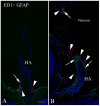
Macrophages surround the hyaloid artery. Double labeling with the macrophage marker ED1 (red) and GFAP (green) in 20-day-old wild type and Nuc1 rat eyes. A. In the wild type rat, some GFAP-positive cells (arrow) are seen at the base of the regressing hyaloid artery (HA). Clusters of ED1-positive cells (arrowheads) are also observed at the base of the involuting hyaloid artery. B. In the Nuc1 rat, the persistent hyaloid artery is surrounded by a layer of GFAP-positive astrocytes (arrows), with abundant ED1-positive cells (arrowheads) in the vicinity of the hyaloid artery. A fragment of GFAP-positive cells (arrow) is also observed in the vitreous, presumably from a tangential section of the hyaloid artery, surrounded by ED1-positive cells (arrowhead). Scale bar = 50 μm, n = 5 wild type, n = 5 Nuc1 homozygote. Reproduced with permission from European Journal of Cell Biology.
Fig. 7
Fig. 7
Bit1 expression by astrocytes at the optic nerve head in 2-month old normal and Nuc1 homozygous rats. Confocal microscopy indicated that in wild type optic nerve (panels a, b, c), Bit1 (red) and GFAP (green) are co-expressed (yellow, c). Interestingly, in the Nuc1 homozygous optic nerve (Panels d, e, f) Bit1 (red) and GFAP (green) show more intense co-expression than in wild type. Adapted with permission from Cell Death and Disease.
Fig. 8
Fig. 8
Effects of anoikis induction on WT and Nuc1 astrocytes. a. Astrocytes were isolated from the optic nerve of wild type and Nuc1 rats and cultured on poly-HEMA plates to induce anoikis. After 5 days on poly-HEMA, Nuc1 astrocytes show significantly higher survival than wild type. Cell death assays performed by Cell Death Detection ELISAPLUS kit after 3 and 5 days of anoikis induction show approximately 20% and 50% less cell death in Nuc1 cultures compared to wild type, respectively. Results were plotted as absorbance at 405 nm with a reference wavelength of 490 nm. Experiments were done in triplicate. P-values were calculated using Student's t-test (*P = 0.001). b. To measure proliferation, the MTS assay was performed with Nuc1 and wild type astrocytes after 5 days of anoikis induction, followed by culture under normal conditions for 0, 4 and 7 days. At 7 days, significant increase in cell proliferation was observed in Nuc1 cultures compared to wild type. The data represent the mean values of absorbance at λ = 490 nm, which is proportional to the number of viable cells. Experiments were done in triplicate. P-values were calculated using Student's t-test (*P = 0.05). c. When Bit1 was knocked-down in wild type astrocytes (western blot in inset) using Bit1 specific siRNA, cell death was decreased following anoikis induction as described above. The data represent the mean values of absorbance at λ = 490 nm, which is proportional to the number of viable cells. Experiments were done in triplicate. P-values were calculated using Student's t-test (*P = 0.05). Adapted with permission from Cell Death and Disease.
Fig. 9
Fig. 9
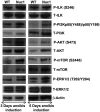
Signaling pathways in WT and Nuc1 astrocytes. To identify pathways activated in the wild type and the Nuc1 astrocytes after anoikis induction, western-blotting analyses were performed for total and phospho PI3K, total and phospho ILK, total and phospho AKT, total and phospho mTOR, total and phospho ERK1/2 with β-actin as loading control. After 3 and 5 days of anoikis induction, Nuc1 astrocytes show a robust increase in the phosphorylated (activated) forms of PI3K, AKT, mTOR, ILK and ERK1/2 compared to wild type. Adapted with permission from Cell Death and Disease.
Fig. 10
Fig. 10
γ-Secretase activity in WT and Nuc1 astrocytes. Post-mitochondrial supernatants from WT and Nuc1 astrocytes were layered on 2.5–30% iodixanol gradients and subjected to ultracentrifugation. Western blots identified LAMP1-positive lysosomes in fractions 3, 4, and 5; Rab5-positive endosomes in fractions 6, 7, and 8; and Golgi in fractions 9 and 10 [37]. a. ELISA was performed to quantify β-amyloid (Aβ40), which is produced by the action of γ-secretase, after incubating above fractions overnight at 37 °C, with or without L-685,458 (γ-secretase inhibitor). In WT astrocytes (blue), γ-secretase activity, as measured by Aβ40 level, was detected in all fractions, with the Golgi fractions (9 and 10) having somewhat lower activity. In Nuc1 astrocytes (red), γ-secretase activity was significantly decreased (approximately 50%) in the endolysosomal compartments (fractions 3–8), whereas the activity in Golgi was not significantly different from WT. b. Over-expression of βA3/A1-crystallin in astrocytes cultured from Nuc1 rats rescued γ-secretase activity in endolysosomal compartments, while empty vector had no effect (relative to vehicle). Statistical analysis was performed by a two-tailed Student's t-test: *P < 0.05. **P < 0.01. Adapted with permission from Nature Communications.
Fig. 11
Fig. 11
Impaired V-ATPase activity in astrocytes lacking functional βA3/A1-crystallin. a. V-ATPase activity was measured by acridine orange fluorescence in WT and Nuc1 astrocytes after intravesicular H+ uptake was initiated by the addition of Mg-ATP. Increase in acridine orange fluorescence (V-ATPase activity) upon addition of ATP was significantly greater in WT astrocytes than in Nuc1 astrocytes. b. In Nuc1 astrocytes, over-expression of βA3/A1-crystallin rescued activity to N80% of normal level (*P = 0.020). Empty vector had no effect. c. Measurement of endolysosomal pH in WT and Nuc1 astrocytes was performed using a fluid phase fluorescent probe, FITC-dextran. 3 h after incubation of WT and Nuc1 astrocytes with FITC-dextran the fluorescence emission was measured at 520 nm with excitation at 450 nm and 495 nm. The fluorescence excitation ratio at 495 nm and 450 nm (I495 nm/I450 nm) was calculated, and endolysosomal pH in WT and Nuc1 astrocytes was measured to be ∼4.5 and 5.7, respectively (*P = 0.031). The elevated pH in Nuc1 homozygous astrocytes was restored to near normal by over-expression of βA3/A1-crystallin (*P = 0.026). Empty vector had no effect. In all panels, graphs show mean values and error bars represent s.d. from a triplicate experiment representative of at least three independent experiments. Statistical analysis was performed by a two-tailed Student's t-test: *P < 0.05. Adapted with permission from Nature Communications.
Fig. 12
Fig. 12
Intracellular processing and degradation of Notch receptor. a. WT and Nuc1 astrocytes transfected with myc-tagged full length Notch1 receptor were monitored for receptor clearance when co-cultured with astrocytes overexpressing Jagged1. Top panels show myc labeling (red) after transfection. Labeling decreased quickly in WT astrocytes, with little remaining after 3 h. Degradation was minimal in the Nuc1 astrocytes. Scale bar = 20 μm. b. Cathepsin D activity was decreased in Nuc1 astrocytes to about 30% of the level in WT. Over-expression of βA3/A1-crystallin in Nuc1 astrocytes increased cathepsin D activity to 75% of WT level (*P = 0.037). Graph shows mean values and error bars represent s.d. from a triplicate experiment representative of at least three independent experiments. Statistical analysis was performed by a two-tailed Student's t-test: **P < 0.01; *P < 0.05. Adapted with permission from Nature Communications.
Fig. 13
Fig. 13
Impaired Notch signaling decreases VEGF secretion in astrocytes a. VEGF quantikine ELISA to detect levels of VEGF secreted into the medium showed significant reduction (∼85%) in Nuc1 cells compared to WT cells. b. Treatment with the Notch inhibitor DAPT, significantly reduced secreted levels of VEGF in both WT (∼60%) and Nuc1 (∼75%) astrocytes compared to the respective vehicle-treated controls. c. Likewise, adenoviral Cre-recombinase-mediated knockdown of Cryba1 in Cryba1fl/fl mouse astrocytes produced a similar reduction in VEGF secretion. Moreover, inhibition of Notch with DAPT further decreased VEGF secretion in both cultures. Error bars indicate s.d.; *P < 0.05. Adapted with permission from Scientific Reports.
Fig. 14
Fig. 14
βA3/A1-crystallin modulates the phosphorylation of STAT3 in astrocytes. a. Analysis of p-STAT3 levels by ELISA revealed 5 fold induction of p-STAT3 in the presence of IL-6 and ∼76% reduction in the presence of Stattic compared to the vehicle control. Stattic significantly decreased the levels of p-STAT3 in the presence of IL-6 (∼85%) compared to the astrocytes treated with IL-6 alone. b. ELISA data revealed that IL-6 treatment induced p-STAT3 by 4.5 fold in Cryba1 KD astrocytes compared to vehicle-treated astrocytes. Stattic decreased the level of p-STAT3 by ∼80% when treated alone and by ∼85% when treated in combination with IL-6 compared to vehicle-treated and IL-6 treated Cryba1 KD astrocytes, respectively. Error bars indicate s.d.; *P < 0.05. Adapted with permission from Scientific Reports.
Fig. 15
Fig. 15
STAT3 regulates the expression of Cryba1 in astrocytes. a. Schematic diagram of the mouse Cryba1 gene showing the transcription start site (TSS) and the two STAT3 binding sites, one 2750 bp upstream of the start site (promoter region) and the other 1942 bp from the TSS in Intron 2. Exons are shown in boxes. b. Treatment of WT astrocytes with IL-6 resulted in 4.5 fold increase in the expression of Cryba1 and Stattic treatment decreased the expression of Cryba1 by ∼51% compared to vehicle treated astrocytes. In astrocytes treated with both IL-6 and Stattic the expression of Cryba1 was reduced by ∼73% compared to astrocytes treated with IL-6 alone. Error bar indicate s.d.; *P < 0.05. Reproduced with permission from Scientific Reports.
Fig. 16
Fig. 16
A schematic representation of signaling pathways and cellular processes modulated by βA3/A1-crystallin in astrocytes. It is likely that the protein regulates multiple processes and pathways in astrocytes by regulating V-ATPase and IGF-II. Solid lines denote links supported by experimental evidence and dashed lines are hypothetical. Abbreviations used: IGF-II, insulin-like growth factor-II; PI3K, phosphatidylinositol-3-kinase; ILK, integrin-linked kinase; mTOR, mechanistic target of rapamycin; V-ATPase, vacuolar-type H+-ATPase; NICD, Notch intracellular domain, p-STAT3, phosphorylated Signal transducers and activators of transcription 3.

Similar articles

Cited by

References

    1. Goldberg MF. Persistent fetal vasculature (PFV): an integrated interpretation of signs and symptoms associated with persistent hyperplastic primary vitreous (PHPV). LIV Edward Jackson Memorial Lecture. Am J Ophthalmol. 1997;124:587–626. - PubMed
    1. Zhang C, Ashaghi L, Gongora C, Patek B, Hose S, Ma B, Aghsaei-Fard M, Brako L, Singh K, Goldberg MF, Handa JT, Lo WK, Eberhart CG, Zigler JS, Jr, Sinha D. A developmental defect in astrocytes inhibits programmed regression of the hyaloid vasculature in the mammalian eye. Eur J Cell Biol. 2011;90(5):440–448. - PMC - PubMed
    1. Sinha D, Valapala M, Patek B, Bhutto I, Zhang C, Hose S, Fang Y, Cano M, Stark WJ, Lutty GA, Zigler JS, Jr, Wawrousek E. βA3/A1-crystallin is required for proper astrocytes template formation and vascular remodeling in the retina. Transgenic Res. 2012;21(5):1033–1042. - PMC - PubMed
    1. Zhang C, Gehlbach P, Gongora C, Cano M, Fariss R, Hose S, Nath A, Green WR, Goldberg MF, Zigler JS, Sinha D. A potential role for β-and γ-crystallins in the vascular remodeling of the eye. Dev Dyn. 2005;234:36–47. - PubMed
    1. Ito M, Yoshioka M. Regression of the hyaloid vessels and pupillary membrane of the mouse. Anat Embryol. 1999;200:403–411. - PubMed

Publication types

MeSH terms