VEGF expression, microvessel density and dendritic cell decrease in thyroid cancer
- PMID: 26019537
- PMCID: PMC4433839
- DOI: 10.1080/13102818.2014.909151
VEGF expression, microvessel density and dendritic cell decrease in thyroid cancer
Abstract
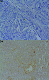
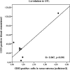
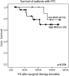
Thyroid cancer is one of the five most common cancers in the age between 20 and 50 years. Many factors including the potent angiogenic vascular endothelial growth factor (VEGF) and different dendritic cell types are known to be related to thyroid tumourogenesis. The study was performed to address the expression of VEGF and microvessel density in thyroid cancers and to evaluate the effect of VEGF expression in thyroid tumour cells on the dendritic cells. We investigated 65 patients with different types of thyroid carcinomas: papillary (PTC), oncocytic (OTC), follicular (FTC) and anaplastic (ATC), immunohistochemically with antibodies against VEGF, CD1a, CD83, S100 and CD31. Our results suggest that the expression of VEGF is significantly more often in PTC than ATC (92.3% vs. 60.0%, p = 0.025). The microvessel density marked with CD31 in the tumour border of PTC was significantly higher as compared to FTC (p = 0.039), but not to ATC and OTC (p = 0.337 and 0.134). We found that CD1a- and CD83-positive cells were dispersed with variable density and in OC CD31+ vessel numbers were positively correlated with CD83+ dendritic cells in tumour stroma (R = 0.847, p = 0.016). We did not find statistically significant associations of the survival of patients with PTC after the surgical therapy with VEGF expression and MVD. In conclusion we may state that VEGF expression in tumour cells of thyroid cancer can induce neovascularization and suppress dendritic cells.
Keywords: CD31; VEGF; dendritic cell; microvessel density; prognosis; thyroid cancer.
Figures


Similar articles
-
Relationships between Lymph Node Metastasis and Expression of CD31, D2-40, and Vascular Endothelial Growth Factors A and C in Papillary Thyroid Cancer.Clin Exp Otorhinolaryngol. 2012 Sep;5(3):150-5. doi: 10.3342/ceo.2012.5.3.150. Epub 2012 Aug 27. Clin Exp Otorhinolaryngol. 2012. PMID: 22977712 Free PMC article.
-
The expression of vascular endothelial growth factor and the type 1 vascular endothelial growth factor receptor correlate with the size of papillary thyroid carcinoma in children and young adults.Thyroid. 2000 Apr;10(4):349-57. doi: 10.1089/thy.2000.10.349. Thyroid. 2000. PMID: 10807064
-
Loss of heterozygosity of the long arm of chromosome 7 in follicular and anaplastic thyroid cancer, but not in papillary thyroid cancer.J Clin Endocrinol Metab. 1999 Sep;84(9):3235-40. doi: 10.1210/jcem.84.9.5986. J Clin Endocrinol Metab. 1999. PMID: 10487693
-
Vascular endothelial growth factor (VEGF), VEGF receptors expression and microvascular density in benign and malignant thyroid diseases.Int J Exp Pathol. 2007 Aug;88(4):271-7. doi: 10.1111/j.1365-2613.2007.00533.x. Int J Exp Pathol. 2007. PMID: 17696908 Free PMC article.
-
Angiogenesis in non-small cell lung cancer: the prognostic impact of neoangiogenesis and the cytokines VEGF and bFGF in tumours and blood.Lung Cancer. 2006 Feb;51(2):143-58. doi: 10.1016/j.lungcan.2005.09.005. Epub 2005 Dec 19. Lung Cancer. 2006. PMID: 16360975 Review.
Cited by
-
Combined Effects of Baicalein and Docetaxel on Apoptosis in 8505c Anaplastic Thyroid Cancer Cells via Downregulation of the ERK and Akt/mTOR Pathways.Endocrinol Metab (Seoul). 2018 Mar;33(1):121-132. doi: 10.3803/EnM.2018.33.1.121. Endocrinol Metab (Seoul). 2018. PMID: 29589394 Free PMC article.
-
The potential role of reprogrammed glucose metabolism: an emerging actionable codependent target in thyroid cancer.J Transl Med. 2023 Oct 18;21(1):735. doi: 10.1186/s12967-023-04617-2. J Transl Med. 2023. PMID: 37853445 Free PMC article. Review.
-
Superb microvascular imaging compared with contrast-enhanced ultrasound to assess microvessels in thyroid nodules.J Med Ultrason (2001). 2020 Apr;47(2):287-297. doi: 10.1007/s10396-020-01011-z. Epub 2020 Mar 3. J Med Ultrason (2001). 2020. PMID: 32125575
-
Novel treatments for anaplastic thyroid carcinoma.Gland Surg. 2020 Jan;9(Suppl 1):S28-S42. doi: 10.21037/gs.2019.10.18. Gland Surg. 2020. PMID: 32055496 Free PMC article. Review.
-
Expression of vascular endothelial growth factor in follicular cell-derived lesions of the thyroid: Is NIFTP benign or precancerous?Turk J Surg. 2022 Mar 28;38(1):60-66. doi: 10.47717/turkjsurg.2022.5318. eCollection 2022 Mar. Turk J Surg. 2022. PMID: 35873744 Free PMC article.
References
-
- Shushanov S. Bronstein M. Adelaide J. Jussila L. Tchipysheva T. Jacquemier J. Stavrovskaya A. Birnbaum D. Karamysheva A. VEGFc and VEGFR3 in human thyroid pathologies. Int J Cancer. 2000;86:47–52. - PubMed
-
- Karaca Z. Tanriverdi F. Unluhizarci K. Ozturk F. Gokahmetoglu S. Elbuken G. Cakir I. Bayram F. Kelestimur F. VEGFR1 expression is related to lymph node metastasis and serum VEGF may be a marker of progression in the follow-up of patients with differentiated thyroid carcinoma. Eur J Endocrinol. 2011;164:277–284. doi: 10.1530/EJE-10-0967. - DOI - PubMed
-
- Robinson CJ. Stringer SE. The splice variants of vascular endothelial growth factor (VEGF) and their receptors. J Cell Sci. 2001;114:853–865. - PubMed
-
- Pepper MS. Ferrara N. Orci L. Montesano R. Vascular endothelial growth factor (VEGF) induces plasminogen activator inhibitor-1 in microvascular endothelial cells. Biochem Piophys Res Commun. 1991;181:902–906. - PubMed
-
- Keck PJ. Hauser SD. Krivi G. Sanzo K. Warren T. Feder J. Connolly DT. Vascular permeability factor, an endothelial cell mitogen related to PDGF. Science. 1989;246:1309–1312. - PubMed
LinkOut - more resources
Full Text Sources
Other Literature Sources
Research Materials
