

Clinico pathological study of adult dermatomyositis: Importance of muscle histology in the diagnosis
- PMID: 26019418
- PMCID: PMC4445196
- DOI: 10.4103/0972-2327.150603
Clinico pathological study of adult dermatomyositis: Importance of muscle histology in the diagnosis
Abstract
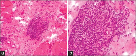
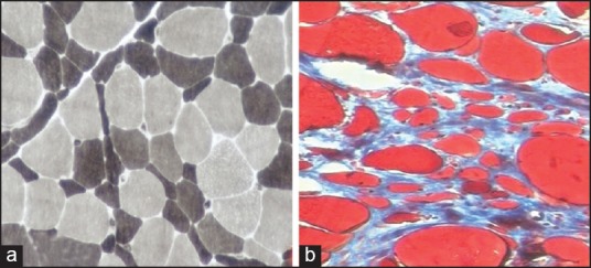
Aims: To study the histological features on muscle biopsy and correlate them with clinical features, other laboratory data in adult patients to make a diagnosis of dermatomyositis (DM), applying the European Neuromuscular center (ENMC) criteria.
Materials and methods: Adult patients who fulfilled clinical, laboratory, and muscle biopsy findings according to ENMC criteria for DM during the period 2010-2013 were included in the study. Cryostat sections of muscle biopsy were reviewed with emphasis on Perifascicular atrophy (PFA), perivascular/endomysial inflammation. Muscular dystrophies and metabolic myopathies were excluded by appropriate immunohistochemistry and special stains.
Results: The diagnosis of adult DM was made in 45 patients out of 170 clinically suspected idiopathic inflammatory myopathies. These included 33 definite, 4 probable, 7 possible sine dermatitis, and 1 amyopathic DM. All patients with definite DM had typical rash and proximal muscle weakness and muscle biopsy showed PFA with or without inflammation. Thirteen patients had quadriparesis, neck muscle weakness, dysphagia/dysphonia at presentation. Patients with probable DM had rash and showed perivascular/endomysial inflammation with no PFA. Possible DM sine dermatitis showed PFA with perivascular/endomysial infiltrates. One patient of amyopathic DM had typical heliotrope rash and characteristic skin biopsy.
Conclusions: Histological features are important for the diagnosis of DM. Relying on PFA for diagnosis of definite DM underestimates the true frequency of DM.
Keywords: Dermatomyositis; perifascicular atrophy; perivascular inflammation.
Conflict of interest statement
Figures

Similar articles
-
Redefining dermatomyositis: a description of new diagnostic criteria that differentiate pure dermatomyositis from overlap myositis with dermatomyositis features.Medicine (Baltimore). 2014 Nov;93(24):318-332. doi: 10.1097/MD.0000000000000222. Medicine (Baltimore). 2014. PMID: 25500701 Free PMC article.
-
A systematic review of adult-onset clinically amyopathic dermatomyositis (dermatomyositis siné myositis): a missing link within the spectrum of the idiopathic inflammatory myopathies.J Am Acad Dermatol. 2006 Apr;54(4):597-613. doi: 10.1016/j.jaad.2005.10.041. Epub 2006 Jan 23. J Am Acad Dermatol. 2006. PMID: 16546580 Review.
-
Edematous myositis: a clinical presentation first suggesting dermatomyositis diagnosis.Brain Pathol. 2020 Sep;30(5):867-876. doi: 10.1111/bpa.12844. Epub 2020 Jun 15. Brain Pathol. 2020. PMID: 32323412 Free PMC article.
-
RIG-I expression in perifascicular myofibers is a reliable biomarker of dermatomyositis.Arthritis Res Ther. 2017 Jul 24;19(1):174. doi: 10.1186/s13075-017-1383-0. Arthritis Res Ther. 2017. PMID: 28738907 Free PMC article.
-
[Inflammatory myopathies: diagnosis and classifications].Presse Med. 2009 Jul-Aug;38(7-8):1141-63. doi: 10.1016/j.lpm.2009.01.013. Epub 2009 Mar 17. Presse Med. 2009. PMID: 19282137 Review. French.
Cited by
-
Infliximab is effective in the treatment of ulcerative colitis with dermatomyositis: A case report.World J Gastroenterol. 2020 Dec 14;26(46):7425-7435. doi: 10.3748/wjg.v26.i46.7425. World J Gastroenterol. 2020. PMID: 33362394 Free PMC article.
-
Anti-MDA5 Antibody-Positive Interstitial Pneumonia with Autoimmune Features Presenting as Amyopathic Hypodermatitic Dermatomyositis: A Case Report.Case Rep Dermatol. 2021 Apr 19;13(1):222-229. doi: 10.1159/000515245. eCollection 2021 Jan-Apr. Case Rep Dermatol. 2021. PMID: 34054457 Free PMC article.
References
-
- Dalakas MC. Polymyositis, dermatomyositis and Inclusion-body myositis. N Engl J Med. 1991;325:1487–98. - PubMed
-
- Dalakas MC, Hohlfeld R. Polymyositis and dermatomyositis. Lancet. 2003;362:971–82. - PubMed
-
- Hoogendijk JE, Amato AA, Lecky BR, Choy FH, Lundberg IE, Rose MR, et al. 119th ENMC international workshop: Trial design in adult idiopathic inflammatory myopathies, with the exception of inclusion-body myositis, 10-12 October 2003, Naarden, The Netherlands. Neuromuscul Disord. 2004;14:337–45. - PubMed
-
- Lazarou IN, Guerne PA. Classification, diagnosis and management of idiopathic inflammatory myopathies. J Rheumatol. 2013;40:550–64. - PubMed
LinkOut - more resources
Full Text Sources
Other Literature Sources
