. 2015 Jun 4;522(7554):106-110.
doi: 10.1038/nature14492. Epub 2015 May 27.

The hypoxic cancer secretome induces pre-metastatic bone lesions through lysyl oxidase

Affiliations

The hypoxic cancer secretome induces pre-metastatic bone lesions through lysyl oxidase

Thomas R Cox et al. Nature. .

Retraction in

Abstract

Tumour metastasis is a complex process involving reciprocal interplay between cancer cells and host stroma at both primary and secondary sites, and is strongly influenced by microenvironmental factors such as hypoxia. Tumour-secreted proteins play a crucial role in these interactions and present strategic therapeutic potential. Metastasis of breast cancer to the bone affects approximately 85% of patients with advanced disease and renders them largely untreatable. Specifically, osteolytic bone lesions, where bone is destroyed, lead to debilitating skeletal complications and increased patient morbidity and mortality. The molecular interactions governing the early events of osteolytic lesion formation are currently unclear. Here we show hypoxia to be specifically associated with bone relapse in patients with oestrogen-receptor negative breast cancer. Global quantitative analysis of the hypoxic secretome identified lysyl oxidase (LOX) as significantly associated with bone-tropism and relapse. High expression of LOX in primary breast tumours or systemic delivery of LOX leads to osteolytic lesion formation whereas silencing or inhibition of LOX activity abrogates tumour-driven osteolytic lesion formation. We identify LOX as a novel regulator of NFATc1-driven osteoclastogenesis, independent of RANK ligand, which disrupts normal bone homeostasis leading to the formation of focal pre-metastatic lesions. We show that these lesions subsequently provide a platform for circulating tumour cells to colonize and form bone metastases. Our study identifies a novel mechanism of regulation of bone homeostasis and metastasis, opening up opportunities for novel therapeutic intervention with important clinical implications.


Conflict of interest statement

Author Information The authors declare no potential conflicts of interest.

Figures

Extended Data Figure 1
Extended Data Figure 1. LOX is hypoxia regulated and strongly associated with osteotropism and metastasis.
a, Retrospective analysis of our patient cohort including only ER-negative patients showed that the hypoxic signature is not significantly associated with liver relapse (P=0.98), brain relapse (P=0.17) or lung relapse (P=0.13). b, Log2 expression levels under conditions of hypoxia (1% O2) and normoxia (21% O2) for secreted proteins from the MDA-MB-231 parent and MDA-MB-231 Bone Tropic (BT) 1833 cell line. Data representative of 4 repeats, 2x label-free repeats, and 2x SILAC (standard and reverse-label) repeats. Acquisition performed on the Orbitrap Q-Exactive (Thermo Fisher Scientific). c, Overlaps between repeats of global secretome analysis in MDA-MB-231 parent and MDA-MB-231 Bone Tropic (BT) cells grown in normoxic (21% O2) and hypoxic (1% O2) conditions from label-free and SILAC approaches. d, Immunoblotting for LOX in MDA parent and 1833 Bone Tropic (BT) subclone under conditions of hypoxia (1% O2) and normoxia (21% O2) confirming expression levels seen in proteomic and transcriptomic analyses. Scans of original western blots available as Supplementary Information.
Extended Data Figure 2
Extended Data Figure 2. Extended patient data analysis.
a, Across all breast cancer patients, the expression of LOX is associated with metastasis formation (P=0.023) and in particular with ER-Negative breast cancer patients (P=0.0029). b, An additional Kruskall-Wallis test between reported bone relapse, relapse elsewhere and no relapse patients with an additional contrast test wherein all pairwise groups were considered shows that in all patients, LOX expression is associated with bone relapse compared to no relapse (P=0.0389). This also pertains to ER-negative patients (P=0.0126) but not ER-positive patients (P=0.9537). c, Cox-regression using log2-LOX expression data was used to estimate the Hazard Ratio (HR) in two analyses. One analysis was performed using the no-relapse patients and the bone relapse patients (data belonging to Fig.1f) and the second analysis including all patients (data belonging to Extended Data Fig.2a). LOX expression is associated with increased Hazard Ratio particularly in ER-negative patients in both analyses.
Extended Data Figure 3
Extended Data Figure 3. Additional patient data analysis in a supporting patient cohort.
a, ROC curve analysis shows LOX expression may be indicative of metastatic dissemination of ER-negative breast cancer (AUC 0.77, P<0.0001) but not ER-positive patients (AUC 0.55, P<0.1504). b, In an alternative second patient dataset (Van de Vijver et al. [pubmed ID: 12490681]) reporting data on 295 lymph node negative (LNN) patients who did not receive adjuvant therapy, with available site of relapse, LOX is significantly higher expressed in bone relapse ER-negative patients, compared to other groups confirming data from the original dataset.
Extended Data Figure 4
Extended Data Figure 4. Hypoxia-induced tumour-derived LOX stimulates osteolytic lesion formation in the absence of tumour cells.
a, Immunoblotting of 4T1 mammary carcinoma line stably expressing either a scrambled (scr) or shLOX vector which leads to a significant decrease in levels of detectable LOX. Scans of original western blots available as Supplementary Information. b, Micro-CT scanning and reconstruction with structural analysis shows decreases in trabecular bone volume (as a percentage of total bone volume) (n=3 mice per group). c, decreases in trabecular number (per mm) (n=3 mice per group) and d, decreases in trabecular thickness in tibiae of mice bearing 4T1scr mammary fatpad tumours over time (n=3 mice per group). e, Micro-CT analysis of mouse tibiae shows increases in focal osteolytic lesions in 4T1scr tumour bearing mice develop over time (n=3 mice per group). (a-e) P-values derived from unpaired parametric two-tailed t-test (*P<0.05**, P<0.01, ***P<0.001). f, Representative immunohistochemical staining for pimonidazole (Hypoxyprobe) in 4μm section of 4T1 orthotopic mammary carcinoma, 3 weeks post-implantation shows hypoxia (brown staining) as a salient feature of tumours. Scale bar = 250μm. g, Bioluminescent imaging of luciferase signal 2 weeks post explant of samples taken from primary tumour, lung, bone marrow (femur and tibia) and distant skin samples at 1 – 5 weeks post primary tumour implant. Selection was under 500ug/mL zeocin for the Luciferase expression cassette (n=3 mice per timepoint). h, Quantification of (g) as a percentage of positive luciferase expressing explants from various sites following 4T1 tumour implant shows tumour cells do not begin to arrive in the bone until 3 weeks. i, Q-RTPCR detection of luciferase expressing 4T1 tumour cells in secondary organs confirms explant culture experiments (n=3 mice per timepoint).
Extended Data Figure 5
Extended Data Figure 5. Effects of LOX modulation on circulating sera levels, primary tumour growth and osteolytic lesion formation.
a, Enzyme-linked immunosorbent assay (ELISA) for LOX in the sera of 4T1scr and 4T1shLOX tumour bearing mice (n: ELISA signal [AU] in mouse sera: 3 mice per group) shows decreased levels of circulating LOX upon genetic silencing at the primary tumour. Data shown is mean ± SEM. P-values derived from unpaired parametric two-tailed t-test (*P<0.05). b, Growth curves as determined by calliper measurement for orthotopic 4T1scr and 4T1shLOX mammary tumours show no difference between primary tumour growth (n: mice; 3 per group). c, Injection of hypoxic CMs from SW480 human colorectal cancer cells stably expressing one of; empty vector control (EV), full-length LOX (+LOX), or a catalytically inactive full-length LOX (+mutLOX)(K320A) confirms a LOX-dependent mechanism of focal osteolytic lesion generation in a second human model of cancer. (n: mice; 8 per group). Data shown is mean ± SEM. P-values derived from unpaired parametric two-tailed t-test (**P<0.01, ***P<0.001). d, C-terminal telopeptide (CTX) ELISA (Ratlaps) on sera of mice injected intraperitoneally biweekly with rLOX for 3 weeks. CTX is a telopeptide that can be used as a biomarker in the serum to measure the rate of bone turnover (n: ng/mL circulating CTX-I in mouse sera; 5 mice per group). Data shown is mean ± SEM. P-values derived from unpaired parametric two-tailed t-test (*P<0.05, **P<0.01, ***P<0.001).
Extended Data Figure 6
Extended Data Figure 6. LOX modulates osteoclasts and osteoblast behaviour independent of RANKL.
a, ELISA for RANKL in the conditioned media (CM) of osteoclast cultures shows no detectable levels of RANKL in MCSF alone (-ve control) and rLOX cultures excluding the likelihood of autocrine production by cells in response to rLOX (n: ELISA signal [AU]; data is from 3x independent experimental repeats in all groups). Data shown is mean ± SEM. P-values derived from unpaired parametric two-tailed t-test. (*P<0.05) b, All proteins detected by Mass Spectrometry analysis in the rLOX preparations (based on MaxQuant 1.5 peptide ID score of 50 and a minimum of 2 unique MS peptide observations). c, Examples of nuclear localisation of NFATc1 following addition of rLOX in the presence and absence of the LOX antibody; Green – NFATc1, Red - Phalloidin , Blue – DAPI). d, Representative Alizarin Red S plate showing mineralisation ability (calcium deposits as detected by Alizarin Red S staining) of primary calvarial mouse osteoblasts following treatment with dexamethasone (DEX) (positive control) or rLOX ± LOX ab; quantification shown in Fig.3g e, High LOX containing hypoxic 4T1scr CM significantly reduces cell proliferation of the human osteoblast like SaOS-2 cell line which can be partially blocked by treatment with anti-LOX antibody (n: normalised cell number per well; control 24 wells; 4T1scr CM 49 wells; 4T1scr CM + LOX Ab 51 wells). Data collected over 3 independent experimental repeats. Data shown in mean ± SEM. P-values derived from unpaired parametric two-tailed t-test. (*P<0.05, **P<0.01, ***P<0.001). f, Mineralisation ability (calcium deposits as detected by Alizarin Red S staining) is increased in the human osteoblast like SaOS-2 cell line in response to high LOX expressing hypoxic 4T1scr CM, the effects of which can be attenuated using the anti-LOX antibody. (n: Alizarin Red S staining per well, data taken from 3 independent repeats; control 18 wells; 4T1scr CM 9 wells; 4T1scr CM + LOX Ab 9 wells). Data shown is mean ± SEM. P-values derived from unpaired parametric two-tailed t-test. (*P<0.05, **P<0.01, ***P<0.001).
Extended Data Figure 7
Extended Data Figure 7. Additional in vivo analysis of lesion formation and primary tumour growth.
a, 4T1scr tumour bearing mice treated with our LOX antibody show a decrease in osteoclast perimeter in tibial bones in support of LOX as a modulator of osteoclastogenesis shown in Fig.3 (n: mice; 4T1scr Tumour + IgG 5, 4T1scr Tumour + LOX Ab 7). Data shown is mean ± SEM. P-values derived from unpaired parametric two-tailed t-test. (*P<0.05) b, Weekly tumour volumetric measurements for 4T1scr tumour bearing mice treated with either zoledronic acid (0.6mg/kg intraperitoneally) or vehicle (PBS), show that when administered alone zoledronic acid does not affect primary 4T1scr primary tumour growth in vivo (n: mice; 4 in all groups). c, Pearson correlation shows a moderate correlation between lesion number as determined by micro-CT analysis and luciferase signal (radiance [p/s/cm2/sr]) from 4T1Luc tumour cells within the bone (r = 0.58, 95% CI 0.2778 – 0.7834, P = 0.0009 [two-tailed]).
Figure 1
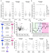
Figure 1. Tumour-secreted LOX is a critical player in ER– breast cancer bone metastasis.
a, Retrospective analysis of 344 LNN primary breast cancers. The hypoxic signature is associated with relapse in all patients, and specifically ER– patients, but not ER+ patients. b, Further analysis indicates the hypoxic signature is specifically associated with bone relapse. c, Schematic overview of quantitative stable isotope labelling by amino acids in cell culture SILAC and label-free global proteomic secretome analysis approaches. d, LOX is more than 1.5 fold upregulated in bone tropic (BT) vs parental cells and the most hypoxia regulated (>2.25 fold) protein associated with osteotropism. A full list is available in Supplementary Information. e, Log2 median centered expression of LOX mRNA in MDA-MB-231 parental and subclone lines (n=3 probesets per cell line) (# indicates 1833 ‘BT’ clone used). P-values derived from unpaired parametric two-tailed t-tests (*P<0.05, **P<0.01). f, LOX expression specifically associates with bone relapse in ER– breast cancer patients but not ER+ patients. (a,b,f) P-values derived from a 2-tailed Mann-Whitney test.
Figure 2
Figure 2. Osteolytic lesion formation in ER– mammary carcinoma models is LOX dependent.
a, Representative 2D cross-sections of tibia from control (top) and tumour-bearing (bottom) mice 3-weeks post orthotopic implantation showing lesions (arrowheads) and loss of trabecular structure (asterisks). b, Micro-CT analysis of osteolytic lesions in tumour-bearing and tumour-free, CM conditioned mice at 3 weeks (n: mice; control 3; 4T1scr Tumour 5; Control Injected 5; 4T1scr CM 5) c, Loss of cortical bone volume in 4T1scr tumour-bearing and tumour-free CM injected models at 3 weeks (n: mice; control 4; 4T1scr Tumour 3; Control Injected 4; 4T1scr CM 6). d, Representative 3D reconstructions of tibiae showing tumour driven osteolytic lesions (arrowheads). e, LOX silencing decreases focal osteolytic lesion formation (n: mice; control 5; 4T1scr Tumour 8; 4T1 shLOX Tumour 5). f, LOX inhibition decreases osteolytic lesion formation in tumour-bearing models (n: mice; control 5; 4T1scr Tumour + IgG 13; 4T1scr Tumour + LOX Ab 14) and g, in tumour-free CM injection models (n: mice; control 5; 4T1scr CM 5; 4T1shLOX CM 5) h, SW480 human CRC lines with stably manipulated LOX expression (EV, +LOX or +mutLOX) confirms LOX-dependency (n=8 mice per condition). i, Exogenous recombinant LOX (rLOX) drives osteolytic lesion formation in Nude and BALB/c models (n: mice; Nude control 8; Nude rLOX 8; BALB/c control 7; BALB/c rLOX 6). (b,c,e,f,g,h,i) Data shown are mean ± SEM. P-values derived from unpaired parametric one-tailed t-tests. (*P<0.05, **P<0.01, ***P<0.001).
Figure 3
Figure 3. Tumour-secreted LOX modulates osteoclasts and osteoblasts in vitro and in vivo.
a, rLOX (in the absence of RANKL) stimulates osteoclastogenesis. b, c, rLOX generated osteoclasts exhibit high resorptive ability; arrow = osteoclast, arrowheads = resorption tracks. (a-c)(n: osteoclast count from 12 independent osteoclast assays per group). d, rLOX induces nuclear localization of the master transcription factor NFATc1 in the absence of RANKL (n: osteoclast NFATc1 nuclear intensity; control 12; +RANKL 196, +rLOX 191 across 3 independent experimental repeats [donors] from 16 fields of view per donor). e, LOX antibody treatment blocks NFATc1 localization. f, Catalase treatment blocks rLOX-induced nuclear localization of NFATc1 (e,f represents data from 32 measurements of NFATc1 nuclear intensity from each of 3 independent donors [96 total]) g, rLOX added to primary mouse calvarial osteoblasts increases mineralization ability (DEX = dexamethasone) (n: alizarin red S intensity; 16 wells per group across 2 independent experimental repeats) h, 4T1scr mammary tumours decrease osteoblast number compared to shLOX (n: mice; Control 10; 4T1scr Tumour 7; 4T1 shLOX Tumour 9) and i, increase osteoclast number on the endocortical surface of bone (per mm bone perimeter)(n: mice; Control 10; 4T1scr Tumour 8; 4T1 shLOX Tumour 9). j, Representative images of osteoblasts (arrows) and osteoclasts (*) in sections of bone. k, 4T1scr tumour bearing mice treated with the anti-LOX antibody show similar effects on osteoblast l, osteoclast number (n: mice; 4T1scr Tumour + IgG 5; 4T1scr Tumour + LOX Ab 7). m, Representative sections of bone from 4T1scr tumour bearing mice with or without anti-LOX antibody or IgG control). (a,c-i,k,l) Data shown is mean ± SEM. P-values derived from unpaired parametric two-tailed t-tests. (*P<0.05, **P<0.01, ***P<0.001).
Figure 4
Figure 4. LOX-mediated lesions are osteoclast-driven and enhance circulating tumour cell colonisation.
a, Representative 3D reconstructions of tibiae from tumour bearing mice with or without BP treatment b, Tibial bone loss is abrogated in tumour bearing mice treated with bisphosphonate (n: mice; Control 5; 4T1scr Tumour 4; 4T1scr Tumour + BP 4) c, Similar effects are observed in CM conditioned models treated with bisphosphonates (n=5 mice all groups) d, Quantification of e, Whole body IVIS imaging of intracardially injected 4T1Luc tumour cells following conditioning with 4T1scr or 4T1shLOX CM. White boxes – tumour burden analysis region of interest (n: mice; 4T1scr CM+IgG 8; 4T1scr CM+LOXAb 8; 4T1shLOX CM+IgG 10) f, Micro-CT lesion analysis of mice after intracardiac injection following pre-conditioning (n: mice; 4T1scr CM+IgG 6; 4T1scr CM+LOXAb 8; 4T1shLOX CM+IgG 8) g, Representative whole body IVIS imaging of 4T1Luc tumour cells at 1 week and 5 weeks after intracardiac injection. Mice were conditioned with hypoxic 4T1scr CM with and without simultaneous treatment with bisphosphonate. White boxes – tumour burden analysis region of interest. h, Log2 quantitation of (g) (n=5 mice all groups) i, Schematic of LOX mediated effects on bone homeostasis in vivo. (b-d,f,h) Data shown is mean ± SEM. P-values derived from unpaired parametric two-tailed t-tests. (*P<0.05, **P<0.01, ***P<0.001).

Comment in

  • Cancer: Opening LOX to metastasis.
    Erez N. Erez N. Nature. 2015 Jun 4;522(7554):41-2. doi: 10.1038/nature14529. Epub 2015 May 27. Nature. 2015. PMID: 26017311 No abstract available.
  • Metastasis: LOX does some prepping.
    Villanueva MT. Villanueva MT. Nat Rev Cancer. 2015 Jul;15(7):384. doi: 10.1038/nrc3976. Epub 2015 Jun 18. Nat Rev Cancer. 2015. PMID: 26084391 No abstract available.
  • Cancer: LOX does some prepping.
    Villanueva MT. Villanueva MT. Nat Rev Drug Discov. 2015 Jul;14(7):458-9. doi: 10.1038/nrd4674. Nat Rev Drug Discov. 2015. PMID: 26129796 No abstract available.

Similar articles

Cited by

References

    1. Chan DA, Giaccia AJ. Hypoxia, gene expression, and metastasis. Cancer Metastasis Rev. 2007;26:333–339. - PubMed
    1. Jin L, et al. Differential secretome analysis reveals CST6 as a suppressor of breast cancer bone metastasis. Cell Res. 2012;22:1356–1373. - PMC - PubMed
    1. Erler JT, et al. Hypoxia-induced lysyl oxidase is a critical mediator of bone marrow cell recruitment to form the premetastatic niche. Cancer Cell. 2009;15:35–44. - PMC - PubMed
    1. Kaplan RN, et al. VEGFR1-positive haematopoietic bone marrow progenitors initiate the pre-metastatic niche. Nature. 2005;438:820–827. - PMC - PubMed
    1. Blanco MA, et al. Global secretome analysis identifies novel mediators of bone metastasis. Cell Res. 2012;22:1339–1355. - PMC - PubMed

Publication types

MeSH terms

Substances