. 2015 Jun;38(6):528-34.
doi: 10.14348/molcells.2015.0026. Epub 2015 May 27.

The Histone Methyltransferase Inhibitor BIX01294 Inhibits HIF-1α Stability and Angiogenesis

Affiliations

The Histone Methyltransferase Inhibitor BIX01294 Inhibits HIF-1α Stability and Angiogenesis

Su Young Oh et al. Mol Cells. 2015 Jun.

Abstract

Hypoxia-inducible factor (HIF) is a key regulator of tumor growth and angiogenesis. Recent studies have shown that, BIX01294, a G9a histone methyltransferase (HMT)-specific inhibitor, induces apoptosis and inhibits the proliferation, migration, and invasion of cancer cells. However, not many studies have investigated whether inhibition of G9a HMT can modulate HIF-1α stability and angiogenesis. Here, we show that BIX01294 dose-dependently decreases levels of HIF-1α in HepG2 human hepatocellular carcinoma cells. The half-life of HIF-1α, expression of proline hydroxylase 2 (PHD2), hydroxylated HIF-1α and von Hippel-Lindau protein (pVHL) under hypoxic conditions were decreased by BIX01294. The mRNA expression and secretion of vascular endothelial growth factor (VEGF) were also significantly reduced by BIX01294 under hypoxic conditions in HepG2 cells. BIX01294 remarkably decreased angiogenic activity induced by VEGF in vitro, ex vivo, and in vivo, as demonstrated by assays using human umbilical vein endothelial cells (HUVECs), mouse aortic rings, and chick chorioallantoic membranes (CAMs), respectively. Furthermore, BIX01294 suppressed VEGF-induced matrix metalloproteinase 2 (MMP2) activity and inhibited VEGF-induced phosphorylation of VEGF receptor 2 (VEGFR-2), focal adhesion kinase (FAK), and paxillin in HUVECs. In addition, BIX01294 inhibited VEGF-induced formation of actin cytoskeletal stress fibers. In conclusion, we demonstrated that BIX01294 inhibits HIF-1α stability and VEGF-induced angiogenesis through the VEGFR-2 signaling pathway and actin cytoskeletal remodeling, indicating a promising approach for developing novel therapeutics to stop tumor progression.

Keywords: BIX01294; G9a HMT inhibitor; HIF-1α; angiogenesis.


Figures
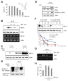
Fig. 1.
Fig. 1.
BIX01294 inhibits hypoxia-induced HIF-1α stability and VEGF expression. (A) HepG2 cells were treated with different concentrations of BIX01294. After 24 h, cell viability was measured by the MTT assay. (B) HepG2 cells were exposed to hypoxic conditions for 24 h in the presence of 0.1–1 μM BIX01294. Expression of HIF-1α was measured by Western blot and PCR. (C) BIX01294 (1 μM)-treated HepG2 cells were exposed to hypoxic conditions for 24 h in the presence or absence of MG132 (100 μM, 6 h) and cell lysates were analyzed by Western blot. (D) HepG2 cells were exposed to hypoxic conditions in the presence of BIX01294 (1 μM) and MG132 (100 μM) for 6 h. The cell lysates were imunoprecipitated with an anti-HIF-1α antibody. Immunoprecipitates were subsequently immunoblotted with an anti-ubiquitin antibody. (E) HepG2 cells were exposed to hypoxic conditions for 24 h in the presence or absence of 1 μM BIX01294. Expression of PHD2, pVHL and hydroxylated-HIF-1α was measured by Western blot. (F) HepG2 cells were exposed to hypoxic conditions for 24 h in the presence of BIX01294 and then treated with cycloheximide (CHX, 100 μM) for the durations indicated. HIF-1α protein levels were determined by Western blot. (G) HepG2 cells were exposed to hypoxic conditions in the presence or absence of BIX01294 for 24 h and then VEGF expression levels were determined by PCR. (H) ELISA was used to measure the levels of secreted VEGF protein in conditioned media. Beta-actin was used for an internal control. **p < 0.01 versus hypoxic control.
Fig. 2.
Fig. 2.
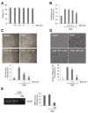
BIX01294 inhibits angiogenesis. (A) HUVECs were exposed to different concentrations of BIX01294. After 24 h, cell viability was measured by the MTT assay. (B) A BrdU-based cell proliferation assay was performed 24 h after treatment with BIX01294. (C) HUVECs were loaded onto the Matrigel-coated plates and treated with VEGF (20 ng/ml) in the presence or absence of BIX01294. Microphotographs were taken after 8 h. **p < 0.01 versus VEGF control. (D) HUVECs were plated onto 24-well Transwell membranes coated with collagen and treated with BIX01294 or vehicle in the presence of VEGF (20 ng/ml) for 24 h. Cells that migrated into the bottom part of the membrane were stained with hematoxylin and eosin. *p < 0.05; **p < 0.01 versus VEGF control. (E) Zymograms of serum-free conditioned medium of HUVECs treated with BIX01294 (1 μM) for 24 h. A clear zone of gelatin digestion indicates the presence of MMP-2.
Fig. 3.
Fig. 3.
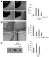
Effect of BIX01294 on the growth of new blood vessels. (A) Chick embryo aortas in Matrigel were treated with VEGF (20 ng/ml) in the absence or presence of BIX01294 for 24 h. Representative aortic rings were photographed. Three independent experiments were performed in triplicate. **p < 0.01 versus VEGF control. (B) Thermonox cover slips with BIX01294 or retinoic acid (RA, 1 μg) were loaded onto CAMs. After 3 days, a fat emulsion was injected under CAMs for better visualization of the vessels. The images shown are representative photographs of the chick CAM assay. Percentages of eggs positive for new vessel formation among all eggs tested were calculated. **p < 0.01 versus control. (C) Mice were injected with Matrigel containing VEGF (2.5 μg/ml) with or without BIX01294 (0.6 μg/ml) into the flank of C57BL6/J mice. After 7 days, the plugs were removed and blood vessels density was quantified by Image J software and represented as a bar diagram. **p < 0.01 versus VEGF control.
Fig. 4.
Fig. 4.
BIX01294 suppresses VEGFR-2 and FAK-mediated signaling pathways. (A) HUVECs were treated with various concentrations of BIX01294 and stimulated with VEGF (20 ng/ml) for 15 min. Activation of VEGFR-2, FAK, and paxillin was determined by Western blot. (B) HUVECs were stimulated with VEGF (20 ng/ml) in the presence or absence of BIX01294, and then filamentous actin was visualized by immunofluorescent staining using Alexa Fluor 488-conjugated phalloidin.

Similar articles

Cited by

References

    1. Abedi H, Zachary I. Vascular endothelial growth factor stimulates tyrosine phosphorylation and recruitment to new focal adhesions of focal adhesion kinase and paxillin in endothelial cells. J. Bio. Chem. 1997;272:15442–15451. - PubMed
    1. Bardos JI, Ashcroft J. Negative and positive regulation of HIF-1: a complex network. Biochimica et Biophysica Acta. 2005;1755:107–120. - PubMed
    1. Cabrita MA, Jones LM, Quizi JL, Sabourin LA, Makay BC, Addison CL. Focal adhesion kinase inhibitors are potent anti-angiogenic agents. Mol. Oncol. 2011;5:517–526. - PMC - PubMed
    1. Carroll VA, Ashcroft M. Role of hypoxia-inducible factor (HIF)-1α versus HIF-2α in the regulation of HIF target genes in response to hypoxia, insulin-like growth factor-I, or loss of von hippel-lindau function: implications for targeting the HIF pathway. Cancer Res. 2008;15:6264–6270. - PubMed
    1. Chang Y, Zhang X, Horton JR, Upadhyay AK, Spannhoff A, Liu J, Snyder JP, Bedford MT, Cheng X. Structural basis for G9a-like protein lysine methyltransferase inhibition by BIX-01294. Nat. Struct. Mol. Biol. 2009;16:312–317. - PMC - PubMed

Publication types

MeSH terms