. 2015 Jul 13;43(12):6062-74.
doi: 10.1093/nar/gkv497. Epub 2015 May 24.

Spring loading a pre-cleavage intermediate for hairpin telomere formation

Affiliations

Spring loading a pre-cleavage intermediate for hairpin telomere formation

Danica Lucyshyn et al. Nucleic Acids Res. .

Abstract

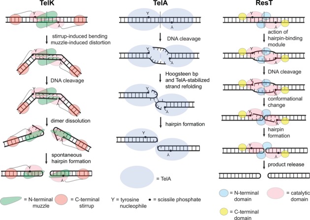
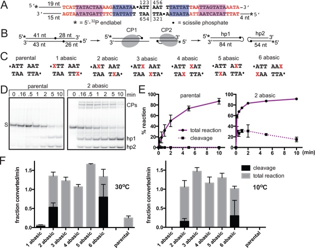
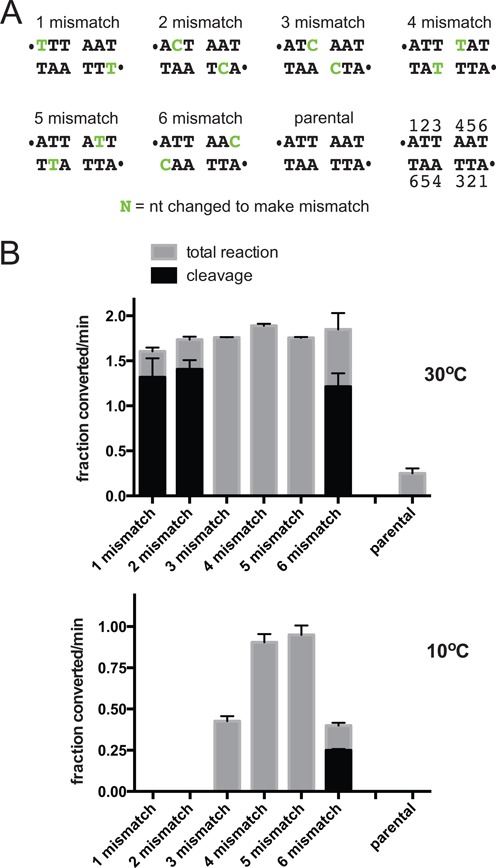
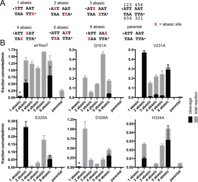
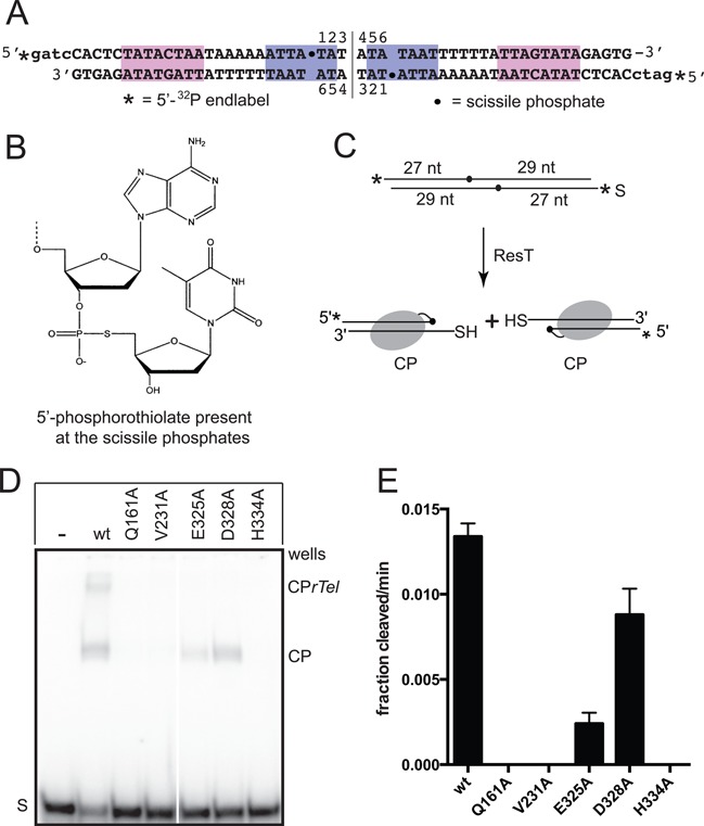
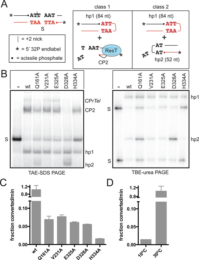
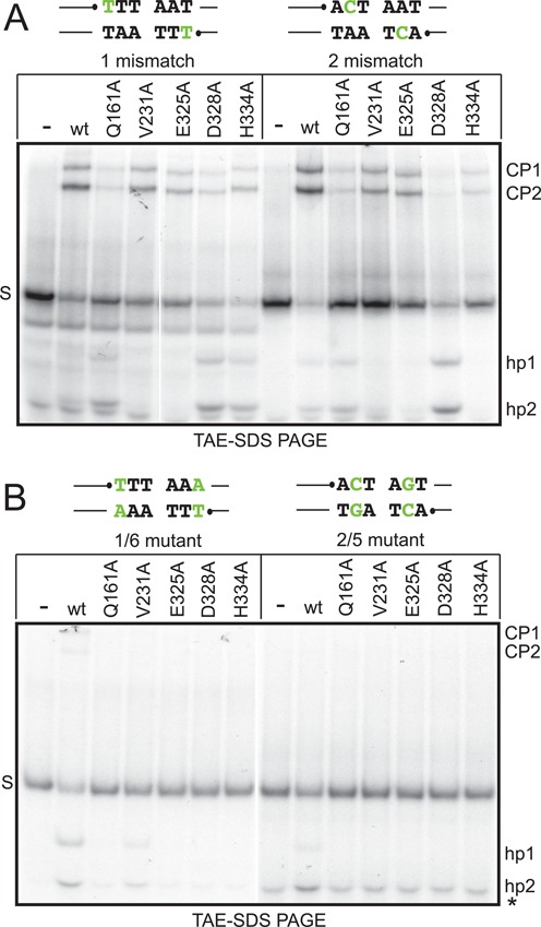
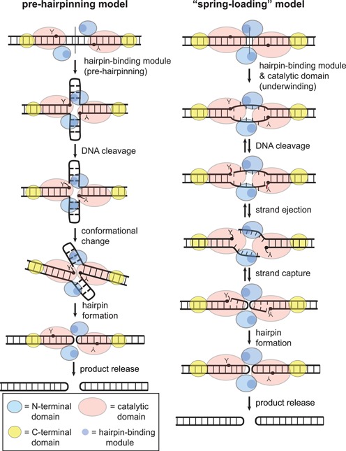
The Borrelia telomere resolvase, ResT, forms the unusual hairpin telomeres of the linear Borrelia replicons in a process referred to as telomere resolution. Telomere resolution is a DNA cleavage and rejoining reaction that proceeds from a replicated telomere intermediate in a reaction with mechanistic similarities to that catalyzed by type IB topoisomerases. Previous reports have implicated the hairpin-binding module, at the end of the N-terminal domain of ResT, in distorting the DNA between the scissile phosphates so as to promote DNA cleavage and hairpin formation by the catalytic domain. We report that unwinding the DNA between the scissile phosphates, prior to DNA cleavage, is a key cold-sensitive step in telomere resolution. Through the analysis of ResT mutants, rescued by substrate modifications that mimic DNA unwinding between the cleavage sites, we show that formation and/or stabilization of an underwound pre-cleavage intermediate depends upon cooperation of the hairpin-binding module and catalytic domain. The phenotype of the mutants argues that the pre-cleavage intermediate promotes strand ejection to favor the forward reaction and that subsequent hairpin capture is a reversible reaction step. These reaction features are proposed to promote hairpin formation over strand resealing while allowing reversal back to substrate of aborted reactions.


Figures

Figure 1.
Figure 1.
Models of telomere resolution derived from different systems. Schemata of the key steps in the reaction for TelK, TelA and ResT are shown. TelK: binding to the substrate DNA induces a large bend in the DNA that features a displacement of the helical path at the dimer interface, severely buckling the basepairs between the scissile phosphates. This proposed pre-cleavage intermediate ‘spring loads’, or stores the energy needed to drive, upon subsequent DNA cleavage, dimer dissolution and spontaneous hairpin formation. The energy stored in the pre-cleavage intermediate, and stable binding of TelK to the final hairpin telomere products, are factors hypothesized to drive the reaction forward. Support for this model is derived from the phenotype of a deletion mutant of the stirrup domain and from the observed non-turnover of the enzyme. Deletion of the stirrup domain, which stabilizes the DNA bend by interacting with the substrate DNA at the outer extremes of the rTel, left TelK competent to reversibly cleave the substrate but not to form the hairpins (3,17). TelA: post-cleavage stabilization of strand refolding, via TelA–DNA contacts and Hoogsteen basepairing across the dimer axis, and stable binding of TelA to the final hairpin telomere products are factors hypothesized to drive the reaction forward. The whole reaction occurs in the context of a dimer of TelA. Support for this model comes from the dimeric TelA-hp product complex, the behavior of TelA mutants in sidechains implicated in stabilizing the refolding strands and from the sensitivity of telomere resolution to rTel sequence changes that would interfere with the stabilizing Hoogsteen base pair formation (16). ResT: initial substrate binding and dimerization engages the hairpin-binding module at the end of the N-terminal domain of ResT to distort the DNA between the scissile phosphates allowing DNA cleavage by the catalytic domain. This distortion has been variously visualized as pre-formation of the hairpins (23) or more simply as DNA unwinding (15). An uncharacterized strand refolding ensues that captures the hairpin conformation after strand sealing; this occurs in the context of an intact dimer of ResT (15). Hairpin telomere products are released after both hp telomeres are formed (15). Support for this proposed model is derived from the behavior of hairpin-binding module mutants and from studies of product release in reactions with bead-immobilized rTels (15,23).
Figure 2.
Figure 2.
Missing base modifications between the scissile phosphates of the substrate rTel alleviate the cold-sensitivity of telomere resolution. (A) The model Type 2 rTel used in this study. To aid the annealing of the oligonucleotides into an rTel, the normal dyad symmetry of native rTels was broken by having distinct sequences on the left and right sides in positions where the sequence of different Type 2 rTels vary (red letters). The right hand halfsite sequence is derived from the left telomeres of lp28-2 and lp36. The left hand halfsite sequence is derived from the right telomere of lp28-4 (25). The magenta and blue/shaded boxes highlight regions of conserved sequence. To allow differentiation of products from the left versus right side of the rTel 15 bp of nontelomeric sequence was added to the left side. The substrate nucleotide numbering noted for the unmodified parental substrate follows the convention reported in the TelA–DNA and TelK–DNA structures. The vertical line between nucleotides 3 and 4 denotes the dyad symmetry axis of the rTel. (B) Schematic representation of a telomere resolution reaction with 5′-32P endlabeled substrate. DNA chain lengths, from the substrate ends to the scissile phosphates, are indicated in the substrate; chain lengths in the hairpin telomere products are shown. Cleavage products (CP), in which ResT is covalently linked to the cleaved DNA via a phosphotyrosyl bond, can be visualized in reactions where hairpin telomere formation fails or is inefficient. (C) Sequence between the scissile phosphates of the parental substrate and the range of tested rTels with missing base (abasic) sites in symmetrically located positions between the scissile phosphates. The abasic modifications are represented in the sequence by the red/shaded X's and the scissile phosphates by dots. (D) Representative 8% PAGE 1× TAE/0.1% SDS gels of timecourse reactions with ResT, parental rTeland the 2 abasic rTel detailed in (C). S, represents the mobility in the gel of the substrate; CPs, mark the gel migration position of the cleavage products (when detectable); hp1 & hp2, mark the gel position of the hairpin telomere products detailed in (B). (E) The left graph shows % reaction (DNA cleavage and total reaction) versus reaction time is plotted for a telomere resolution reaction with wild type ResT and parental rTel incubated at 30°C (left graph). In the reaction shown the cleavage intermediates do not accumulate and are not detected on the gel so total reaction is equivalent to the rate of hairpin telomere formation. When cleavage intermediates do accumulate the rate of their formation was determined separately and the total reaction becomes the summed rate of DNA cleavage plus hp telomere formation. The right graph shows % total reaction versus reaction time of a telomere resolution reaction with wild type ResT and the 2 abasic rTel incubated at 30°C. Shown are the mean and standard deviation of at least 3 independent replicates. Initial rates shown in (F) were determined from individual % reaction versus time plots by determination of the slope of the initial linear portion of the curves; means and standard deviations for the initial rates were determined from at least three independent timecourses. (F) Comparison of the initial rates of DNA cleavage and total reaction (DNA cleavage + hp telomere formation) of the parental rTel and the abasic modifications shown in A and C) in reactions incubated at the standard reaction temperature of 30°C compared to incubations at 10°C. The initial rates are expressed as the fraction substrate converted/min (1.0 being 100% conversion). Shown is the mean and standard deviation of at least three independent replicates. The asterisk above the one abasic data indicates that the slow observed DNA cleavage was aberrant producing, predominantly, substrate cleaved on only one strand.
Figure 3.
Figure 3.
The effect of DNA mismatches, between the scissile phosphates of the substrate, on the cold-sensitivity of telomere resolution. (A) Sequence between the scissile phosphates of the parental substrate and the range of tested rTels with mismatches in symmetrically located positions between the scissile phosphates. The parental rTel and substrate nucleotide numbering is shown. The sequence changes that introduce the mismatches are represented in the sequence by the green/shaded letters and the scissile phosphates by dots. (B) Comparison of the initial rates of DNA cleavage and total reaction (DNA cleavage + hp telomere formation) of the parental rTel and the substrates with mismatches shown in (A) in reactions incubated at the standard reaction temperature of 30°C versus 10°C. The initial rates are expressed as the fraction substrate converted/min (1.0 being 100% conversion). Shown is the mean and standard deviation of at least three independent replicates.
Figure 4.
Figure 4.
The pattern of activity of ResT mutants defective for telomere resolution assayed with substrates with missing base modifications. (A) Sequence between the scissile phosphates of the parental substrate and the range of tested substrate rTels with missing base (abasic) sites positioned in symmetric locations on the top and bottom strands (details as in Figure 2C). (B) Comparison of the initial rates of DNA cleavage and total reaction (DNA cleavage + hp telomere formation) of wild-type ResT and the indicated mutants assayed on the parental Type 2 rTel and the array of abasic rTels detailed in (A). The x-axis presents the substrates assayed and the y-axis the initial rates of DNA cleavage (black bars) and the total reaction rate (DNA cleavage + hp formation, gray bars). The initial rates are expressed as the fraction substrate converted/min (1.0 being 100% conversion). Normally, cleavage and hp formation are concerted, so no cleavage intermediates accumulate; in these cases the total reaction rate is just the rate of hp formation. In some cases, the missing base modifications allowed DNA cleavage with only slow, subsequent, hp telomere formation; this leads to the accumulation of cleavage intermediates (see Figure 2B schematic). Representative data and details of the initial rate determinations are shown in Supplementary Figure S4. The asterisk above the 1 abasic data (wt ResT and D328A reactions) indicates that the observed slow DNA cleavage was aberrant producing, predominantly, substrate cleaved on only one strand. All initial rates were determined from a minimum of three independent replicates, and the error bars represent the standard deviation. Note that the scaling of the y-axes are not necessarily the same from one ResT variant to another.
Figure 5.
Figure 5.
Assessing the cleavage competence of the mutants. (A) The OPS rTel used. The sequence of the rTel is derived from the left telomere of lp17. The magenta and blue/shaded boxes highlight regions of conserved sequence. The OPS rTel is dyad symmetric. This necessitates construction by annealing halfsites followed by ligation and gel purification (see Supplemental Material and Methods, Supplementary Table S1 and (23)). The substrate nucleotide numbering is noted. The vertical line between nucleotides 3 and 4 denotes the dyad symmetry axis of the rTel. The 5′-32P endlabels are indicated by asterisks, and the scissile phosphate are indicated by dots. (B) Schematic representation of the basestep surrounding the scissile phosphates in a suicide rTel harboring 5′-phosphorothiolate moieties (OPS). (C) Schematic of a replicated telomere cleavage assay performed with 5′-32P endlabeled OPS rTel. DNA chain lengths, from the substrate ends to the scissile phosphates, are indicated in the substrate. Cleavage products, in which ResT is covalently linked to the cleaved DNA, accumulate due to the inability of the resulting 5′-sulfhydryl groups to participate in strand joining reactions to generate hp telomeres or to regenerate substrate. S, represents the OPS rTel; CP, represents the cleavage products. (D) 8% PAGE 1X TAE/0.1% SDS gel analysis of reactions assayed with wild-type ResT and the telomere resolution deficient ResT mutants using an OPS-modified suicide rTel. Reactions containing 75 nM ResT, and 5.25 nM 5′-32P endlabeled OPS rTel were incubated at 30°C for 30 min. The ResT variant added is noted in the gel-loading key above the gel. S, represents the mobility in the gel of the OPS rTel; CP, represents the gel migration position of the cleavage products detailed in A); CPrTel, marks the gel migration position of a cleavage product in which only one strand has been cleaved. Note that the efficiency of OPS rTel utilization by ResT is less efficient than that of the unmodified ‘parental’ rTel used elsewhere in this study. (E) Comparison of the cleavage rate of the mutants compared to the cleavage rate of wild-type ResT. The initial rates are expressed as the fraction substrate cleaved/min (1.0 being 100% conversion). All rates are determined from a minimum of three independent replicates; the error bars represent the standard deviation.
Figure 6.
Figure 6.
The pattern of activity of ResT mutants, defective for telomere resolution, assayed with a substrate nicked near the substrate's symmetry axis. (A) Schematic of the +2 nick rTel, sequence as depicted in Figure 2A. The two classes of reaction outcome observed are summarized schematically. In class 1, most of the substrate is cleaved normally, hp1 forms but hp2 formation is blocked or slow, resulting in the accumulation of cleavage product (CP2) in which ResT is covalently attached to the cleaved half site. In class 2, the nicked rTel is cleaved normally and both hairpin telomeres (hp1 and hp2) form. hp2, when it can form, does so despite being 2 nt shorter than normal, due to the diffusion away, after cleavage, of the dinucleotide 3′ to the top strand scissile phosphate and despite the fact that the resulting 4 nt strand lacks self-complementarity. (B) Summary 8% TAE–SDS and TBE–urea PAGE gels are shown of reactions of wild type and mutant ResT variants with the +2 nick Type 2 rTel. The reactions shown were incubated at 30°C for 30 min. Only free DNA strands enter the TBE–urea PAGE gels; the cleavage products with ResT covalently linked to the 5′-32P endlabeled strands do not enter these gels. S, represents the mobility in the gel of the substrate; CP2, marks the gel migration position of the cleavage product from the right side of the substrate; CPrTel, marks the gel migration position of a cleavage product in which only the top strand has been cleaved; hp1 and hp2, mark the gel position of the hairpin telomere products. (C) Comparison of the initial rates of total substrate usage of wild type and mutant ResT variants tested in reactions with nicked Type 2 rTel incubated at 30°C. All rates are determined from a minimum of three independent replicates; the error bars represent the standard deviation. (D) Comparison of the initial rate of total substrate usage of wild type ResT tested in reactions incubated at 10°C versus 30°C. No reaction was detectable for the mutants assayed at 10°C.
Figure 7.
Figure 7.
Assessing the fidelity of hairpin telomere formation with substrates with mismatches that inhibit hp formation. (A) 8% PAGE 1× TAE/0.1% SDS gel analysis of telomere resolution reactions incubated at 30°C for 10 min using Type 2 rTels with 1 mismatch and 2 mismatch modifications. The sequence of the modified rTels is shown above the gel with the nucleotide changed to create the mismatches represented in green/shaded script. The ResT variants assayed are indicated in the loading key above the gel. Gel labels are as described for Figure 6 except CP1 represents the cleavage product derived from the left side of the substrate. See Figure 2A and B for substrate and reaction details. (B) 8% PAGE 1× TAE/0.1% SDS gel analysis of telomere resolution reactions incubated at 30°C for 10 min using Type 2 rTels with 1/6 AT to TA and 2/5 TA to CG mutations that return full basepairing to the substrates but maintain the sequence change present in the mismatched substrates used in (A). The sequence of the mutant rTels is shown above the gel with the nucleotides changed to create the mutations represented in green/shaded script. Gel labels are as described previously except *, indicates unannealed oligo present in unreacted substrate.
Figure 8.
Figure 8.
Comparison of pre-hairpinning and spring-loading models of ResT-mediated telomere resolution. The pre-hairpinning model, initially reported, proposed a distortion of the substrate DNA between the scissile phosphates that preforms hairpins prior to DNA cleavage (23). Similarity of the last ∼20 amino acids of the N-terminal domain to IS4 family transposases that excise transposons via a DNA hairpin intermediate, prompted this area of the ResT to be called the hairpin-binding module. In this model the hairpin-binding module creates the pre-hairpinned structure and the catalytic domain is restricted to DNA cleavage and rejoining activities. Most mutants in the hairpin-binding module display a cold-sensitivity that is rescued by heteroduplexing the central two base pairs of the rTel. Proposition of the pre-hairpinning model was motivated by the finding that hairpin-binding module mutants were cleavage defective rather than just defective for hp telomere formation (23). This model does not predict reversibility of the reaction after DNA cleavage. The spring-loading model, based on this report, similarly hypothesizes a distortion of the substrate DNA between the scissile phosphates that promotes DNA cleavage. Here the proposed distortion is an underwinding of the DNA. Breaking the 1/6 and 2/5 base pairs promotes subsequent strand ejection once the DNA is cleaved. The hypothesized underwound or ‘spring loaded’ pre-cleavage intermediate is formed by cooperation of the hairpin-binding module and the catalytic domain. The underwound conformation promotes strand ejection and the forward direction of the reaction. Our discovery of hp formation fidelity mutants suggests the presence of a reversible hp capture step in the reaction. The Q161, D328 and H334 residues are used to read out hp capture.

Similar articles

Cited by

References

    1. Ravin N.V., Strakhova T.S., Kuprianov V.V. The protelomerase of the phage-plasmid N15 is responsible for its maintenance in linear form. J. Mol. Biol. 2001;312:899–906. - PubMed
    1. Hertwig S., Klein I., Lurz R., Lanka E., Appel B. PY54, a linear plasmid prophage of Yersinia enterocolitica with covalently closed ends. Mol. Microbiol. 2003;48:989–1003. - PubMed
    1. Huang W.M., Joss L., Hsieh T., Casjens S. Protelomerase uses a topoisomerase IB/Y-recombinase type mechanism to generate DNA hairpin ends. J. Mol. Biol. 2004;337:77–92. - PubMed
    1. Huang W.M., DaGloria J., Fox H., Ruan Q., Tillou J., Shi K., Aihara H., Aron J., Casjens S. Linear chromosome-generating system of Agrobacterium tumefaciens C58: protelomerase generates and protects hairpin ends. J Biol Chem. 2012;287:25551–25563. - PMC - PubMed
    1. Picardeau M., Lobry J.R., Hinnebusch B.J. Physical mapping of an origin of bidirectional replication at the centre of the Borrelia burgdorferi linear chromosome. Mol. Microbiol. 1999;32:437–445. - PubMed

Publication types

MeSH terms

LinkOut - more resources