. 2015 Sep 30;43(17):e112.
doi: 10.1093/nar/gkv550. Epub 2015 May 24.

A modular open platform for systematic functional studies under physiological conditions

Affiliations

A modular open platform for systematic functional studies under physiological conditions

Christopher B Mulholland et al. Nucleic Acids Res. .

Abstract

Any profound comprehension of gene function requires detailed information about the subcellular localization, molecular interactions and spatio-temporal dynamics of gene products. We developed a multifunctional integrase (MIN) tag for rapid and versatile genome engineering that serves not only as a genetic entry site for the Bxb1 integrase but also as a novel epitope tag for standardized detection and precipitation. For the systematic study of epigenetic factors, including Dnmt1, Dnmt3a, Dnmt3b, Tet1, Tet2, Tet3 and Uhrf1, we generated MIN-tagged embryonic stem cell lines and created a toolbox of prefabricated modules that can be integrated via Bxb1-mediated recombination. We used these functional modules to study protein interactions and their spatio-temporal dynamics as well as gene expression and specific mutations during cellular differentiation and in response to external stimuli. Our genome engineering strategy provides a versatile open platform for efficient generation of multiple isogenic cell lines to study gene function under physiological conditions.


Figures

Figure 1.
Figure 1.
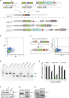
Generation of MIN-tagged cell lines. (A) Schematic overview of MIN-tag insertion into the Dnmt1 locus via CRISPR/Cas assisted gene editing. The MIN-tag donor harbors the attP site and homology to the genomic sequence 5′ and 3′ of the start codon. Integration is facilitated by double strand breaks created by Cas9 directed to the target sequence by a specific gRNA. Restriction enzyme recognition sites used for screening in this study are indicated above the attP sequence. (B) Schematic overview of the surrogate reporter used to enrich for cells expressing a functional Cas9 complex. The respective Cas9 target sequence (tSeq) is placed downstream of mRFP followed by a stop codon and an out-of-frame GFP ORF. This surrogate reporter is transfected into the cells together with a vector expressing Cas9 and a U6 driven gRNA expression cassette. (C) Cells that express a functional Cas9 complex can then be identified by expression of GFP and enriched via FACS. (D) Screening PCRs followed by restriction digest with HincII or SacII of all generated MIN-tagged cell lines. (N) and (C) refer to N- and C-terminal tagging, respectively.
Figure 2.
Figure 2.
Application of the anti-MIN monoclonal antibody. (A) DNA sequence of the attP site and corresponding translated MIN peptide sequence (orange). (B) Fluorescence micrographs of wt mESCs, Dnmt1attp/attp cells and of a mixed culture (1:10) of wt and Dnmt1attP/attP cells stained with the anti-MIN antibody. DAPI is used as DNA counterstain. Scale bars represent 5 μm. (C) IP experiments performed with anti-MIN and anti-DNMT1 antibody in Dnmt1attP/attP cell extracts (input (I), flow through (FT), bound (B)). (D) Co-IP of DNMT3B in wt and Dnmt3battP/attP cells using the anti-MIN antibody. DNMT3B co-precipitated SNF2H in Dnmt3battP/attP cells as determined by western blot.
Figure 3.
Figure 3.
Bxb1-mediated insertion of functional cassettes into the Dnmt1 locus. (A) Schematic outline of the strategy and vectors used to create knockout, GFP knockin and cDNA knockin functionalizations of the Dnmt1attP/attP cell line. cDNAs can be cloned into the attB-GFP-Stop-Poly(A) vector using the 8-cutters AsiSI and NotI. (B) FACS plot depicting the gating and sorting of mESCs to enrich for cells positive for integration of the knockout cassette (2.05% of parent population) based on GFP expression. (C) The Bxb1 surrogate reporter consists of a constitutive CMV promoter followed by an attP site. If Bxb1 and attB donor plasmid containing GFP is present in the cell, recombination of the donor into the reporter leads to expression of GFP. The Bxb1 surrogate reporter can be used to enrich for successful recombination events by FACS. (D) Gel electrophoresis of the multiplex PCR for wt, Dnmt1attP/attP (attP/attP), Dnmt1KO/KO (KO/KO), Dnmt1cDNA/cDNA (cDNA/cDNA) and Dnmt1GFP/GFP (GFP/GFP) as well as 1:1 mixtures with Dnmt1attP/attP genomic DNA, to control for amplification biases. Blue arrows indicate expected sizes of the non-recombined (attP) and recombined allele (attL). (E) DNA methylation levels at the major satellite repeats of Dnmt1KO/KO cells compared to wt and Dnmt1attP/attP cells. (F) Western blot analysis of DNMT1 expression levels in wt, Dnmt1attP/attP and Dnmt1KO/KO cells generated by Bxb1-mediated insertion of a knockout cassette. (G) Western blot analysis of DNMT1 and GFP expression in Dnmt1attP/attP and homozygous GFP-knockin cells (Dnmt1GFP/GFP) generated by Bxb1-mediated insertion. (H) Western blot analysis of DNMT1 and GFP expression in Dnmt1attP/attP and Dnmt1cDNA/cDNA cells expressing a GFP-Dnmt1 minigene from the endogenous promoter.
Figure 4.
Figure 4.
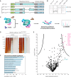
Study of TET1 regulation. (A) Schematic representation of the Tet1 cDNA constructs used for Bxb1-mediated recombination into Tet1attP/attP cells. (B) Western blot analysis of TET1 expression in Tet1attP/attP cell line and its derivatives expressing GFP-TET1Δ1–1363 (Δ1–1363), GFP-TET1Δ833–1053 (1Δ833–1053) and GFP-TET1Δ833–1363 (Δ833–1363). Note that fusion to GFP increases the MW of TET1 constructs by 29 kDa. (C) Schematic representation of the BioID approach as described by Roux etal. (27). (D) SDS-PAGE analysis of a BioID pulldown experiment using the Tet1BirA*/BirA* cell line. Cells were cultured either without (control) or with 50 μM biotin (+biotin). C: Cytoplasm, I: Crude nuclei input, FT: Flowthrough, B: Bound, W1-W3: Wash. (E) Volcano plot of proteins identified in the streptavidin pulldown of the TET1-BioID experiment, quantified with the MaxQuant Label-Free-Quantification algorithm (32). The x-axis reflects the difference in protein abundance in the BioID pull-down compared to the negative control while the y-axis shows the logarithmized P-value of a student's t-test. Significantly enriched proteins are highlighted in pink (FDR = 0.01, S0 = 3, indicated by black line (40)). Experiments were performed in duplicates. (F) GO term enrichment of proteins identified as significant in BioID.
Figure 5.
Figure 5.
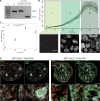
Spatio-temporal dynamics of DNMT3B during epiblast differentiation. (A) Gel electrophoresis of the multiplex screening PCR for wt, Dnmt3battP/attP and Dnmt3bGFP/GFP. Blue arrows indicate expected sizes of the non-recombined (attP) and recombined allele (attL). (B) Evaluation of GFP signals during live cell imaging of Dnmt3bGFP/GFP cells. The graph depicts mean gray values of nuclear GFP signals. Error bars represent standard deviations (n > 81). Lower panels show Z-projections of Dnmt3bGFP/GFP cells representative of the indicated time frame. Scale bar represents 10 μm. (C) Quantitative real-time PCR of Dnmt3b mRNA levels in wt and Dnmt3battP/attP cells during epiblast differentiation. (D) 3D-SIM nuclear mid-sections of anti-MIN (green) and anti-H3K4me3 (red) antibody distributions 30 and 60 h after induction of EpiLC differentiation combined with DAPI counterstaining (gray) in Dnmt3battP/attP cells. Lower panels represent 7× magnifications of selected boxed regions. Scale bars represent 3 μm and 500 nm in insets.
Figure 6.
Figure 6.
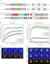
Protein dynamics of DNMT3B and its isoforms during epiblast differentiation. (A) Schematic representation of the Dnmt3b genomic loci in the Dnmt3bGFP/GFP and the Dnmt3bmCh-3b1/GFP-3b6 cell lines. (B) Quantitative evaluation of FRAP experiments (average of 11–14 cells) comparing GFP-DNMT3B with GFP-DNMT3B6 and mCh-DNMT3B1 in Dnmt3bGFP/GFP and the Dnmt3bmCh-3b1/GFP-3b6 cell lines differentiated for 35 h. Error bars represent standard error of the mean. (C) Quantitative evaluation of FRAP experiments (average of 10–12 cells) as in (B) with cells treated with 5-azadC 12 h before imaging. (D and E) Representative images of FRAP experiments performed in (B) and (C), respectively. White circles indicate the bleach ROI with a diameter of 2 μm.
Figure 7.
Figure 7.
The MIN-tag strategy. (A) Schematic outline of the genome engineering strategy. Small homology donors are used to insert serine integrase (attP) sites in-frame after the ATG codon of target genes via CRISPR/Cas assisted HR. The attP site is translated as a novel epitope tag suitable for IF and IP with the specific monoclonal antibody. The attP site is also recognized by the serine integrase Bxb1 and used for specific and directional integration of attB-carrying functional cassettes into the tagged gene locus. All derivatives are subjected to their endogenous gene regulation ensuring that subsequent studies are performed at physiological expression levels. (B) Timeline for generation of MIN-tagged genes and subsequent modification by Bxb1-mediated recombination. MIN-tagged cell lines can be generated within 2–3 weeks. These cell lines can then be modified within another 2–3 weeks to generate multiple isogenic cell lines with different functional modifications.

Similar articles

Cited by

References

    1. Cong L., Ran F.A., Cox D., Lin S., Barretto R., Habib N., Hsu P.D., Wu X., Jiang W., Marraffini L.A., et al. Multiplex genome engineering using CRISPR/Cas systems. Science. 2013;339:819–823. - PMC - PubMed
    1. Haurwitz R.E., Jinek M., Wiedenheft B., Zhou K., Doudna J.A. Sequence- and structure-specific RNA processing by a CRISPR endonuclease. Science. 2010;329:1355–1358. - PMC - PubMed
    1. Mali P., Yang L., Esvelt K.M., Aach J., Guell M., DiCarlo J.E., Norville J.E., Church G.M. RNA-guided human genome engineering via Cas9. Science. 2013;339:823–826. - PMC - PubMed
    1. Sampson T.R., Saroj S.D., Llewellyn A.C., Tzeng Y.-L., Weiss D.S. A CRISPR/Cas system mediates bacterial innate immune evasion and virulence. Nature. 2013;497:254–257. - PMC - PubMed
    1. Kuscu C., Arslan S., Singh R., Thorpe J., Adli M. Genome-wide analysis reveals characteristics of off-target sites bound by the Cas9 endonuclease. Nat. Biotechnol. 2014;32:677–683. - PubMed

Publication types