

Fibrosis in the lens. Sprouty regulation of TGFβ-signaling prevents lens EMT leading to cataract
- PMID: 26003864
- PMCID: PMC4654713
- DOI: 10.1016/j.exer.2015.02.004
Fibrosis in the lens. Sprouty regulation of TGFβ-signaling prevents lens EMT leading to cataract
Abstract
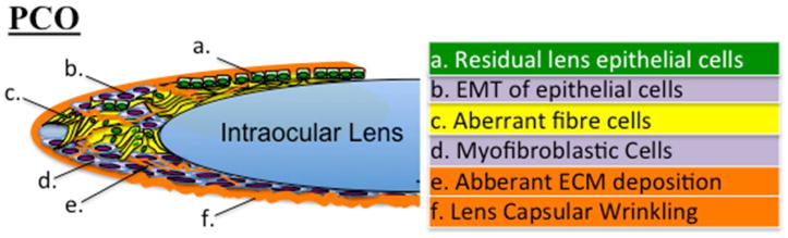
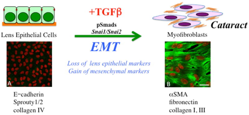
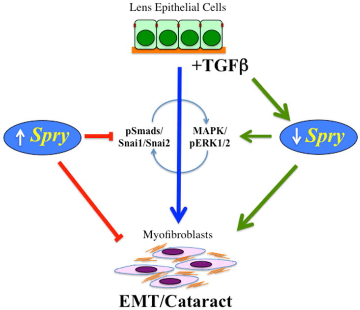
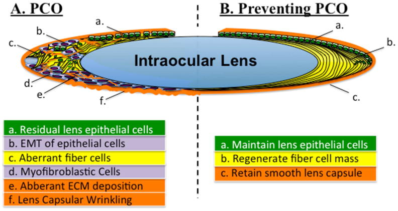
Cataract is a common age-related condition that is caused by progressive clouding of the normally clear lens. Cataract can be effectively treated by surgery; however, like any surgery, there can be complications and the development of a secondary cataract, known as posterior capsule opacification (PCO), is the most common. PCO is caused by aberrant growth of lens epithelial cells that are left behind in the capsular bag after surgical removal of the fiber mass. An epithelial-to-mesenchymal transition (EMT) is central to fibrotic PCO and forms of fibrotic cataract, including anterior/posterior polar cataracts. Transforming growth factor β (TGFβ) has been shown to induce lens EMT and consequently research has focused on identifying ways of blocking its action. Intriguingly, recent studies in animal models have shown that EMT and cataract developed when a class of negative-feedback regulators, Sprouty (Spry)1 and Spry2, were conditionally deleted from the lens. Members of the Spry family act as general antagonists of the receptor tyrosine kinase (RTK)-mediated MAPK signaling pathway that is involved in many physiological and developmental processes. As the ERK/MAPK signaling pathway is a well established target of Spry proteins, and overexpression of Spry can block aberrant TGFβ-Smad signaling responsible for EMT and anterior subcapsular cataract, this indicates a role for the ERK/MAPK pathway in TGFβ-induced EMT. Given this and other supporting evidence, a case is made for focusing on RTK antagonists, such as Spry, for cataract prevention. In addition, and looking to the future, this review also looks at possibilities for supplanting EMT with normal fiber differentiation and thereby promoting lens regenerative processes after cataract surgery. Whilst it is now known that the epithelial to fiber differentiation process is driven by FGF, little is known about factors that coordinate the precise assembly of fibers into a functional lens. However, recent research provides key insights into an FGF-activated mechanism intrinsic to the lens that involves interactions between the Wnt-Frizzled and Jagged/Notch signaling pathways. This reciprocal epithelial-fiber cell interaction appears to be critical for the assembly and maintenance of the highly ordered three-dimensional architecture that is central to lens function. This information is fundamental to defining the specific conditions and stimuli needed to recapitulate developmental programs and promote regeneration of lens structure and function after cataract surgery.
Keywords: EMT; Fibrosis; Lens epithelium; Lens regeneration; Myofibroblasts; RTK antagonists; Sprouty; TGFβ.
Crown Copyright © 2015. Published by Elsevier Ltd. All rights reserved.
Figures
Similar articles
-
Negative regulation of TGFβ-induced lens epithelial to mesenchymal transition (EMT) by RTK antagonists.Exp Eye Res. 2015 Mar;132:9-16. doi: 10.1016/j.exer.2015.01.001. Epub 2015 Jan 7. Exp Eye Res. 2015. PMID: 25576668
-
Sprouty and Spred temporally regulate ERK1/2-signaling to suppress TGFβ-induced lens EMT.Exp Eye Res. 2022 Jun;219:109070. doi: 10.1016/j.exer.2022.109070. Epub 2022 Apr 9. Exp Eye Res. 2022. PMID: 35413282
-
Transforming growth factor-beta-induced epithelial-mesenchymal transition in the lens: a model for cataract formation.Cells Tissues Organs. 2005;179(1-2):43-55. doi: 10.1159/000084508. Cells Tissues Organs. 2005. PMID: 15942192 Review.
-
Sprouty is a negative regulator of transforming growth factor β-induced epithelial-to-mesenchymal transition and cataract.Mol Med. 2012 Jul 18;18(1):861-73. doi: 10.2119/molmed.2012.00111. Mol Med. 2012. PMID: 22517312 Free PMC article.
-
Roles of TGF β and FGF Signals in the Lens: Tropomyosin Regulation for Posterior Capsule Opacity.Int J Mol Sci. 2018 Oct 9;19(10):3093. doi: 10.3390/ijms19103093. Int J Mol Sci. 2018. PMID: 30304871 Free PMC article. Review.
Cited by
-
TGF-β/Smad Signalling Activation by HTRA1 Regulates the Function of Human Lens Epithelial Cells and Its Mechanism in Posterior Subcapsular Congenital Cataract.Int J Mol Sci. 2022 Nov 20;23(22):14431. doi: 10.3390/ijms232214431. Int J Mol Sci. 2022. PMID: 36430917 Free PMC article.
-
Intrinsic and extrinsic regulatory mechanisms are required to form and maintain a lens of the correct size and shape.Exp Eye Res. 2017 Mar;156:34-40. doi: 10.1016/j.exer.2016.04.009. Epub 2016 Apr 21. Exp Eye Res. 2017. PMID: 27109030 Free PMC article. Review.
-
The myofibroblast, biological activities and roles in eye repair and fibrosis. A focus on healing mechanisms in avascular cornea.Eye (Lond). 2020 Feb;34(2):232-240. doi: 10.1038/s41433-019-0684-8. Epub 2019 Nov 25. Eye (Lond). 2020. PMID: 31767967 Free PMC article.
-
GDF-15 Attenuates the Epithelium-Mesenchymal Transition and Alleviates TGFβ2-Induced Lens Opacity.Transl Vis Sci Technol. 2024 Jul 1;13(7):2. doi: 10.1167/tvst.13.7.2. Transl Vis Sci Technol. 2024. PMID: 38949633 Free PMC article.
-
Endoplasmic reticulum stress regulates epithelial‑mesenchymal transition in human lens epithelial cells.Mol Med Rep. 2020 Jan;21(1):173-180. doi: 10.3892/mmr.2019.10814. Epub 2019 Nov 12. Mol Med Rep. 2020. PMID: 31746423 Free PMC article.
References
-
- Assinder S, Beniamen D, Lovicu FJ. Co-suppression of Sprouty (SPRY) and Sprouty-related (SPRED) negative regulators of FGF signalling in prostate cancer: A working hypothesis. In “Signal Transduction Inhibitors as Promising Anticancer Agents”. Biomedical Research International. 2014 in press, Nov, 2014.
-
- Awasthi N, Guo S, Wagner BJ. Posterior capsular opacification: a problem reduced but not yet eradicated. Archives of Ophthalmology. 2009;127:555–62. - PubMed
-
- Baker JC, Harland RM. From receptor to nucleus: the Smad pathway. Current Opinion in Genetics & Development. 1997;7:467–73. - PubMed
-
- Bakin AV, Rinehart C, Tomlinson AK, Arteaga CL. p38 mitogen-activated protein kinase is required for TGFbeta-mediated fibroblastic transdifferentiation and cell migration. J Cell Sci. 2002;115:3193–206. - PubMed
-
- Beiran I, Scharf J, Tamir A, Miller B. Influence of systemic diseases and environmental factors on age at appearance, location and type of acquired cataract. Metabolic, Pediatric & Systemic Ophthalmology. 1994;17:34–7. - PubMed
Publication types
MeSH terms
Substances
Grants and funding
LinkOut - more resources
Full Text Sources
Other Literature Sources
Research Materials
Miscellaneous
