Review
. 2015 Jun;16(6):329-44.
doi: 10.1038/nrm3999. Epub 2015 May 20.

Live to die another way: modes of programmed cell death and the signals emanating from dying cells

Affiliations
Review

Live to die another way: modes of programmed cell death and the signals emanating from dying cells

Yaron Fuchs et al. Nat Rev Mol Cell Biol. 2015 Jun.

Abstract

All life ends in death, but perhaps one of life's grander ironies is that it also depends on death. Cell-intrinsic suicide pathways, termed programmed cell death (PCD), are crucial for animal development, tissue homeostasis and pathogenesis. Originally, PCD was almost synonymous with apoptosis; recently, however, alternative mechanisms of PCD have been reported. Here, we provide an overview of several distinct PCD mechanisms, namely apoptosis, autophagy and necroptosis. In addition, we discuss the complex signals that emanate from dying cells, which can either trigger regeneration or instruct additional killing. Further advances in understanding the physiological roles of the various mechanisms of cell death and their associated signals will be important to selectively manipulate PCD for therapeutic purposes.


Conflict of interest statement

Competing interests statement

The authors declare no competing interests.

Figures

Figure 1
Figure 1. The core of the apoptotic machinery is conserved
Functional homologous of apoptotic proteins in C. elegans, Drosophila melanogaster and mammals uncover evolutionary conservation of the apoptotic pathway. A. In C. elegans, EGL-1 (BH3-only protein homolog) binds and inhibits CED-9 (BCL-2-family homolog), resulting in the release of CED-4 (APAF1 homolog) from the CED-9–CED-4 complex. This enables the elimination of cells by CED-3 (caspase). B. In Drosophila, the IAP antagonists Reaper, Hid and Grim (RHG) mediate the degradation of DIAP1, thus liberating Drice and Dcp-1. In addition, this enables Dronc (Caspase-9 homolog) to interact with dArk and form the apoptosome that efficiently activates executioner caspases. The p35 protein is a specific inhibitor of executioner caspases and the activation of the apoptosome might be regulated by the Bcl-2 family members Debcl and Buffy (hence depicted by a dashed line). C. In mammals, the crucial decision as to whether a cell commits to apoptosis is regulated by the fine interplay between the anti-apoptotic BCl-2 subfamily of proteins and the pro-apoptotic BH3-only subfamily of proteins. During apoptosis, BH3-only proteins facilitate a BAX- and BAK-dependent release of cytochrome c from the mitochondria, which binds to APAF1 and gives rise to the apoptosome. In parallel, IAP antagonists including DIABLO, HTRA2 and ARTS translocate from mitochondria and release caspases from their negative regulation by IAPs. In particular, Caspase-9 is liberated from XIAP and activated by the apoptosome, triggering executioner caspases-3 and -7. Homologous proteins are similarly depicted in the three panels.
Figure 2
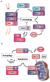
Figure 2. Crosstalk between autophagy- and apoptosis-related proteins
The BH3 domain of the autophagy-related protein Beclin 1 (BECN1) enables the formation of a BCL-2–BCL-XL–BECN1 complex that localizes to the ER and inhibits autophagy. Competitive interaction by the BH3-only protein BNIP3 and phosphorylation by the death-associated protein kinase (DAPK) regulate the dissociation of this complex and drive autophagy. In addition, BECN1 has also been found to be a substrate of caspase-3. B. The autophagy-related protein ATG5 can undergo calpain-dependent cleavage, which switches its function from pro-survival to pro-death. The C-terminal cleavage gives rise to a pro-apoptotic product that translocates to the mitochondria, binds BCL-XL and induces apoptosis. ATG5 can also induce apoptosis by binding to FADD, and it seems that apoptotic features such as phosphatidylserine exposure and membrane blebbing depend upon autophagy for the provision of energy in the form of ATP.
Figure 3
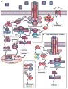
Figure 3. TNF-mediated survival, apoptosis and necroptosis
A. TNF ligation results in recruitment of RIPK1, TRADD, cIAP1 and cIAP2, and TRAF2 and TRAF5 to the TNFR1 to form complex I. cIAP1 and cIAP2 mediate Lys63-linked ubiquitylation of RIPK1, enabling docking of TAK1 (transforming-growth-factor-β-activated-kinase-1) and its binding partners TAB2 and TAB3. The signal is then perpetuated to IKK ((IκBα (inhibitor of kappa B) kinase)), which degrades IκBα, the cytoplasmic inhibitor of canonical NFκB. Once liberated from IκBα, NFκB translocates to the nucleus where it drives the transcription of pro-survival genes as well as feedback antagonists. Two pro-survival NFκB target genes are A20 and FLIP which promote association of RIPK1 with cytoplasmic RIPK3, TNF-receptor-associated-death-domain (TRADD), FADD, caspase-8 and the NF-κB target FLIP to form the death-inducing signaling complex (DISC). FLIPL can hetrodimerize with Caspase-8 and facilitate the cleavage and degradation of CYLD, RIP1K and RIPK3. However, the DISC also enables homo-dimerization and catalytic activation of caspase-8, which activates caspase-3 and caspase-7 to induce apoptosis. When caspase-8 is deleted or inhibited, RIPK1 interacts with RIPK3, resulting in the formation of the necrosome, an interaction that can be inhibited by necrostatin 1 (nec-1). RIPK3 recruits and phosphorylates MLKL, leading to the formation of oligomers that translocate to the plasma membrane. Once at the plasma membrane, MLKL forms membrane-disrupting pores, regulating both Na+ and Ca2+ influx, resulting in membrane rupture. B. cIAPs also function as negative regulators of the non-cannonical NF-kB pathway. In resting cells, a complex encompassing cIAP1, cIAP2, TRAF2 and TRAF3 targets and degrades NIK. Upon ligation of TNFR1, the TRAF2-TRAF3-cIAP1/2 complex is recruited to the receptor, changing the substrate specificity from NIK to the components of the complex. This results in increased NIK levels, phosphorylation of IKKα and p100, proteolytic processing of p100 to p52, and translocation to the nucleus. When cIAPs are depleted the canonical NFκB pathway is inhibited whereas the non-canonical NFκB pathway is not. Under these conditions, the large (~2-MDa) ripoptosome is assembled and, similarly to complex IIa, can stimulate caspase-8-mediated apoptosis.
Figure 4
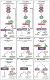
Figure 4. Apoptosis-induced proliferation
Apoptotic cells can secrete mitogens that stimulate growth and regeneration. Caspase targets and mitogenic factors are depicted in pink, and question marks indicate uncertainty. A. In Drosophila, p35 inhibits executioner caspases and retains the cells in an “undead” state. Under these conditions, p53 and JNK are activated and promote the release of Wg (Wnt ortholog) and Dpp (TGFb/BMP homolog), which promotes hyperplastic overgrowth through apoptosis-induced proliferation (AiP). B. In Drosophila, in a natural non p35-dependent system, apoptosis induces tissue regeneration by the secretion of Wg. C. In Drosophila, differentiated neurons induce apoptosis-induced compensatory proliferation via Hedghog (Hh), stimulating non-neuronal cell proliferation. In this scenario, the signal is downstream of Drice and Dcp-1 and not Dronc. D. In Hydra, head regeneration is a caspase-dependent process and apoptotic cells secrete Wnt3 to induce apoptosis-induced compensatory proliferation. E. In newts and Planaria, apoptotic cells and caspase activity are seen at the amputation site; however, it remains to be seen whether the apoptotic cells are responsible for the secretion of Wnt and Hh. F. In Xenopus, tail amputation leads to caspase activity, and inhibition of caspase-3 and caspase-9 inhibits proliferation and regeneration. It is unclear whether this form of compensatory proliferation is mediated by Wnt. G. In zebrafish, fin amputation results in ROS activity, which induces caspase and JNK activation. It remains to be established if FGF20, Wnt and sdf1 are secreted from apoptotic cells to regulate apoptosis-induced compensatory proliferation. H. In mice, liver injury results in ROS production and secretion of IL-11 from hepatocytes, which facilitates apoptosis-induced compensatory proliferation. G. In mice, wound repair and liver regeneration depend on caspase-3 and caspase-7. Caspase-3 mediates the proteolysis of iPLA2, producing PGE2, which is known to stimulate stem cell proliferation, wound repair and regeneration.
Figure 5
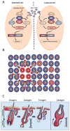
Figure 5. Apoptosis-induced apoptosis
A. Expression of the IAP antagonists Hid or Reaper in ‘doomed’ cells (left panel) can cause apoptosis in distant cells (right panel). The apoptotic signal is propagated by Eiger (a TNF homolog), which can cross compartmental borders (marked by a dashed line), to activate JNK and instruct apoptosis in distant cells. Presumably, the production of Eiger depends on JNK (depicted by a question mark). B. Schematic representation of apoptosis-induced apoptosis. Cells illustrated in red represent the initial apoptotic focal point. These cells emit Eiger in Drosophila and TNF in mice, which result in secondary apoptosis (pink cells). (C right panel) Schematic diagram depicting the hair follicle cycle. Hair follicles cycle between phases of growth (anagen), destruction (catagen) and rest (telogen). During catagen, apoptosis eliminates the lower two-thirds of the hair follicle. Post-catagen, the hair follicle enters the resting telogen phase, followed by a new cycle of hair growth (anagen). (C left panel) Apoptosis-induced apoptosis regulates the hair follicle cycle. During catagen, a primary apoptotic event (red cells), induces a wave of secondary apoptosis (pink cells) that is required for the cohort elimination of transient hair follicle cells.

Similar articles

Cited by

References

    1. Vogt C. Untersuchungen über die Entwicklungsgeschichte der Geburtshelferkröte (Alytes obstetricans) Solothurn: Jent und Gassman. 1842:130.
    1. Lockshin RA, Williams CM. Programmed cell death. II Endocrine potentiation of the breakdown of the intersegmental muscles of silkmoths. J Insect Physiol Dev. 1964;10:643–64.
    1. Tata JR. Requirement for RNA and protein synthesis for induced regression of the tadpole tail in organ culture. Dev Biol. 1966;13:77–94. - PubMed
    1. Kerr JF, Wyllie AH, Currie AR. Apoptosis: a basic biological phenomenon with wide-ranging implications in tissue kinetics. Br J Cancer. 1972;26:239–57. - PMC - PubMed
    1. Ellis HM, Horvitz HR. Genetic control of programmed cell death in the nematode C. elegans. Cell. 1986;44:817–29. - PubMed

Publication types