. 2015 Jul;8(4):788-98.
doi: 10.1161/CIRCHEARTFAILURE.114.001963. Epub 2015 May 18.

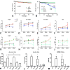
Smad3 Signaling Promotes Fibrosis While Preserving Cardiac and Aortic Geometry in Obese Diabetic Mice

Affiliations

Smad3 Signaling Promotes Fibrosis While Preserving Cardiac and Aortic Geometry in Obese Diabetic Mice

Anna Biernacka et al. Circ Heart Fail. 2015 Jul.

Abstract

Background: Heart failure in diabetics is associated with cardiac hypertrophy, fibrosis and diastolic dysfunction. Activation of transforming growth factor-β/Smad3 signaling in the diabetic myocardium may mediate fibrosis and diastolic heart failure, while preserving matrix homeostasis. We hypothesized that Smad3 may play a key role in the pathogenesis of cardiovascular remodeling associated with diabetes mellitus and obesity.

Methods and results: We generated leptin-resistant db/db Smad3 null mice and db/db Smad3+/- animals. Smad3 haploinsufficiency did not affect metabolic function in db/db mice, but protected from myocardial diastolic dysfunction, while causing left ventricular chamber dilation. Improved cardiac compliance and chamber dilation in db/db Smad3+/- animals were associated with decreased cardiomyocyte hypertrophy, reduced collagen deposition, and accentuated matrix metalloproteinase activity. Attenuation of hypertrophy and fibrosis in db/db Smad3+/- hearts was associated with reduced myocardial oxidative and nitrosative stress. db/db Smad3 null mice had reduced weight gain and decreased adiposity associated with attenuated insulin resistance, but also exhibited high early mortality, in part, because of spontaneous rupture of the ascending aorta. Ultrasound studies showed that both lean and obese Smad3 null animals had significant aortic dilation. Aortic dilation in db/db Smad3 null mice occurred despite reduced hypertension and was associated with perturbed matrix balance in the vascular wall.

Conclusions: Smad3 mediates diabetic cardiac hypertrophy, fibrosis, and diastolic dysfunction, while preserving normal cardiac geometry and maintaining the integrity of the vascular wall.

Keywords: TGF-β; diabetes mellitus; diabetic cardiomyopathies; fibrosis; obesity.


Figures

Figure 1
Figure 1
db/db mice exhibit cardiac hypertrophy and fibrosis associated with activation of TGF-β/Smad. A. db/db mice had significantly higher LV mass than corresponding lean WT animals. B. db/db and WT animals had comparable LVEDD between 1-2 months of age and exhibited only subtle increases in chamber dimensions at 4-6 months. C. LVEDD:LVmass ratio was markedly lower in db/db animals reflecting predominant hypertrophic remodeling. D. 6 month-old db/db hearts were fibrotic showing a marked increase in collagen content (measured with a hydroxyproline assay). E. There was a trend towards increased TGF-β1 mRNA expression in db/db hearts. F. Expression of p-Smad2 was increased in db/db myocardium (*p<0.05, **p<0.01 vs. WT). (echocardiography WT n=25-73/group, db/db n=14-44/group; collagen n=8/group; mRNA n=15/group; western blotting n=8/group).
Figure 2
Figure 2
Smad3 loss increases mortality in both lean and db/db mice and is associated with attenuated weight gain and reduced adiposity. A. WT mice, db/db mice, Shet and dbShet mice had comparable survival curves. B. When compared with WT animals, Smad3 knockout mice (SKO) had higher mortality (**p<0.01). In comparison to db/db, db/db Smad3 -/- (dbSKO) animals had higher mortality (**p<0.01). All dbSKO mice died before 6 months of age. C. SKO mice had significantly lower body weight than corresponding lean WT animals (**p<0.01). D-E: Reduced weight in SKO mice was due to reductions in both lean weight (D) and fat content (E). F. 2 month-old SKO mice had lower fat % content than WT animals (**p<0.01); however at 4 and 6 months, fat % was not significantly different between WT and SKO. Shet animals had increased fat % content at 6 months of age (^^p<0.01 vs. WT). G. db/db mice had a marked increase in body weight at 2-6 months of age (^^p<0.01 vs. age-matched WT mice). In comparison to db/db, dbSKO mice had markedly lower body weight (**p<0.01). H-I. Reduced weight gain in dbSKO mice was due to reductions in both lean weight and fat content (*p<0.05, **p<0.01 vs. db/db). At 6 months of age, dbShet mice had a statistically significant increase in fat content when compared with db/db animals (^^p<0.01 vs. db/db). J. Percent fat content was markedly increased in db/db animals (^^p<0.01 vs. corresponding WT). However, Smad3 loss in dbSKO animals was associated with reduced percent fat content only at 4 months of age, suggesting that lower body weight in the absence of Smad3 was predominantly related to stunted growth. K. db/db and dbShet mice had significantly increased plasma glucose levels when compared with WT animals (^^p<0.01 vs. WT); dbSKO mice had attenuated hyperglycemia. L. Insulin levels were also markedly increased in db/db and dbShet animals (^p<0.05 vs. WT); however dbSKO mice had comparable insulin levels with lean WT controls. M. HOMA-IR was also significantly higher in db/db and in dbShet mice when compared with corresponding WT controls (^p<0.05, ^^p<0.01 vs. WT); in contrast, in dbSKO mice, HOMA IR was comparable with WT animals suggesting attenuated insulin resistance. Survival: WT n=86, Shet n=175, SKO n=70, db/db n=32, dbShet n=55, dbSKO n=17; weight and adiposity: WT n=14, db/db n=16, dbShet n=25, dbSKO n=19 (2mo) n=9 (4mo), Shet n=16, SKO n=19 (2mo) n=17 (4mo); metabolic analysis: n=6.
Figure 3
Figure 3
Smad3 heterozygosity in db/db mice is associated with increased chamber dilation and a modest reduction in ejection fraction. Systolic function, dilative and hypertrophic remodeling were compared between lean WT mice and corresponding Shet and SKO animals (A-E), and between db/db, dbShet and dbSKO mice (F-J) using echocardiography. Panels K-O show comparison of echocardiographic endpoints between WT, db and dbShet animals at 6 and 12 months of age (at these timepoints comparison with SKO and dbSKO mice is not possible due to their early mortality) A-E: Shet and SKO mice had modest chamber dilation with preserved systolic function. A: At 4 months of age, Shet mice had higher LVEDD than WT animals (**p<0.01). B: LVEDV was significantly higher in Shet animals at 4 months of age. C: When adjusted to body weight, SKO animals also exhibited an increase in LVEDD:BW ratio. D. Ejection fraction was comparable between groups at 2 and 4 months of age. E: No statistically significant differences in LV mass were noted. F: dbShet mice had a significantly higher LVEDD than db/db animals. G: dbSKO mice had significantly increased LVEDD:body weight ratio. H. LVEDV was significantly higher in dbShet mice at 4 months of age. I. When compared with age-matched WT animals, db/db mice had increased ejection fraction (^^p<0.01 vs WT). When compared with db/db animals, dbShet mice and dbSKO animals had a modest, but significant reduction in ejection fraction (**p<0.01 vs db/db). J-K: db/db mice had increased LV mass and higher cardiomyocyte size than age-matched lean controls (^p<0.05, ^^p<0.01 vs. WT). dbSKO mice had attenuated LV hypertrophy, suggested by reduced LV mass (J) and decreased cardiomyocyte size (K) (*p<0.05, **p<0.01 vs. db/db). L-N: At 6-12 months of age, dbShet mice had persistent chamber dilation (*p<0.05, **p<0.01 vs. age-matched db/db) without significant progression of dilative remodeling O. Ejection fraction was significantly lower in dbShet mice at 6 and 8 months of age (**p<0.01 vs. db/db); however progressive systolic dysfunction was not observed. P: In comparison to WT animals, db/db mice had significantly higher LV mass at 6-8 months of age (^^p<0.01 vs. WT). dbShet mice and db/db mice had comparable LV mass at 6 and 12 months of age (2 mo of age: WT n=14, Shet=26, SKO n=10, db/db n=16, dbShet n=15, dbSKO n=19; 4mo of age: WT n=45, Shet n=27, SKO n=6, db/db n=42, dbShet n=16, dbSKO n=10, Shet n=16) (^p<0.05, ^^p<0.01 vs. age-matched WT)..
Figure 4
Figure 4
dbShet mice exhibit attenuated diastolic dysfunction associated with reduced matrix deposition and increased MMP activity. A. Pressure:volume relations in 12 month-old animals demonstrated that db/db mice exhibit a shift of the curve to the left and that dbShet hearts had improved compliance. B. Left ventricular chamber stiffness constant was increased in db/db mice. Partial loss of Smad3 reduced the chamber stiffness constant in db/db animals (*p<0.05, **p<0.01). C-D. Improved diastolic function in dbShet hearts was associated with reduced collagen content, assessed through Sirius red staining (C) and through a hydroxyproline assay (D). E-F. Reduced collagen in dbShet hearts was not due to differences in collagen transcription. G-I. dbShet animals had increased myocardial MMP9 (H) and MMP2 (I) activity, assessed with zymography (G). Accentuated MMP activity in dbShet hearts was not due to increased MMP2 (J) or MMP9 (K) transcription.
Figure 5
Figure 5
A. Smad3 loss attenuates oxidative and nitrosative stress in WT and db/db hearts. DCFDA assay showed that db/db hearts had increased ROS levels when compared with age-matched WT hearts (*p<0.05 vs WT). Partial or complete loss of Smad3 markedly reduced myocardial ROS levels in WT and in db/db animals (^p<0.05, ^^p<0.01 vs. WT; **p<0.01 vs. db/db, n=6/group). B-C. 3-nitrotyrosine expression was markedly increased in db/db hearts (**p<0.01 vs WT). Smad3 loss in db/db mice resulted in attenuated 3-nitrotyrosine expression (**p<0.01 vs. db/db).
Figure 6
Figure 6
SKO and dbSKO mice had aortic dilation and a high incidence of spontaneous rupture. A. Hemothorax in a 2 month-old SKO mouse that died spontaneously. B. Dilated ascending aorta in a dbSKO animal. C. Casts of the aorta show dilation and aneurysm formation (arrows) in a 4 month-old dbSKO and a 2 month-old SKO mouse. D. Ultrasound showed that at 2 months of age, the ascending aorta was significantly dilated in dbShet and in dbSKO mice (*p<0.05, **p<0.01 vs. db/db). E. At 4 months of age, lean Shet and SKO mice had increased aortic diameter (*p<0.05, **p<0.01 vs. WT). dbShet and dbSKO exhibited increased aortic dimensions in comparison to db/db animals (**p<0.01 vs. db/db). (WT n=10, Shet n=11, SKO n=16; db/db n=14, dbShet n=13, dbSKO n=9). F-I. Aortic dilation in SKO and dbSKO mice was not due to increased systemic blood pressure. When compared with 4-month old WT animals, age-matched Shet and SKO mice had lower systolic (F) and diastolic (G) blood pressure (*p<0.05, **p<0.01 vs. WT). 4 month-old db/db mice had higher systolic (H) and diastolic (I) blood pressure than lean WT animals (^^p<0.01 vs. WT). Smad3 loss in db/db mice protected from increase in systemic blood pressure (H, I) (**p<0.01). (WT n=8, db/db n=6; dbShet, dbSKO, SKO, Shet n=5).

Similar articles

Cited by

References

    1. Marwick TH. Diabetic heart disease. Heart. 2006;92:296–300. - PMC - PubMed
    1. Abel ED, Litwin SE, Sweeney G. Cardiac remodeling in obesity. Physiol Rev. 2008;88:389–419. - PMC - PubMed
    1. Ho JE, Lyass A, Lee DS, Vasan RS, Kannel WB, Larson MG, Levy D. Predictors of New-Onset Heart Failure: Differences in Preserved Versus Reduced Ejection Fraction. Circ Heart Fail. 2013;6:279–286. - PMC - PubMed
    1. Rubler S, Dlugash J, Yuceoglu YZ, Kumral T, Branwood AW, Grishman A. New type of cardiomyopathy associated with diabetic glomerulosclerosis. Am J Cardiol. 1972;30:595–602. - PubMed
    1. Asbun J, Villarreal FJ. The pathogenesis of myocardial fibrosis in the setting of diabetic cardiomyopathy. J Am Coll Cardiol. 2006;47:693–700. - PubMed

Publication types

MeSH terms