

MicroRNA-19a regulates lipopolysaccharide-induced endothelial cell apoptosis through modulation of apoptosis signal-regulating kinase 1 expression
- PMID: 25982447
- PMCID: PMC4446110
- DOI: 10.1186/s12867-015-0034-8
MicroRNA-19a regulates lipopolysaccharide-induced endothelial cell apoptosis through modulation of apoptosis signal-regulating kinase 1 expression
Abstract
Background: MicroRNAs, small non-encoding RNAs that post-transcriptionally modulate expression of their target genes, have been implicated as critical regulatory molecules in endothelial cells.
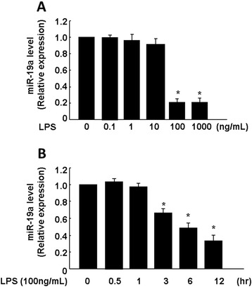
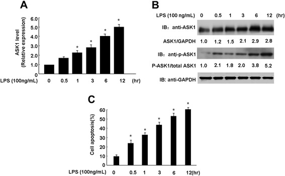
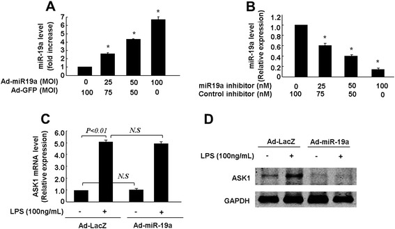
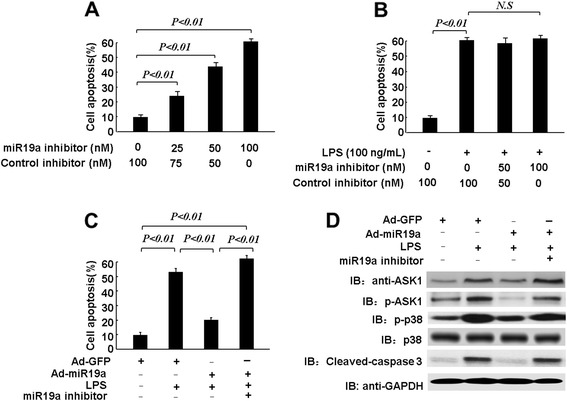
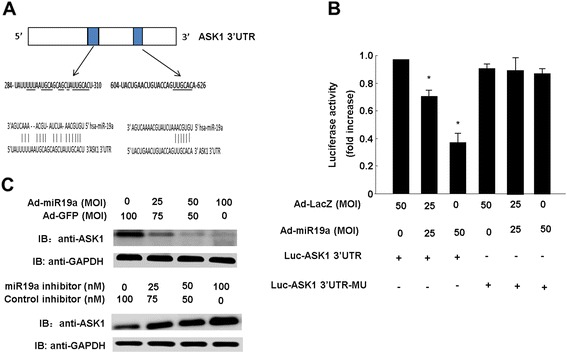
Results: In the present study, we found that overexpression of miR-19a protects endothelial cells from lipopolysaccharide (LPS)-induced apoptosis through the apoptosis signal-regulating kinase 1 (ASK1)/p38 pathway. Quantitative real-time PCR demonstrated that the expression of miR-19a in endothelial cell was markedly down-regulated by LPS stimulation. Furthermore, LPS-induced apoptosis was significantly inhibited by over-expression of miR-19a. Finally, both a luciferase reporter assay and western blot analysis showed that ASK1 is a direct target of miR-19a.
Conclusions: MiR-19a regulates ASK1 expression by targeting specific binding sites in the 3' untranslated region of ASK1 mRNA. Overexpression of miR-19a is an effective method to protect against LPS-induced apoptosis of endothelial cells.
Figures
Similar articles
-
miR-106a Increases Granulosa Cell Viability and Is Downregulated in Women With Diminished Ovarian Reserve.J Clin Endocrinol Metab. 2018 Jun 1;103(6):2157-2166. doi: 10.1210/jc.2017-02344. J Clin Endocrinol Metab. 2018. PMID: 29590425
-
Suppression of Bim by microRNA-19a may protect cardiomyocytes against hypoxia-induced cell death via autophagy activation.Toxicol Lett. 2016 Aug 22;257:72-83. doi: 10.1016/j.toxlet.2016.05.019. Epub 2016 May 21. Toxicol Lett. 2016. PMID: 27220268
-
MicroRNA-495 regulates the proliferation and apoptosis of human umbilical vein endothelial cells by targeting chemokine CCL2.Thromb Res. 2015 Jan;135(1):146-54. doi: 10.1016/j.thromres.2014.10.027. Epub 2014 Nov 6. Thromb Res. 2015. PMID: 25466836
-
miR-206 modulates lipopolysaccharide-mediated inflammatory cytokine production in human astrocytes.Cell Signal. 2015 Jan;27(1):61-8. doi: 10.1016/j.cellsig.2014.10.006. Epub 2014 Oct 23. Cell Signal. 2015. PMID: 25452104
-
Apoptosis signal-regulating kinase 1-mediated signaling pathway regulates lipopolysaccharide-induced tissue factor expression in pulmonary microvasculature.Int Immunopharmacol. 2010 Sep;10(9):1062-7. doi: 10.1016/j.intimp.2010.06.006. Epub 2010 Jun 11. Int Immunopharmacol. 2010. PMID: 20601186
Cited by
-
MiR-19a mediates the negative regulation of the NF-κB pathway in lipopolysaccharide-induced endometritis by targeting TBK1.Inflamm Res. 2019 Mar;68(3):231-240. doi: 10.1007/s00011-019-01213-3. Epub 2019 Jan 23. Inflamm Res. 2019. PMID: 30673803
-
MiR-30b Is Involved in the Homocysteine-Induced Apoptosis in Human Coronary Artery Endothelial Cells by Regulating the Expression of Caspase 3.Int J Mol Sci. 2015 Jul 31;16(8):17682-95. doi: 10.3390/ijms160817682. Int J Mol Sci. 2015. PMID: 26263983 Free PMC article.
-
MicroRNA-19a-3p regulates cell growth through modulation of the PIK3IP1-AKT pathway in hepatocellular carcinoma.J Cancer. 2020 Feb 10;11(9):2476-2484. doi: 10.7150/jca.37748. eCollection 2020. J Cancer. 2020. PMID: 32201518 Free PMC article.
-
miR-19a and miR-20a and Tissue Factor Expression in Activated Human Peripheral Blood Mononuclear Cells.Thrombosis. 2017;2017:1076397. doi: 10.1155/2017/1076397. Epub 2017 Oct 30. Thrombosis. 2017. PMID: 29214079 Free PMC article.
-
MicroRNA-Mediated Down-Regulation of Apoptosis Signal-Regulating Kinase 1 (ASK1) Attenuates the Apoptosis of Human Mesenchymal Stem Cells (MSCs) Transplanted into Infarcted Heart.Int J Mol Sci. 2016 Oct 20;17(10):1752. doi: 10.3390/ijms17101752. Int J Mol Sci. 2016. PMID: 27775615 Free PMC article.
References
-
- Grabarek JB. RNA silencing. Adv Exp Med Biol. 2003;544:145–58. - PubMed
Publication types
MeSH terms
Substances
LinkOut - more resources
Full Text Sources
Other Literature Sources
Molecular Biology Databases
Miscellaneous
