. 2015 May 15;11(5):e1005242.
doi: 10.1371/journal.pgen.1005242. eCollection 2015 May.

Fibroblast Growth Factor 9 Regulation by MicroRNAs Controls Lung Development and Links DICER1 Loss to the Pathogenesis of Pleuropulmonary Blastoma

Affiliations

Fibroblast Growth Factor 9 Regulation by MicroRNAs Controls Lung Development and Links DICER1 Loss to the Pathogenesis of Pleuropulmonary Blastoma

Yongjun Yin et al. PLoS Genet. .

Abstract

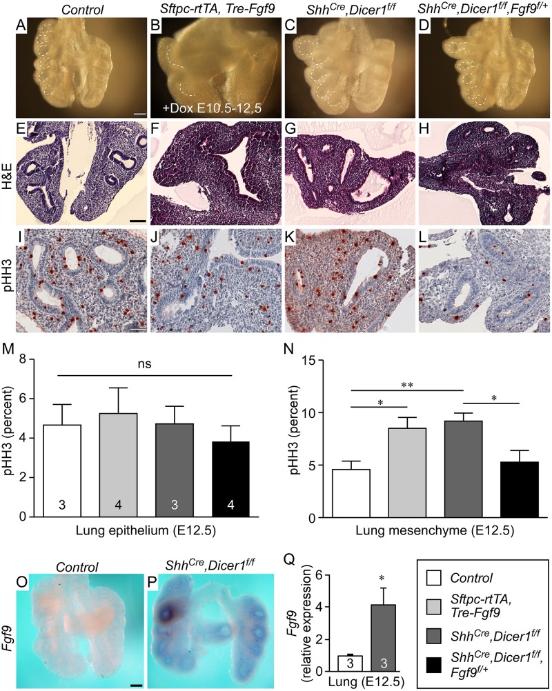
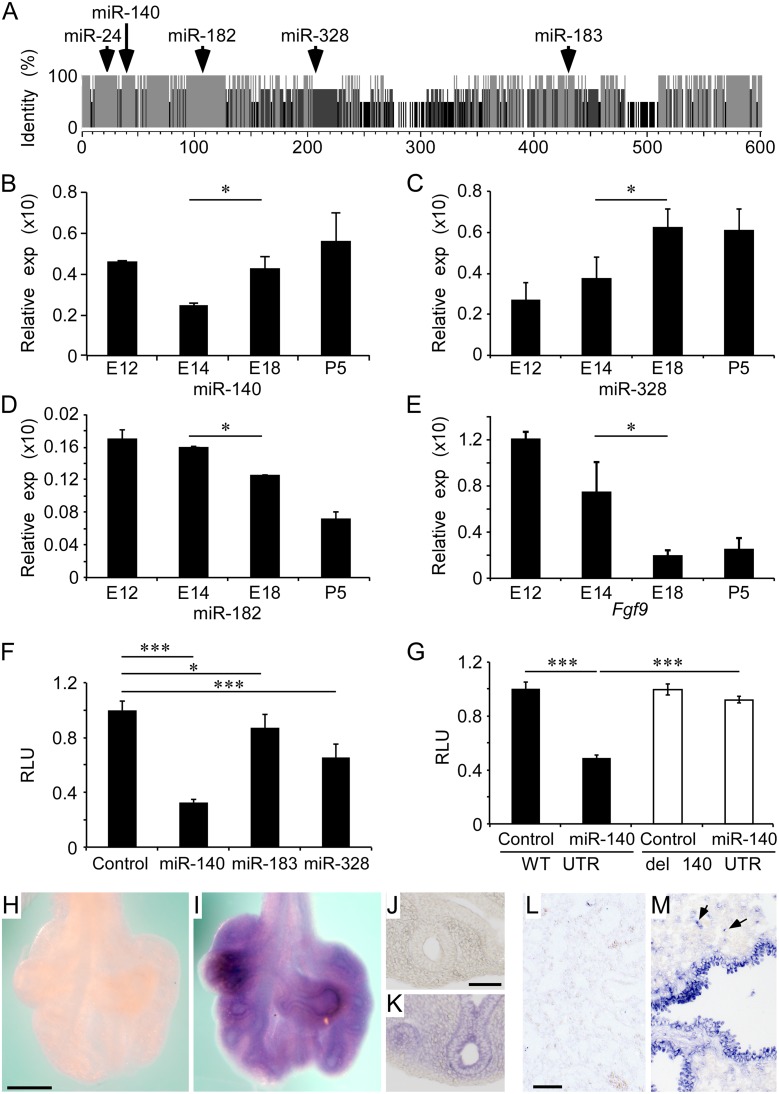
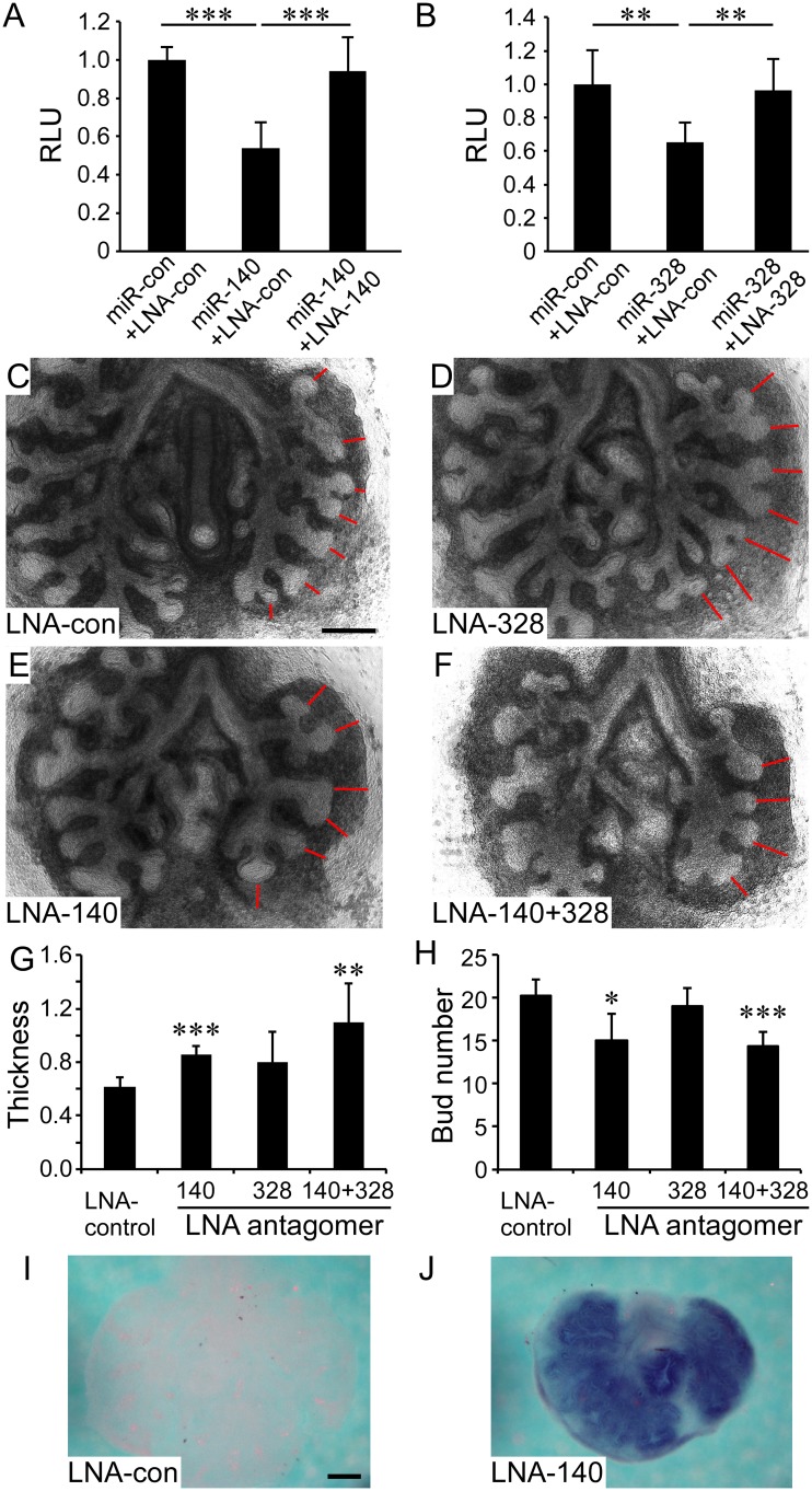
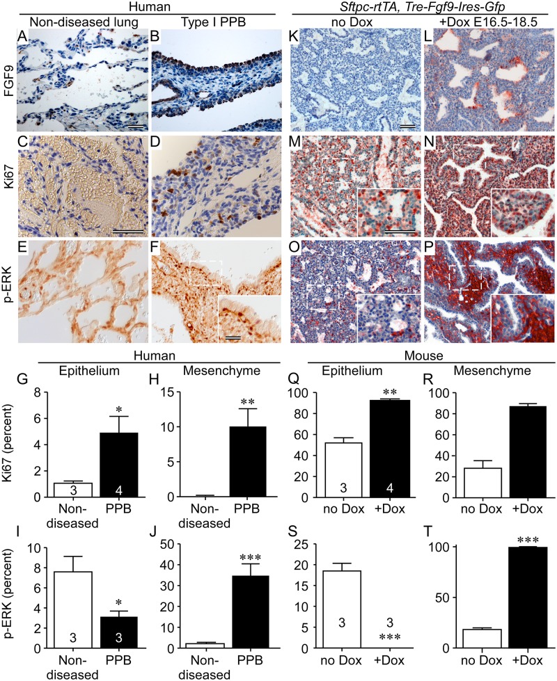
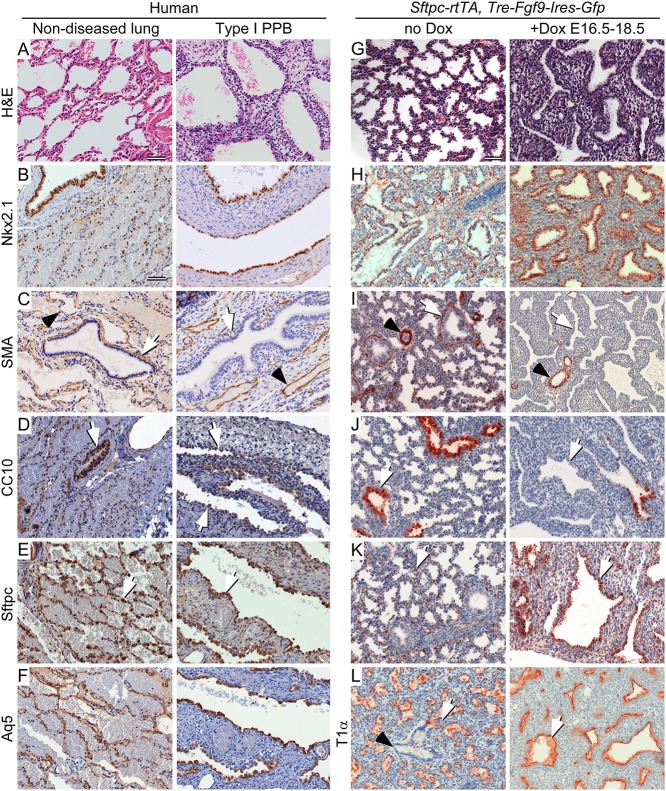
Pleuropulmonary Blastoma (PPB) is the primary neoplastic manifestation of a pediatric cancer predisposition syndrome that is associated with several diseases including cystic nephroma, Wilms tumor, neuroblastoma, rhabdomyosarcoma, medulloblastoma, and ovarian Sertoli-Leydig cell tumor. The primary pathology of PPB, epithelial cysts with stromal hyperplasia and risk for progression to a complex primitive sarcoma, is associated with familial heterozygosity and lesion-associated epithelial loss-of-heterozygosity of DICER1. It has been hypothesized that loss of heterozygosity of DICER1 in lung epithelium is a non-cell autonomous etiology of PPB and a critical pathway that regulates lung development; however, there are no known direct targets of epithelial microRNAs (miRNAs) in the lung. Fibroblast Growth Factor 9 (FGF9) is expressed in the mesothelium and epithelium during lung development and primarily functions to regulate lung mesenchyme; however, there are no known mechanisms that regulate FGF9 expression during lung development. Using mouse genetics and molecular phenotyping of human PPB tissue, we show that FGF9 is overexpressed in lung epithelium in the initial multicystic stage of Type I PPB and that in mice lacking epithelial Dicer1, or induced to overexpress epithelial Fgf9, increased Fgf9 expression results in pulmonary mesenchymal hyperplasia and a multicystic architecture that is histologically and molecularly indistinguishable from Type I PPB. We further show that miR-140 is expressed in lung epithelium, regulates epithelial Fgf9 expression, and regulates pseudoglandular stages of lung development. These studies identify an essential miRNA-FGF9 pathway for lung development and a non-cell autonomous signaling mechanism that contributes to the mesenchymal hyperplasia that is characteristic of Type I PPB.


Conflict of interest statement

The authors have declared that no competing interests exist.

Figures

Fig 1
Fig 1. Dicer1 regulation of lung epithelial development requires Fgf9.
(A-D) Comparison of E12.5 whole mount lung morphology of Control (A), Sftpc-rtTA, Tre-Fgf9-Ires-eGfp lungs induced with doxycycline from E10.5-E12.5 (B), Shh Cre/+, Dicer1 f/f (C) and Shh Cre/+, Dicer1 f/f, Fgf9 f/+ (D). (E-H) H&E stained histological sections of the lungs shown above in panels A-D. (I-L) Representative immunostaining for phospho-histone H3 (pHH3) of the lungs shown in panels A-D. (M and N) Quantification of epithelial (M) and mesenchymal (N) cell proliferation of pHH3 labeled lung tissue in Control; Sftpc-rtTA, Tre-Fgf9-Ires-eGfp lungs induced with doxycycline from E10.5-E12.5; Shh Cre/+, Dicer1 f/f lungs; and Shh Cre/+, Dicer1 f/f, Fgf9 f/+ lungs. For each group, at least 3 individual samples were included, 3 different slides were chosen from each sample, and for each section, three 10x fields were counted for the number of positive cells per 100 cells. (O and P) Whole mount in situ hybridization showing increased expression of Fgf9 in E12.5 Shh Cre/+, Dicer1 f/f lung epithelium (P) compared to control lung (O). (Q) Quantitative RT-PCR showing increased expression of Fgf9 in E12.5 Shh Cre/+, Dicer1 f/f lung (n = 3) epithelium compared to control lung (n = 3). *P<0.05; **P<0.01; ns, not significant. Scale bars: A, 200 μm; E, 100 μm; I, 50 μm; O, 200 μm. Sample numbers (n) are indicated in data bars.
Fig 2
Fig 2. Epithelial Dicer1 regulation of mesenchymal Wnt/β-Catenin signaling and epithelial differentiation requires Fgf9.
(A-C) Expression of Wnt2a in E12.5 lung showing increased expression in Shh Cre/+, Dicer1 f/f distal lung mesenchyme (B) compared to the Control (A). Inactivation of one allele of Fgf9 in Shh Cre/+, Dicer1 f/f lung epithelium (C) reduces Wnt2a expression to levels observed in control lungs. (D-F) The downstream target of Wnt signaling, Lef1, was increased in Shh Cre/+, Dicer1 f/f lung mesenchyme (E) compared to the Control (D). Inactivation of one allele of Fgf9 in Shh Cre/+, Dicer1 f/f lung epithelium (F) reduces Lef1 expression to levels observed in control lungs. (G-I) Expression of Sftpc in E12.5 lung showing decreased expression in Shh Cre/+, Dicer1 f/f distal lung mesenchyme (H) compared to the Control (G). Inactivation of one allele of Fgf9 in Shh Cre/+, Dicer1 f/f lung epithelium (I) results in increased Sftpc expression. (J) Quantitative PCR analysis of E12.5 Control lung and Shh Cre/+, Dicer1 f/f rescued with one or two Fgf9 floxed alleles showing increased Sftpc expression. * P<0.04. Images shown are representative of three embryos for each genotype. Scale bars: 200 μm.
Fig 3
Fig 3. Regulation of the Fgf9 3' UTR by miRNAs expressed in developing lung epithelium.
(A) Conservation of miRNA binding sites by comparison of Fgf9 3’ UTR sequences from human, chimp, mouse, and pig. Grey, 100%; Dark grey, 75%; Black, 50% sequence identity. The position of specific miRs that are also expressed and regulated during lung development are indicated. (B-D) Relative developmental expression profile at E12.5, E14.5, E18 and P5 (normalized to U6 snRNA or Hprt) of miR-140 (B), miR-328 (C), miR-182 (D), and Fgf9 (E). (F) Repression of luciferase activity by miR-140, miR-183, and miR-328 double stranded miRNA mimics compared to a cel-miR-67 control double stranded miRNA mimic (S3 Fig, S2 Table), when co-transfected in HEK293 cells with a luciferase reporter construct containing a wild type mouse Fgf9 3’ UTR. (G) Repression of the Fgf9 3’ UTR by miR-140 (solid bars) was blocked by mutations that deleted the seed sequences for miR-140 (open bars). (H-M) In situ hybridization to localize expression of miR-140 in E12.5 and E18.5 lung. E12.5 lung was hybridized with a scrambled LNA in situ probe (H) or with the hsa-miR-140-5p LNA probe (I) (S2 Table). J and K are frozen sections from the tissue in H and I, respectively. Histological sections from E18.5 wild type mouse lung were hybridized with a scrambled LNA in situ probe (L) or with hsa-miR-140-5p LNA probe (M). Arrows in (M) indicate patterns that are consistent with expression in type II pneumocytes. All data is derived from at least 3 independent experiments. *P<0.05, *** P<0.001. Scale bars: H, 200 μm; J and L, 50 μm.
Fig 4
Fig 4. MiR-140 and miR-328 regulate in vitro lung development and Fgf9 expression.
(A-B) Validation of tiny LNA antagomers ability to block the activity of miR-140 and miR-328. Repression of the Fgf9 3’ UTR by 10 nM miR-140 (A) or 10 nM miR-328 (B) transfected into HEK293 cells with a luciferase reporter construct containing a wild type mouse Fgf9 3’ UTR was blocked by adding 10 nM of the corresponding tiny LNAs to the culture medium. The control LNA (LNA-con) contains a single mismatch in the LNA-140 sequence. (C-F) E12.5 lung explants were cultured in the presence of 100 nM tiny LNA oligonucleotides (S2 Table) for 48 hr. (C) Control LNA (100 nM), (D) LNA-328, E) LNA-140, and (F) 50 nM of LNA-140 and LNA-328 (total concentration 100 nM). Red lines indicate mesenchymal thickness. (G and H) Quantification of mesenchymal thickness (G) and the epithelial bud number (H) of lung explants in response to treatment with tiny LNA antagomers (n = 4–5 explants per condition). (I and J) Whole mount in situ hybridization showing expression of Fgf9 in E12.5 wild type lung explants cultured in the presence of 100 nM control LNA (I) or LNA-140 (J). Images shown are representative of at least three independent experiments. *P<0.05, **P<0.01, *** P<0.001. Scale bars: 200 μm.
Fig 5
Fig 5. FGF9 overexpression in Type I PPB phenocopies ectopically expressed FGF9 in mouse lung epithelium.
(A, B, K, and L) Immunostaining for FGF9 showing increased expression in Type I PPB-associated lung epithelium and in doxycycline-induced Sftpc-rtTA, Tre-Fgf9-Ires-Gfp mouse lung. Non-diseased human lung and uninduced (no Dox) mouse lung were used as controls. (C, D, M, and N) Mesenchymal and epithelial proliferation in Type I PPB and induced mouse lung identified by immunostaining for Ki67. Inserts show higher magnification of the boxed regions. (E, F, O, and P) Immunostaining showing increased phosphorylated Erk1/2 (p-ERK) in Type I PPB and in induced mouse lung mesenchyme and reduced p-ERK in epithelium. Inserts show higher magnification of the boxed regions. (G, H, Q and R) Quantification of Ki67 immunostaining in C, D, M, and N above, showing increased in proliferation in both epithelial and mesenchymal tissues of Type I PPB (G and H) and Fgf9-induced mouse lung (Q and R). (I, J, S, and T) Quantification of p-ERK immunostaining in E, F, O and P above, showing decreased epithelial p-ERK (I and S) and increased mesenchymal p-ERK (J and T), in Type I PPB and Fgf9-induced mouse lung compared to control tissue. Error bars represent SD. * p<0.05, ** p<0.01, *** p<0.001. (A and B) Five month-old male; (C and D) Three month-old female; (E) Six month-old female; (F) 34 month-old female. Scale bars: A and B, 20 μm; C-P, 50 μm. Sample numbers (n) are indicated on the data bars.
Fig 6
Fig 6. Type I PPB and induced late gestation expression of epithelial FGF9 in mice have similar histopathology and cell differentiation.
(A-F) Comparison of non-diseased human lung (left) and Type I PPB (right). (G-L) Comparison of normal (no Dox) E18.5 mouse lung (left) and mouse lung from Sftpc-rtTA, Tre-Fgf9-Ires-eGfp double transgenic mice induced (+Dox) to overexpress Fgf9 from E16.5 to E18.5 (right). (A and G) H&E stained histological sections. (B and H) Immunostaining for Nkx2.1 to identify lung epithelium. (C and I) Immunostaining for smooth muscle actin (SMA). Peri-bronchiolar SMA immunostaining (white arrow) and perivascular SMA immunoreactivity (black arrowhead) are differentially affected. (D and J) Immunostaining for Club cell secretory protein (CC10) showing reduced expression of CC10 in proximal lung epithelium (white arrow) compared to that of non-diseased human lung and uninduced mouse lung, respectively. (E and K) Immunostaining for surfactant protein C (Sftpc) showing expanded proximal expression (white arrow) in all cystic lung epithelium in human Type I PPB and Fgf9-induced mouse lung. In non-diseased human lung and uninduced mouse lung, Sftpc immunostaining (white arrow) was consistent with expression in Type II pneumocytes. (F and L) Immunostaining for the distal lung Type I pneumocyte marker Aquaporin 5 (Aq5, human) and T1α (mouse). In human, Aq5 was expressed in distal alveoli of non-diseased lung tissue and in Type I PPB associated epithelium. T1α was similarly expressed in distal mouse lung and throughout the epithelium of Fgf9-induced mouse lung (white arrow). (A) Three month-old female. (B-F) Three month-old female. Scale bar: A, 20 μm, B-L, 50 μm.

Similar articles

Cited by

References

    1. Colvin JS, White A, Pratt SJ, Ornitz DM. Lung hypoplasia and neonatal death in Fgf9-null mice identify this gene as an essential regulator of lung mesenchyme. Development. 2001;128:2095–106. - PubMed
    1. White AC, Xu J, Yin Y, Smith C, Schmid G, Ornitz DM. FGF9 and SHH signaling coordinate lung growth and development through regulation of distinct mesenchymal domains. Development. 2006;133(8):1507–17. Epub 2006/03/17. - PubMed
    1. Yin Y, White AC, Huh SH, Hilton MJ, Kanazawa H, Long F, et al. An FGF-WNT gene regulatory network controls lung mesenchyme development. Dev Biol. 2008;319(2):426–36. 10.1016/j.ydbio.2008.04.009 - DOI - PMC - PubMed
    1. Bartel DP. MicroRNAs: genomics, biogenesis, mechanism, and function. Cell. 2004;116(2):281–97. - PubMed
    1. Vasudevan S. Posttranscriptional upregulation by microRNAs. Wiley Interdiscip Rev RNA. 2012;3(3):311–30. Epub 2011/11/11. 10.1002/wrna.121 - DOI - PubMed

Publication types

MeSH terms

Supplementary concepts