Review
. 2015 Jun 2;54(21):3250-68.
doi: 10.1021/acs.biochem.5b00353. Epub 2015 May 20.

The Voltage-Gated Proton Channel: A Riddle, Wrapped in a Mystery, inside an Enigma

Affiliations
Review

The Voltage-Gated Proton Channel: A Riddle, Wrapped in a Mystery, inside an Enigma

Thomas E DeCoursey. Biochemistry. .

Abstract

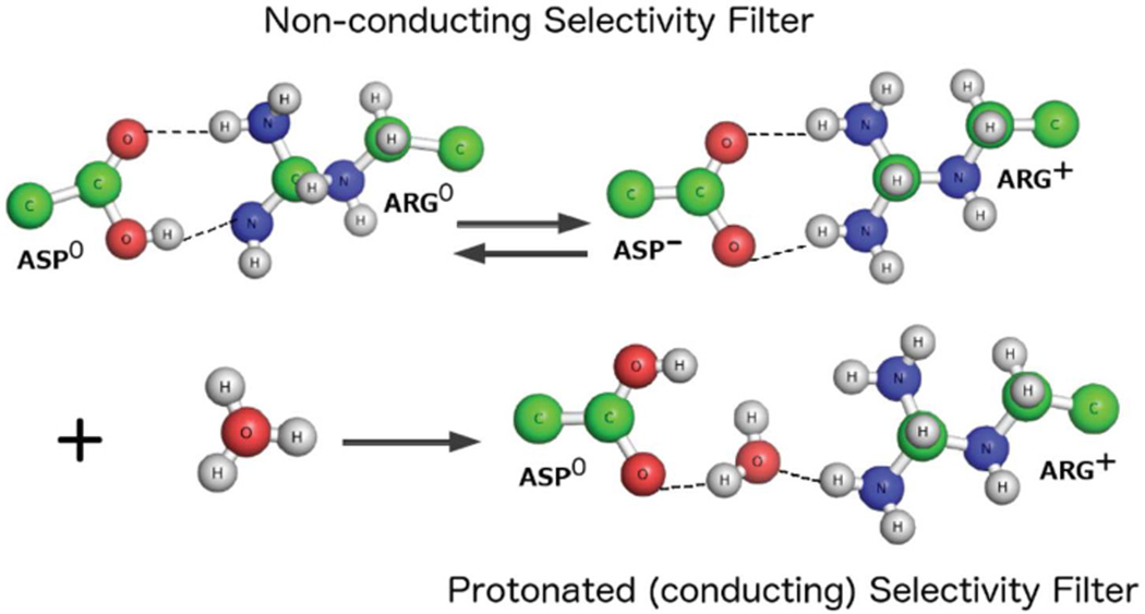
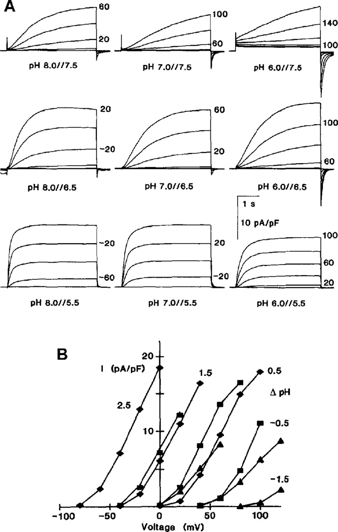
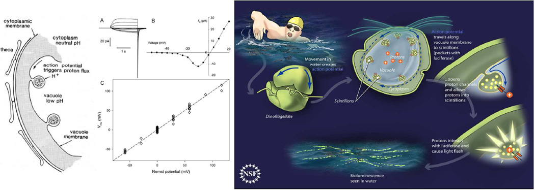
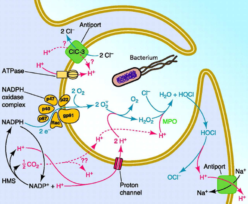
The main properties of the voltage-gated proton channel (HV1) are described in this review, along with what is known about how the channel protein structure accomplishes its functions. Just as protons are unique among ions, proton channels are unique among ion channels. Their four transmembrane helices sense voltage and the pH gradient and conduct protons exclusively. Selectivity is achieved by the unique ability of H3O(+) to protonate an Asp-Arg salt bridge. Pathognomonic sensitivity of gating to the pH gradient ensures HV1 channel opening only when acid extrusion will result, which is crucial to most of its biological functions. An exception occurs in dinoflagellates in which influx of H(+) through HV1 triggers the bioluminescent flash. Pharmacological interventions that promise to ameliorate cancer, asthma, brain damage in ischemic stroke, Alzheimer's disease, autoimmune diseases, and numerous other conditions await future progress.


Figures

Figure 1
Figure 1
Topology of three molecules that contain voltage-sensing domains (VSD): a generic K+ channel, the proton channel, and a voltage-sensitive phosphatase. The upper row shows the orientation of a monomer in the membrane, with the transmembrane helical segments labeled S1, S2, etc. The lower row shows the assembled tetramer, dimer, or monomer, respectively. Ion conduction in the K+ channel occurs through the single central pore (in red) which is comprised of four S5–S6 pairs. HV1 is a dimer in mammals but each monomer has an intrinsic conduction pathway; monomeric constructs retain the major functional properties of the dimer,–. S1–S4 form the VSD that senses membrane potential, which is transduced into channel opening or closing (gating) in “voltage-gated” ion channels, or into changes in the rate of phosphatase activity in the voltage sensing phosphatase (VSP). Reprinted with permission from. Copyright 2010, American Physiological Society.
Figure 2
Figure 2
Fluorescence-based H+ flux assay for vesicles containing hHV1 (dark blue), the VSD of KvAP channels (green), KvAP (dark green), the paddle chimera (cyan), and empty vesicles (red). Valinomycin and protonophore CCCP were added at the indicated time points. Vesicles loaded with K+ were placed in a low K+ buffer. Valinomycin allows K+ efflux which drives H+ influx, detected by a fluorescent dye. Reprinted with permission from. ©James Anthony Letts, 2014.
Figure 3
Figure 3
A maximum likelihood phylogenetic tree from a multiple sequence alignment of the VSD portion of 37 HV1s. Branch lengths are proportional to the distance between sequences. Eight genes confirmed to be HV1 by direct electrophysiological measurement are starred; the rest are predicted to be HV1. Two more genes not shown have been confirmed recently by voltage-clamp, from a bioluminescent dinoflagellate Lingulodinium polyedrum, and Nicoletia phytophila [personal communication from G. Chaves, C. Derst, B. Musset]. Reprinted with permission from. ©2011 by The National Academy of Sciences of the USA.
Figure 4
Figure 4
Transmembrane domain of the human voltage gated proton channel, hHV1. The four helical segments are color-coded (S1 red, S2 yellow, S3 green, S4 blue); the extracellular end is at the top. Acidic and basic residues are labeled; dashed lines indicate salt bridges predicted in molecular dynamics simulations of a homology model to stabilize the open channel. D112 is crucial to proton selectivity. ©. Originally published in the Journal of General Physiology. doi: 10.1085/jgp.201210856
Figure 5
Figure 5
Unrooted phylogram of 122 VSDs from several classes of VSD-containing molecules. The branch length shows the degree of difference from neighbors. Despite including only the VSD (S1–S4) and excluding the pore-forming S5–S6 domain, all K+ channels separated onto one branch, Na+ and Ca2+ channels onto another branch. A third branch includes voltage-sensing phosphatases, HV1, and c15orf27, molecules of unknown function. Reprinted with permission from. ©2011, Nature Publishing Group.
Figure 6
Figure 6
The Asp residue that is crucial to proton selectivity in hHV1 can be shifted from position 112 to 116 in the S1 helix. The diagrams show S1 and S4 segments schematically in the open hHV1 channel. The three S4 Arg residues (R1-R3 in blue) are numbered from the outside (R205, R208, and R211). Yellow indicates Val, red Asp, and gray all positions where Asp did not produce functioning channels. The families of voltage-clamp currents (in 10 mV increments up to the voltage indicated) above each diagram illustrate H+-selective currents in WT, no current in D112V, and H+-selective currents in the D112V/V116D double mutant, all at pHo 7.0 and pHi 5.5. ©. Originally published in the Journal of General Physiology. doi: 10.1085/jgp.201311045
Figure 7
Figure 7
Proton selectivity occurs because H3O+ is uniquely able to break the Asp-Arg connection in the selectivity filter, opening its own conduciton pathway. Quantum calculations reveal that Asp and Arg interact in the hHV1 selectivity filter via two hydrogen bonds, with stable optimized configurations with Asp (top left) or Arg (top right) protonated. Introducing a hydronium ion, H3O+ into either (lower row) results in protonation of Asp producing a neutral water molecule that mediates interactions between side chains. Computed G values are negative, indicating a favorable forward reaction, assuming dielectric constants of either 4 or 30 to reflect low or high solvent accessibility. Figure generously provided by Karin Mazmanian.
Figure 8
Figure 8
Predicted aqueous accessibility (blue surface) of the pore of the closed mouse HV1 channel, based on its crystal structure. Dashed lines show membrane surface; the top is the extracellular end. [From]
Figure 9
Figure 9
Maximum conductance of hHV1 mutants in which Asp has been replaced by the indicated amino acid. Acidic, basic, or neutral side chains are indicated by red, aqua, or blue colors. The more hydrophobic the side chain, the smaller the conductance. Hydrophobicity, defined as in, increases to the left. Data are from or unpublished data from the same authors.
Figure 10
Figure 10
The voltage at which channels open is determined by the pH gradient, pH. (A) Families of proton currents recorded in rat alveolar epithelial cells at several pH, indicated as pHo//pHi, during depolarizing voltage pulses shown in 20 mV increments. The peak currents from these families are plotted in B, with diamonds for pHi 5.5, squares for pHi 6.5, and triangles for pHi 7.5. @. Originally published in the Journal of General Physiology. doi: 10.1085/jgp.105.6.861
Figure 11
Figure 11
Proton channels trigger the flash in bioluminescent dinoflagellates. The diagram on the left and the cartoon on the right illustrate J.W. Hastings’ original proposal. Movement of water initiates an action potential that travels along the vacuolar membrane (the tonoplast) and invades the scintillon membrane where it opens voltage gated proton channels. H+ readily enters the luciferase-containing scintillon due to the enormous driving force; the vacuole pH is 3.5–4.5,. The resulting influx of H+ activates the acid-activated luciferase, contained in the scintillon, as well as releasing luciferin from its binding protein. Both events enable the enzyme luciferase to catalyze the oxidation of its substrate luciferin to an excited species that releases a photon, resulting in the flash. Reprinted with permission from. ©2001 by Elsevier. The first gene for a dinoflagellate proton channel (or ion channel of any kind) was obtained from a cDNA library from Karlodinium veneficum. When expressed in mammalian cells the gene product (kHV1) produced depolarization-activated currents (A) that reversed near the Nernst potential for H+ (EH) and were thus proton selective (C). Unlike any other proton channel identified to date, kHV1 opens well below EH and thus conducts inward current, as evident in the current-voltage curve in B. This property is ideally suited to the proposed purpose, because in the tonoplast, HV1 would orient with the vacuole being topologically extracellular. HV1 from any other species would prefer to extrude acid. To trigger the flash, HV1 must open and allow inward current, precisely as observed. In addition to enabling H+ flux into the scintillon, these proton channels may also mediate the action potential that triggers the flash. Reprinted with permission from. ©2011 by The National Academy of Sciences of the USA. Artwork by Zina Deretsky, National Science Foundation.
Figure 12
Figure 12
A symbiotic relationship exists between HV1 and NOX2, the phagocyte NADPH oxidase enzyme complex. When a bacterium is engulfed into the nascent phagosome, NOX2 rapidly assembles in the phagosome membrane (the membrane is depicted as a tan double line) and becomes extremely active, producing a “respiratory burst.” This enzyme produces bactericidal reactive oxygen species (ROS) by extracting an electron from NADPH inside the cell, and transferring it across a redox chain to reduce extracellular oxygen to superoxide anion, from which other ROS are produced. Because NOX2 moves electrons across the membrane it is electrogenic and tends to depolarize the phagosome membrane, while leaving behind a proton in the cell tends to acidify the cytoplasm. HV1 in phagocytes,– acts as a countercharge, limiting depolarization,,–. At the same time, H+ efflux through HV1 restores cytoplasmic pH. Two other consequences of HV1 activity during the “respiratory burst” are to limit the osmotic consequences of charge compensation and to provide the necessary substrate for ROS production in the phagosomes. Roughly 95% of the charge compensation required is provided by HV1, and ROS production is attenuated by up to 75% in HV1 knock-out mice,,–,, or rats. Reprinted with permission from. Copyright 2010, American Physiological Society.
Figure 13
Figure 13
Currents in 10-mV increments up to 80 mV before (A) and after (B) PMA (phorbol myristate acetate) stimulation. Phosphorylation of HV1 by the PKC activator, PMA results in faster activation, slower deactivation, larger currents, and activation at more negative voltages. All changes increase the activity and response of HV1. Reprinted with permission from. ©2008 by The National Academy of Sciences of the USA.
Figure 14
Figure 14
Enhanced gating response of hHV1 in a B lymphocyte from a patient with chronic lymphocytic leukemia. Control is the gH derived from a family of proton currents in a resting cell. After PMA stimulation, the current amplitude increased, activation (turn-on) became faster, and as evident in the graph, the gH turned on at voltages at least 20 mV more negative. The PKC inhibitor, GF109203X (GFX), reversed all of these changes, confirming they were due to phosphorylation of the channel. Reprinted with permission from. ©2014 by The National Academy of Sciences of the USA.
Figure 15
Figure 15
Weak base inhibitors are weak inhibitors of hHV1. Families of currents during identical 12-s pulses in 10-mV increments up to +100 mV from an inside-out patch of membrane, in the absence or presence of 1 mM chlorpromazine (CPZ). The cationic form of CPZ was proposed to inhibit from the internal solution. Unpublished data by V.V. Cherny and T.E. DeCoursey.
Figure 16
Figure 16
Complex interaction of 400 nM 2GBI with the F150A mutant of hHV1 is seen in dimer (A), but not in monomer (B). Pulse pairs to +140 mV were applied separated by a variable interval at −80 mV. Current decay during the pulses reflects block by internally-applied drug, with the time course of recovery reflected in the peak current during the second pulse of the pair. From doi.org/10.1016/j.neuron.2012.11.013 ©2013 by Elsevier user license.

Similar articles

Cited by

References

    1. de Grotthuss CJT. Mémoire sur la décomposition de l'eau et des corps qu'elle tient en dissolution à l'aide de l'électricité galvanique. Annales de Chimie LVIII. 1806:54–74.
    1. de Grotthuss CJT. Memoir on the decomposition of water and of the bodies that it holds in solution by means of galvanic electricity. 1805. Biochim. Biophys. Acta. 2006;1757:871–875. - PubMed
    1. Markovitch O, Chen H, Izvekov S, Paesani F, Voth GA, Agmon N. Special pair dance and partner selection: elementary steps in proton transport in liquid water. J. Phys. Chem. B. 2008;112:9456–9466. - PubMed
    1. Danneel H. Notizüber Ionengeschwindigkeiten. Zeitschrift für Elektrochemie und angewandte physikalische Chemie. 1905;11:249–252.
    1. Ramsey IS, Moran MM, Chong JA, Clapham DE. A voltage-gated proton-selective channel lacking the pore domain. Nature. 2006;440:1213–1216. - PMC - PubMed

Publication types

LinkOut - more resources