Genomic regulation of senescence and innate immunity signaling in the retinal pigment epithelium
- PMID: 25963977
- PMCID: PMC4450138
- DOI: 10.1007/s00335-015-9568-9
Genomic regulation of senescence and innate immunity signaling in the retinal pigment epithelium
Abstract
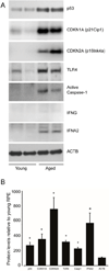
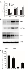
The tumor suppressor p53 is a major regulator of genes important for cell cycle arrest, senescence, apoptosis, and innate immunity, and has recently been implicated in retinal aging. In this study we sought to identify the genetic networks that regulate p53 function in the retina using quantitative trait locus (QTL) analysis. First we examined age-associated changes in the activation and expression levels of p53; known p53 target proteins and markers of innate immune system activation in primary retinal pigment epithelial (RPE) cells that were harvested from young and aged human donors. We observed increased expression of p53, activated caspase-1, CDKN1A, CDKN2A (p16INK4a), TLR4, and IFNα in aged primary RPE cell lines. We used the Hamilton Eye Institute (HEI) retinal dataset ( www.genenetwork.org ) to identify genomic loci that modulate expression of genes in the p53 pathway in recombinant inbred BXD mouse strains using a QTL systems biology-based approach. We identified a significant trans-QTL on chromosome 1 (region 172-177 Mb) that regulates the expression of Cdkn1a. Many of the genes in this QTL locus are involved in innate immune responses, including Fc receptors, interferon-inducible family genes, and formin 2. Importantly, we found an age-related increase in FCGR3A and FMN2 and a decrease in IFI16 levels in RPE cultures. There is a complex multigenic innate immunity locus that controls expression of genes in the p53 pathway in the RPE, which may play an important role in modulating age-related changes in the retina.
Figures


Similar articles
-
Ectopic AP4 expression induces cellular senescence via activation of p53 in long-term confluent retinal pigment epithelial cells.Exp Cell Res. 2015 Nov 15;339(1):135-46. doi: 10.1016/j.yexcr.2015.09.013. Epub 2015 Oct 9. Exp Cell Res. 2015. PMID: 26439195
-
Interferon-gamma induces cellular senescence through p53-dependent DNA damage signaling in human endothelial cells.Mech Ageing Dev. 2009 Mar;130(3):179-88. doi: 10.1016/j.mad.2008.11.004. Epub 2008 Nov 21. Mech Ageing Dev. 2009. PMID: 19071156
-
Transcriptional coactivator with PDZ-binding motif is required to sustain testicular function on aging.Aging Cell. 2017 Oct;16(5):1035-1042. doi: 10.1111/acel.12631. Epub 2017 Jun 14. Aging Cell. 2017. PMID: 28613007 Free PMC article.
-
Emerging roles of p53 and other tumour-suppressor genes in immune regulation.Nat Rev Immunol. 2016 Dec;16(12):741-750. doi: 10.1038/nri.2016.99. Epub 2016 Sep 26. Nat Rev Immunol. 2016. PMID: 27667712 Free PMC article. Review.
-
P53 and The Immune Response: 40 Years of Exploration-A Plan for the Future.Int J Mol Sci. 2020 Jan 15;21(2):541. doi: 10.3390/ijms21020541. Int J Mol Sci. 2020. PMID: 31952115 Free PMC article. Review.
Cited by
-
DeepTACT: predicting 3D chromatin contacts via bootstrapping deep learning.Nucleic Acids Res. 2019 Jun 4;47(10):e60. doi: 10.1093/nar/gkz167. Nucleic Acids Res. 2019. PMID: 30869141 Free PMC article.
-
Age-Related Macular Degeneration: A Disease of Cellular Senescence and Dysregulated Immune Homeostasis.Clin Interv Aging. 2024 May 23;19:939-951. doi: 10.2147/CIA.S463297. eCollection 2024. Clin Interv Aging. 2024. PMID: 38807637 Free PMC article. Review.
-
Does senescence play a role in age-related macular degeneration?Exp Eye Res. 2022 Dec;225:109254. doi: 10.1016/j.exer.2022.109254. Epub 2022 Sep 21. Exp Eye Res. 2022. PMID: 36150544 Free PMC article. Review.
-
Mechanisms of RPE senescence and potential role of αB crystallin peptide as a senolytic agent in experimental AMD.Exp Eye Res. 2022 Feb;215:108918. doi: 10.1016/j.exer.2021.108918. Epub 2022 Jan 2. Exp Eye Res. 2022. PMID: 34986369 Free PMC article.
-
Global Sensory Impairment in Older Adults in the United States.J Am Geriatr Soc. 2016 Feb;64(2):306-313. doi: 10.1111/jgs.13955. J Am Geriatr Soc. 2016. PMID: 26889840 Free PMC article.
References
-
- Baggetta R, De Andrea M, Gariano GR, Mondini M, Ritta M, Caposio P, Cappello P, Giovarelli M, Gariglio M, Landolfo S. The interferon-inducible gene IFI16 secretome of endothelial cells drives the early steps of the inflammatory response. Eur. J. Immunol. 2010;40:2182–2189. - PubMed
Publication types
MeSH terms
Substances
Grants and funding
LinkOut - more resources
Full Text Sources
Other Literature Sources
Medical
Molecular Biology Databases
Research Materials
Miscellaneous

