Fibroblast growth factor-21 enhances mitochondrial functions and increases the activity of PGC-1α in human dopaminergic neurons via Sirtuin-1
- PMID: 25932355
- PMCID: PMC4409609
- DOI: 10.1186/2193-1801-3-2
Fibroblast growth factor-21 enhances mitochondrial functions and increases the activity of PGC-1α in human dopaminergic neurons via Sirtuin-1
Abstract
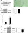
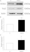
Mitochondrial dysfunctions accompany several neurodegenerative disorders and contribute to disease pathogenesis among others in Parkinson's disease (PD). Peroxisome proliferator-activated receptor γ coactivator-1α (PGC-1α) is a major regulator of mitochondrial functions and biogenesis, and was suggested as a therapeutic target in PD. PGC-1α is regulated by both transcriptional and posttranslational events involving also the action of growth factors. Fibroblast growth factor-21 (FGF21) is a regulator of glucose and fatty acid metabolism in the body but little is known about its action in the brain. We show here that FGF21 increased the levels and activity of PGC-1α and elevated mitochondrial antioxidants in human dopaminergic cells in culture. The activation of PGC-1α by FGF21 occurred via the NAD(+)-dependent deacetylase Sirtuin-1 (SIRT1) subsequent to an increase in the enzyme, nicotinamide phosphoribosyltransferase (Nampt). FGF21 also enhanced mitochondrial respiratory capacity in human dopaminergic neurons as shown in real-time analyses of living cells. FGF21 is present in the brain including midbrain and is expressed by glial cells in culture. These results show that FGF21 activates PGC-1α and increases mitochondrial efficacy in human dopaminergic neurons suggesting that FGF21 could potentially play a role in dopaminergic neuron viability and in PD.
Keywords: Dopaminergic neurons; FGF21; Mitochondria; PGC-1α; Parkinson’s disease; SIRT1.
Figures


Similar articles
-
Peroxisome proliferator-activated receptor-γ (PPARγ) agonist is neuroprotective and stimulates PGC-1α expression and CREB phosphorylation in human dopaminergic neurons.Neuropharmacology. 2016 Mar;102:266-75. doi: 10.1016/j.neuropharm.2015.11.020. Epub 2015 Nov 26. Neuropharmacology. 2016. PMID: 26631533
-
SIRT1 Protects Dopaminergic Neurons in Parkinson's Disease Models via PGC-1α-Mediated Mitochondrial Biogenesis.Neurotox Res. 2021 Oct;39(5):1393-1404. doi: 10.1007/s12640-021-00392-4. Epub 2021 Jul 12. Neurotox Res. 2021. PMID: 34251648
-
Fibroblast growth factor 21 regulates energy metabolism by activating the AMPK-SIRT1-PGC-1alpha pathway.Proc Natl Acad Sci U S A. 2010 Jul 13;107(28):12553-8. doi: 10.1073/pnas.1006962107. Epub 2010 Jun 28. Proc Natl Acad Sci U S A. 2010. PMID: 20616029 Free PMC article.
-
Role of mitochondria in diabetic peripheral neuropathy: Influencing the NAD+-dependent SIRT1-PGC-1α-TFAM pathway.Int Rev Neurobiol. 2019;145:177-209. doi: 10.1016/bs.irn.2019.04.002. Epub 2019 Jun 8. Int Rev Neurobiol. 2019. PMID: 31208524 Free PMC article. Review.
-
Deacetylation of PGC-1α by SIRT1: importance for skeletal muscle function and exercise-induced mitochondrial biogenesis.Appl Physiol Nutr Metab. 2011 Oct;36(5):589-97. doi: 10.1139/h11-070. Epub 2011 Sep 2. Appl Physiol Nutr Metab. 2011. PMID: 21888529 Review.
Cited by
-
Modulation of the Astrocyte-Neuron Lactate Shuttle System contributes to Neuroprotective action of Fibroblast Growth Factor 21.Theranostics. 2020 Jul 9;10(18):8430-8445. doi: 10.7150/thno.44370. eCollection 2020. Theranostics. 2020. PMID: 32724479 Free PMC article.
-
Long-term caloric restriction in ApoE-deficient mice results in neuroprotection via Fgf21-induced AMPK/mTOR pathway.Aging (Albany NY). 2016 Nov 29;8(11):2777-2789. doi: 10.18632/aging.101086. Aging (Albany NY). 2016. PMID: 27902456 Free PMC article.
-
The Roles and Pharmacological Effects of FGF21 in Preventing Aging-Associated Metabolic Diseases.Front Cardiovasc Med. 2021 Mar 31;8:655575. doi: 10.3389/fcvm.2021.655575. eCollection 2021. Front Cardiovasc Med. 2021. PMID: 33869312 Free PMC article. Review.
-
FGF21 activates AMPK signaling: impact on metabolic regulation and the aging process.J Mol Med (Berl). 2017 Feb;95(2):123-131. doi: 10.1007/s00109-016-1477-1. Epub 2016 Sep 27. J Mol Med (Berl). 2017. PMID: 27678528 Review.
-
Diagnostic Performance of Serum Biomarkers Fibroblast Growth Factor 21, Galectin-3 and Copeptin for Heart Failure with Preserved Ejection Fraction in a Sample of Patients with Type 2 Diabetes Mellitus.Diagnostics (Basel). 2021 Aug 30;11(9):1577. doi: 10.3390/diagnostics11091577. Diagnostics (Basel). 2021. PMID: 34573919 Free PMC article.
References
-
- Belluardo N, Wu G, Mudo G, Hansson AC, Pettersson R, Fuxe K. Comparative localization of fibroblast growth factor receptor-1,-2, and-3 mRNAs in the rat brain: in situ hybridization analysis. J Comp Neurol. 1997;379(2):226–246. doi: 10.1002/(SICI)1096-9861(19970310)379:2<226::AID-CNE5>3.0.CO;2-5. - DOI - PubMed
LinkOut - more resources
Full Text Sources
Other Literature Sources
Miscellaneous

